![]() |
miRBase |
![]() |
![]() |
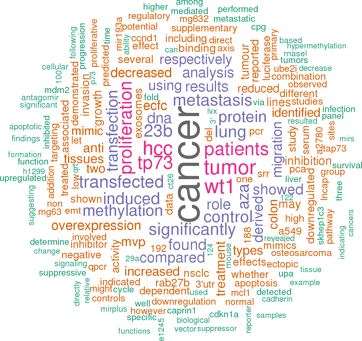
![]() 148 publications mentioning hsa-mir-193a (showing top 100)Open access articles that are associated with the species Homo sapiens and mention the gene name mir-193a. Click the [+] symbols to view sentences that include the gene name, or the word cloud on the right for a summary. |
|
1 |
![]()
Other miRNAs from this paper: hsa-mir-23a, hsa-mir-25, hsa-mir-147a, hsa-mir-7-1, hsa-mir-7-2, hsa-mir-7-3, hsa-mir-214, hsa-let-7i, hsa-mir-124-1, hsa-mir-124-2, hsa-mir-124-3, hsa-mir-130b, hsa-mir-362, hsa-mir-365a, hsa-mir-365b, hsa-mir-369, hsa-mir-338, hsa-mir-425, hsa-mir-429, hsa-mir-483, hsa-mir-572, hsa-mir-147b, hsa-mir-543
Treatment with 5-aza-dC upregulated miR-193a-3p expression, impaired cell proliferation ability, and promoted apoptosis in NSCLC cells via downregulation of one of miR-193a-3p targets, the antiapoptotic myeloid leukemia cell sequence-1 (Mcl-1) [11].
[score:11]
In particular, miR-193a-3p directly targeted JNK-1, a tyrosine kinase, since the ectopic expression of miR-193a-3p determined the dysregulation of cell cycle components including the decrease of CDK4, PIK3CA, and cyclin D1 and the overexpression of p27.
[score:9]
Recently, it has been demonstrated that miR-193a-3p acted as a negative regulator of urokinase-type plasminogen activator (uPA) in breast cancer and HCC cell lines and the high expression of miR-193a by mimics transfection strongly inhibited uPA expression and decreased cell aggressive properties [3, 32, 33].
[score:8]
Altogether, the data obtained by Khoo et al., by using the miRnome studies, in silico miRNA target database analyses combined with proteome arrays and luciferase reporter assays in miR mimic -treated ECFCs, allowed to identify the negative regulatory role of miR-193a-3p in the vascular function of these cells and in the proliferation and migration abilities of these cells via directly targeting the HMGB1 expression (Table 2).
[score:8]
By considering a small cohort of cancer patients (only 36 cases), Yi et al. found that miR-193a-3p is upregulated in 24/36 esophageal squamous cell carcinoma (ESCC) tissues compared with adjacent normal tissues and the downregulation of miR-193a-3p by a synthesized inhibitor decreases migration and proliferation and promotes apoptosis in ESCC cells [25].
[score:8]
Treatment with the inhibitor of DNA methylation 5-azacytidine (5-aza-dC) restored miR-193a-3p expression and reduced its target, the oncogene c-kit.
[score:7]
In addition, miR-193a-3p is found downregulated in transformed cells and its downregulation seemed to be required for cellular transformation in two isogenic mo dels (breast epithelial cells and fibroblasts) [3].
[score:7]
In human thyroid carcinoma WRO cells and MRO cells, the gene silencing of XB130 by stable transfection of short hairpin increased both pri-miR-193a and its mature form (miR-193a-3p), while the ectopic expression of XB130 induced their downregulation [7].
[score:6]
Interestingly, Linc00152 competitively bound miR-193a-3p inducing the upregulation of its ERBB4 target and the consequent activation of AKT signaling pathway which, in turn, conferred resistance [17].
[score:6]
On the contrary, MVP knockout determined miR-193a-3p accumulation in tumor cells triggering the inhibition of cell proliferation and cell cycle G1 arrest due to miR-193a-3p binding to its target, caprin-1 [62].
[score:6]
Similarly, Li et al. revealed that the downregulation of miR-193a-3p was strongly associated with fusion protein AML1/ETO expressed in hematopoietic cells isolated from patients affected by acute myeloid leukemia (AML) with t(8;21).
[score:6]
miR-193a-3p seemed to induce antiapoptotic signals in 5-Fu resistant cells by suppressing SRSF2, a splicing factor that preferentially upregulates the proapoptotic form of caspase 2 (CASP2L) to the antiapoptotic form CASP2S [15].
[score:6]
The overexpression of miR-193a-3p not only downregulated K-Ras but also reverted the whole protein signature associated with the signaling downstream of K-Ras identified by proteomic analysis of lung cancer samples.
[score:6]
The lacking of HNF4 α expression in the liver of young adult mice (Hnf4α-Liv KO) determined the downregulation of some miRs, including miR-193a that is in cluster with miR-365 on the chromosome 11 of Mus musculus [5].
[score:6]
Furthermore, the overexpression of miR-193a-3p inhibited migration, invasion, and epithelial mesenchymal transition in vitro and impaired the formation of metastasis in vivo.
[score:5]
In particular, the different expression levels of these miRNAs (including the downregulation of miR-193a-3p) were identified in HBV patients who developed HCC (preclinical HCC patients) compared to the HBV group that did not develop HCC [57].
[score:5]
Furthermore, the low expression of miR-193a-3p was significantly related to reduced overall survival (OS) and disease-free survival (DFS) of HCC patients indicating its possible prognostic role in this cancer type [14].
[score:5]
In the same clinical context, miR-193a-3p has been also found to directly regulate the expression of DNMT3a, HDAC3, and cyclin D1 consequently blocking the cell cycle progression during granulopoiesis and inducing the differentiation of myeloid precursors [4].
[score:5]
Conversely, miR-193a-3p was expressed at low amount in the proliferative CB-ECFCs with its in vitro ectopic overexpression limiting the proliferation and the cell cycle progression of these cells and consequent reduction of their vascular tubule formation and cell migration.
[score:5]
For hsa-miR-193a-3p, TargetScan 7.1 predicted 293 putative target genes with 168 displaying the highest score (cumulative weighted context++ score < −0.24).
[score:5]
In conclusion, it has been proved that DNA hypermethylation of CpG island associated to MIR193A gene was responsible for miR-193a-3p downmodulation in certain types of cancer which in turn led to increased expression levels of miR-193a-3p targets involved in cell malignant behavior.
[score:5]
Interestingly, miR-193a-3p was reported as downregulated in BRAF mutation with respect to wild-type melanoma [49] suggesting that miR-193a-3p may have a role in BRAF -associated events.
[score:5]
Interestingly, Linc00152 and UCA1 functioned as miRNA sponges and suppressed the endogenous effect of miR-193a-3p by silencing the miR target ERBB4.
[score:5]
In chemoresistant BCa cells, miR-193a-3p decreased the expression of the following targets: SRSF2, PLAU, HIC2 [40], LOXL4 [41], ING-5 [42], HOXC9 [43], and PSEN-1 [44].
[score:5]
As indicated in Figure 2, the expression of miR-193a (without discriminating between miR-193a-3p and miR-193a-5p) has been detected in all tissues, with the exception of the bladder, some brain components (hippocampus, nucleus accumbens, and spinal cord), and cervix (endocervix) where miR-193a expression was not found.
[score:5]
To the best of our knowledge, all published data pointed toward a role of miR-193a-3p as tumor suppressor miRNA (ts-miRNA) in both solid and liquid cancers since it impaired tumor cell aggressive properties by targeting oncogenes.
[score:5]
Moreover, miR-193a-3p repressed cell proliferation of AML cells through the inhibition expression of c-kit, an oncogene encoding a transmembrane glycoprotein belonging to the type III receptor tyrosine kinase family [9].
[score:5]
It has also been shown that miR-193a-3p decreased the abilities of proliferation by the expression inhibition of some TFs, including E2F6, and other genes involved in the growth of several cancer types, for example, K-Ras, ERBB4, and cyclin D1 [3, 8, 27, 28].
[score:5]
Using chromatin immunoprecipitation (ChIP) and siRNA -mediated inhibition, they demonstrated that Max and RXR α bound directly to the miR-193a regulatory region and repressed its transcription in ER-Src-transformed cells [3].
[score:5]
Consistent with the role of ts-miRNA, miR-193a-3p was found downregulated in the majority of primary cancer tissues, such as HCC [14, 47], NSCLC [37, 48], MPM [13], and AML [4, 9].
[score:4]
Further, these data demonstrate hormonal regulation in miRNA (i. e., miR-193a-3p) expression in a human endometrium.
[score:4]
2. The Regulation of miR-193a-3p Expression.
[score:4]
In malignant pleural mesothelioma (MPM), miR-193a-3p was inhibited when compared to normal pleura, but the DNA hypermethylation of miR-193a -associated CpG island was not responsible for the inhibition of miR-193a-3p in MPM cells as verified by MSP [13].
[score:4]
Different factors can contribute to the regulation of MIR193a expression, including TFs, DNA methylation and, at posttranscriptional level, ceRNAs.
[score:4]
Similarly, Pu et al. reported that miR-193a-3p and miR-193a-5p were downregulated in osteosarcoma cells defined as highly tumorigenic and metastatic (MG63.2) in respect to less metastatic parental MG63 cell line.
[score:4]
Furthermore, the authors identified Rab27, a member of the RabGTPase family, as a direct target of miR-193a-3p.
[score:4]
Finally, Pu et al. found that both miR-193a-3p and miR-193a-5p were hypermethylated and downregulated in a metastatic osteosarcoma cell line [12].
[score:4]
In association to miR-124 and miR-147, miR-193a-3p has been shown to coregulate and inhibit G1/S transition and proliferation in breast cancer and glioblastoma cell lines [26].
[score:4]
The findings obtained using a genomic profiling of miRNAs and mRNAs clearly showed that miR-193a-3p is one of the 12 miRs found to be upregulated in the midsecretory phase samples.
[score:4]
In particular, downregulation of miR-193a-3p has been associated with events contributing to the myofiber alterations of patients with myotonic dystrophy type 2 (DM2, OMIM 602688) [22].
[score:4]
Iliopoulos et al. showed that the downregulation of miR-193a-3p was driven by Max, the Myc -associated factor X, and RXR α, a nuclear receptor, both involved in the processes causing cellular transformation of breast epithelial cells.
[score:4]
In particular, Kuokkanen et al. provided strong evidence of the hormonal regulation of miR-193a-3p expression in isolated uterine epithelial cells derived from midreproductive aged women [23].
[score:4]
The direct binding of miR-193a-3p and Mcl-1 has also been demonstrated in human ovarian cancer cell line where the overexpression of miR-193a-3p induced the activation of caspase 3/7 and resulted in apoptotic cell death [31].
[score:4]
Regarding the biological function in physiological contexts, certain types of human normal cells seem to require high expression level of miR-193a-3p when they do not need to proliferate.
[score:3]
When considering the miRNA target genes validated in different cancers, the role of miR-193a-3p in affecting genes with the consequence of promoting apoptosis stood out.
[score:3]
Similarly, miR-193a-3p expression was found increased by DNA hypomethylation in HCC cells presenting resistance to 5-fluorouracil (5-Fu).
[score:3]
In severe disease, the high level of miR-193a-3p into the exosomes led to the reduction of cytoplasmatic miR-193a-3p that in turn promoted the progression of premetastatic cells to metastatic ones.
[score:3]
By considering all experimental validation studies in cancer, genes targeted by miR-193a-3p are involved in several biological processes, including proliferation, apoptosis, migration, and metastasis.
[score:3]
Finally, we highlighted the aberrant expression of miR-193a-3p both at tissue and at circulating levels in several pathological conditions, including cancer, in order to offer novel insights in the role of miR-193a-3p as a diagnostic and prognostic biomarker.
[score:3]
These intriguing data will require further investigations since it is not clear how the overexpression of miR-193a-3p can dictate chemoresistance even if its tumor suppressor functions have been well established in many primary cancers.
[score:3]
Gao et al. demonstrated that the promoter hypermethylation repressed miR-193a-3p expression in acute myeloid leukemia (AML).
[score:3]
The functional annotation analysis conducted using DAVID 6.8 on the candidate target genes of miR-193a-3p highlighted 8 biological KEGG pathways overrepresented as statistically significant (p < 0.05).
[score:3]
6. Gene Annotation Analysis on Predicted and Experimentally Validated miR-193a-3p Targets.
[score:3]
Kwon et al. demonstrated that miR-193a-3p expression was induced by ionizing irradiation in U-251 glioma cells and HeLa cells.
[score:3]
In addition, among the 24 experimentally validated and published miR-193a-3p targets (Table 2), the KEGG pathway enrichment analysis outlines 6 terms (p < 0.05): acute myeloid leukemia, chronic myeloid leukemia, microRNA in cancer, proteoglycans in cancer, Erb signaling pathway, pancreatic cancer, and pathways in cancer.
[score:3]
To provide complete information on the global expression profile of miR-193a-3p in normal tissues, we referred to data deposited in Genome Browser.
[score:3]
5. miR-193a-3p Functions as Tumor Suppressor miRNA in Cancer.
[score:3]
The authors clearly determined the effects of miR-193a-3p on the cell aggressive properties via the targeting of K-Ras.
[score:3]
The expression of miR-193a-3p and miR-193a-5p was positively associated with cellular invasion and migration by assessing human lung cancer cell with high metastatic potential (SPC-A-1sci) previously established from weakly metastatic cell (SPC-A-1) through in vivo selection in NOD/SCID mice [38].
[score:3]
In the present report, we focused on miR-193a-3p because several findings support its role as tumor suppressor miR both in solid and in liquid tumors leading to believe that the detection of this miR at tissue and/or circulating level may be employed as a diagnostic and prognostic biomarker for certain types of tumors.
[score:3]
The ability of miR-193a-3p to promote apoptosis was further demonstrated in MPM xenograft mo dels when targeted by miR mimics delivered using EDV nanocells, bacterially-derived minicells that can be packaged with a variety of cargoes and be delivered to tumors via bispecific antibodies attached on surface [13].
[score:3]
We think that these findings are extremely interesting, but further studies are needed in order to support the detection of circulating miR-193a-3p as a noninvasive biomarker for these diseases.
[score:3]
3. Expression Profile of miR-193a in Human Normal Tissues.
[score:3]
The authors found that overexpression of MVP (major vault protein) transported miR-193a-3p from the tumor cells to exosomes.
[score:3]
As indicated in Figure 3(a), miR-193a-3p seems to be implicated in the activation of drug resistance pathways via repressing different targets.
[score:3]
In summary, the overexpression of miR-193a-3p in cultured normal cells derived from physiological contexts, in particular in PB-ECFC cells and in midsecretory uterine epithelial cells, seemed to limit cell proliferation and cell cycle progression.
[score:3]
Interestingly, Salvi et al. showed that the combination of miR-193a-3p mimics and sorafenib had additional effects on HCC inhibition of cell proliferation and induction of apoptosis suggesting that miR-193a-3p could also play an important role in promoting the sensitivity to sorafenib [32], the only innovative drug used for advanced HCC.
[score:3]
On the other hand, the local delivery of anti-miR-193a-3p molecules could be an effective intervention for local ischemic diseases.
[score:3]
Adipose and breast tissues displayed the highest miR-193a expression level.
[score:3]
Correspondingly, the inhibition of miR-193a-3p by antagomiR transfection in MG63 cells induced invasive properties.
[score:3]
In addition, the overexpression of Linc00152 or UCA1 increased cell growth through modulation of ERBB4 while this effect was attenuated by transfection of miR-193a-3p mimics in both cell lines.
[score:3]
However, the lack of data (KO, KI, and conditional KD) in the development of an organism (i. e., Mus musculus and Danio rerio) carrying the ortholog MIR193A (Gene Card data) has prevented the proposal of hypothesis on the role of miR-193a-3p during development [24].
[score:3]
The abilities of MG63.2 cells to invade and migrate resulted decreased by restoring the miR-193a-3p expression level using transient transfection of miR mimics.
[score:3]
In NSCLC, the expression of miR-193a-3p was negatively correlated to tumor size, lymph node metastasis (LNM), and TNM stages.
[score:3]
Recently, Yu et al. validated miR-193a-3p as a negative regulator of ErbB4, belonging to the ErbB family of tyrosine kinase receptors and the ribosomal protein S6K2, both playing a critical role in cell movement, growth, and development [37].
[score:3]
The alteration of these factors is context dependent and determines the aberrant expression of miR-193a-3p in cancer.
[score:3]
Dysregulation of miR-193a-3p in Cancer Tissues.
[score:2]
From a general point of view, miR-193a-3p had been studied in Homo sapiens and, to the best of our knowledge, no information is available in development.
[score:2]
Several mechanisms, including transcription factors, DNA methylation, and competing endogenous RNAs (ceRNAs), have been reported to be involved in the dysregulation of miR-193a-3p in pathological contexts (Table 1).
[score:2]
Other two factors, hepatocyte nuclear factor α (HNF4 α) and XB130, may also have a relevant role in the regulation of miR-193a-3p.
[score:2]
The elucidation of these mechanisms may allow extending our knowledge on the level of miR-193a-3p dysregulation.
[score:2]
These evidences strongly suggested that the ceRNA regulatory network should be considered as a mechanism involved in the dysregulation of miR-193a-3p.
[score:2]
However, these results do not permit to outline a hypothesis on the functional role in normal myofibers of miR-193a-3p as well as those of the other 10 miRNAs deregulated in DM2 muscle biopsies.
[score:2]
4. Biological Function of miR-193a-3p in Development and in Cell Physiology.
[score:2]
In particular, it has been demonstrated that miR-193a-3p negatively regulated K-Ras in lung cancer cells by binding two 3′UTR sites that have not been reported previously to be mutated in cancer.
[score:2]
These evidences unmistakably suggest a multifactorial regulation of miR-193a-3p at transcriptional or posttranscriptional level with the possibility of a context -dependent activation of specific mechanisms.
[score:2]
In regard to circulating miR-193a-3p, the use of next-generation sequencing and qPCR revealed different levels of this miR in patients with schizophrenia or with Parkinson's disease (PD) when compared with control subjects.
[score:2]
These data indicated that the regulation of miR-193a-3p may be mediated by HNF4 α and XB130 in a healthy liver and thyroid carcinoma, respectively.
[score:2]
Many studies have confirmed that miR-193a-3p has a significant role in the regulation of cancer cell growth.
[score:2]
Consistently with the competing endogenous RNA role of lncRNAs, two different ceRNAs have been found to target miR-193a-3p where the physical association with mature miR-193a-3p has been demonstrated by RNA immunoprecipitation (RIP) and luciferase reporter assays.
[score:2]
miR-193a-3p negatively regulated Mcl-1 and promoted apoptosis by inducing ROS accumulation and DNA damage [30].
[score:2]
In particular, it has been found that miR-193a-3p was one of the 25 miRNAs differentially regulated in CB-ECFC versus PB-ECFC [21].
[score:2]
For skeletal muscle, a miRNA profiling approach combined with bioinformatics analyses and qPCR experimental validation has identified 11 miRNAs including miR-193a-3p involved in the homeostasis of normal myofibers.
[score:1]
miR-193a was also found tumor specifically methylated in patients with non-small-cell lung cancer (NSCLC) [10].
[score:1]
These results would point toward a variable miR-193a dependence on CpG DNA methylation in HCC.
[score:1]
5.4. miR-193a-3p Modulates Drug Resistance in Cancer Cells.
[score:1]
Oh et al. demonstrated that the exosomes containing miR-193a-3p were able to induce differentiation of F11 cells (rat dorsal root ganglion and mouse neuroblastoma hybrid cells).
[score:1]
The findings reported by Greco et al. clearly showed that the level of miR-193a-3p downmodulation contributed to the DM2 miRNA score allowing to distinguish the muscle specimens of DM2 patients from those of controls.
[score:1]
These findings may pave the way to further studies aimed to elucidate the possible use of miR-193a-3p for experimental therapeutic procedures.
[score:1]
Very little is known on the biological function of miR-193a-3p in cell physiology.
[score:1]
miR-193a-3p is highly conserved across several Hominidae (humans, chimpanzees, orangutans, and rhesus) and other mammals (Mus musculus, Bos taurus, and Canis familiars), as indicated in the microRNA viewer database (last update February 28, 2012) [2].
[score:1]
The authors indicated that miR-193a-3p may be used to discriminate between ESCC cases and healthy controls with high sensitivity and specificity in a cohort of 63 patients and 63 controls.
[score:1]
Regarding cancer, circulating miR-193a-3p levels were found increased in many malignancies.
[score:1]
In orthotopic xenograft K-Ras-mutated lung tumor mo dels, miR-193a-3p encapsulated in DOPC nanoliposomes showed a reduction of tumor growth and metastasis at various sites [29].
[score:1]
7. miR-193a-3p as Diagnostic and Prognostic Biomarker.
[score:1]
The MIR193A gene is embedded in a 1556 bp CpG island that counts 196 CpG sites (Figure 1(a)).
[score:1]
Consequentially, the different sequence that characterizes miR-193a-3p and miR-193a-5p determines distinct target sets for each miRNA.
[score:1]
5.1. miR-193a-3p Limits Cancer Cell Proliferation and Impairs Cell Cycle Progression.
[score:1]
Taken together, these evidences strongly suggest a potential role of miR-193a-3p as a metastasis-preventing miRNA.
[score:1]
The pre-miR-193a generates two mature miRNAs, miR-193a-3p and miR-193a-5p, depending on the arm that is processed during miRNA biogenesis (Figure 1(b)).
[score:1]
In addition, the protein profile of SPC-A-1sci cells stably transfected with miR-193a-3p has been determined by using a proteomic approach (iTRAQ and Nano LC-MS/MS) followed by DAVID (database for annotation, visualization, and integrated discovery; http://david.
[score:1]
Interestingly, in a retrospective longitudinal phase 3 biomarker study, a set of 5 serum miRNAs (miR-193a-3p, miR-369-5p, miR-672, miR-429, and let-7i∗) was identified as a specific biomarker for the surveillance and preclinical screening of HCC in a high-risk population of patients infected by hepatitis B virus (HBV).
[score:1]
In addition, the release of lactate dehydrogenase (LDH) from MPM cells transfected with miR-193a-3p could suggest that miR-193a-3p induced cell death at least in part by the induction of necrosis.
[score:1]
Circulating miR-193a-3p Levels in Pathological Conditions.
[score:1]
By comparing two independent miRNA microarrays, one in tissue and one in blood of colorectal cancer patients, Yong et al. identified higher levels of miR-193a-3p (in combination to miR-23a and miR-338-5p) in cancer patients, and the positive correlation was demonstrated between tissue and blood samples [53].
[score:1]
In this pathological context, AML1/ETO acted as a transcriptional repressor by localizing the AML1 binding site on the MIR193a upstream region and recruiting HDAC and DNMTs.
[score:1]
In particular, oncogenic Linc00152 (long intergenic noncoding RNA 152) and LncRNA-UCA1 (urothelial carcinoma -associated 1) competitively bind miR-193a-3p in colon cancer and NSCLC cell lines, respectively [17, 18].
[score:1]
In this review, we focused on the human miR-193a-3p since the increasing number of evidences has described its importance in several biological functions.
[score:1]
Furthermore, miR-193a-3p could also be used to distinguish the HY-stage1 in PD patients from healthy controls [51].
[score:1]
The high-throughput TaqMan low-density array (TLDA) combined with qPCR validation allowed to establish the high level of miR-193a-3p included in two different five-serum miRNA panels, either in renal cell carcinoma (RCC) or in NSCLC patients.
[score:1]
In this regard, the delivery of miR-193a-3p mimics by nanosized particles could represent a novel therapeutic tool for the treatment of cancer since it may hamper tumor aggressive properties in tumor xenograft mo dels by restoring the miR original levels.
[score:1]
Several studies have found that altered DNA methylation occurring in the CpG sites of the miR-193a promoter in different types of tumors.
[score:1]
These results indicated the negative impact of miR-193a-3p on cancer invasion by repressing Rab27B and its downstream pathways in osteosarcoma cells [12].
[score:1]
5.3. miR-193a-3p Impairs Cancer Migration, Invasion, and Metastasis.
[score:1]
For these reasons, they described miR-193a-3p as an oncogenic miRNA in ESCC and suggested further studies to define the controversial role of miR-193a-3p in ESCC.
[score:1]
Interestingly, miR-193a-3p decreased cell cycle progression (G1-S) and cell proliferation in vitro and blocked colony formation in three-dimensional cultures.
[score:1]
The transcription silencing by specific TFs was reported to play a critical role in the inactivation of miR-193a-3p in different pathological contexts.
[score:1]
By analyzing the region of 2000 bp spanning MIR193a, a typical CpG island is identified (chr17:31558803-31560358) in which miR-193a coding sequence is embedded.
[score:1]
This bioinformatics analysis underscored once again the key role of miR-193a-3p in cancer contexts.
[score:1]
However, the miR-193a-3p CpG sites resulted methylated in the differentiated HepG2 cells and the treatment with 5-aza-dC led to miR-193a-3p increasing as observed also by Ma and colleagues [14, 15].
[score:1]
In support of this notion, Yue et al. reported that the reduced activity of miR-193a-3p caused by the sponge effect of Linc00152 was related to oxaliplatin (L-OHP) resistance in colon cancer both in vitro and in vivo (Figure 3(b)).
[score:1]
In this review, we focused on the miR-193a-3p biological and molecular mechanisms both from the physiological and the pathological perspectives.
[score:1]
Furthermore, the transfection with miR-193a-3p mimics in MPM cells reduced Mcl-1 protein level and increased the number of late apoptotic cells.
[score:1]
It was clearly demonstrated by ROC analysis that the 5-miRNA -based panels (miR-193a-3p, miR-362, miR-572, miR-425-5p, and miR-543) had a high sensitivity and specificity in the discrimination of patients with early-stage RCC from healthy controls [54].
[score:1]
By using the same experimental approaches (TLDA followed by qPCR), Wu et al. identified significantly elevated levels of miR-193a-3p in sera from patients with esophageal squamous cell carcinoma (ESCC).
[score:1]
The authors demonstrated that the downmodulation of miR-193a-3p in HCC was not mediated by DNA methylation in a cohort of 30 matched peritumoral and HCC tissues from bioptic samples.
[score:1]
Interestingly, cells derived from a 4D mo del treated with miR-193a-3p showed less proliferation ability than those from untreated mo del.
[score:1]
In addition, the modulation of miR-193a-3p level by the injection of antagomir or agomir molecules in either resistant or sensitive BCa cells reversed the chemoresistance in tumor xenografts nude mice.
[score:1]
Interestingly, MIR193a is found internal to a sequence that displays a high level of enrichment of H3K27Ac, H3K4Me1, and H3K4Me3 histone marks.
[score:1]
5.2. miR-193a-3p Induces Cell Death Mainly by Promoting Apoptosis.
[score:1]
The use of 1,2-dioleoyl-sn-glycero-3-phosphocholine (DOPC) nanoliposomes to deliver miR-193a-3p reduced the number of viable cells and impaired the presence of cancer cells in contralateral lobe that is indicative of metastasis formation in this mo del.
[score:1]
Finally, compelling evidences indicated that miR-193a-3p is detectable not only in primary cancer tissues but also at circulating level (in exosomes or not) in cancer patients indicating the possible diagnostic and prognostic value of miR-193a-3p.
[score:1]
In NSCLC, the effectiveness of the 5-miRNA panel (miR-193a-3p, miR-483-5p, miR-214, miR-25, and miR-7) in discriminating cancer patients from normal subjects was confirmed in a multiethnic and multicentric study in which 438 participants from both China and America were enrolled (221 NSCLC patients, 161 normal controls, and 56 benign nodules) [55].
[score:1]
Further studies are really necessary to verify whether the detection of miR-193a-3p may be helpful for the characterization of these two diseases.
[score:1]
The chromatin remo deling complex formed by AML1/ETO and the DNA hypermethylation triggered the silencing of miR-193a-3p in t(8;21) AML [4].
[score:1]
Teng et al. demonstrated that miR-193a-3p was present into exosomes obtained from tissue and cell culture media and serum, derived from primary mouse colon tumors and human liver metastasis of colon cancer.
[score:1]
The level of circulating miR-193a-3p was reduced after ESCC surgical removal indicating that this miR may have been originally secreted by the tumor cells [56].
[score:1]
It is worth to outline that the functional role of a given miR can be tissue- or tumor-type dependent and that few data indicate a possible oncogenic role of miR-193a-3p in ESCC; nevertheless, we think that only strong clinical evidence, as well as biological studies of the miR mimics on the proliferation of ESCC cell lines will elucidate this possible role of miR-193a-3p in this specific context of human cancer.
[score:1]
[1 to 20 of 148 sentences]
|
2 |
![]()
Other miRNAs from this paper: hsa-mir-15a, hsa-mir-31, mmu-mir-125a, mmu-mir-132, mmu-mir-134, mmu-mir-149, mmu-mir-181a-2, mmu-mir-193a, mmu-mir-200b, hsa-mir-181a-2, hsa-mir-181a-1, hsa-mir-200b, hsa-mir-132, hsa-mir-125a, hsa-mir-134, hsa-mir-149, mmu-mir-200a, mmu-mir-15a, mmu-mir-31, mmu-mir-135b, hsa-mir-200c, mmu-mir-200c, mmu-mir-181a-1, hsa-mir-200a, hsa-mir-135b
Having shown that miR-193a negatively regulates WT1 expression and inhibits metastasis, we directly address the role of WT1 downregulation in the anti-metastasis activity.
[score:10]
MiR-193a was reported to be a suppressive miRNA and overexpression of miR-193a suppressed proliferation and promoted apoptosis via targeting several oncogenes, such as c-KIT [23] and MCL-1 [24].
[score:9]
was performed using Student’s t-test To determine potential target genes by miR-193a, several miRNAs predicting software including miRwalk, TargetScan, and PicTar were used to predict possible target genes.
[score:7]
The further decrease of miR-193a by TGF-β loses the ability of suppressing WT1 and finally enhances the expression of WT1, which leads to the decreased expression of E-cadherin (Additional file 6: Figure S4B).
[score:7]
Because miR-193a expression was more decreased in lung cancer cells with greater migration and invasion abilities, we speculated that TGF-β1 inhibited the expression of miR-193a.
[score:7]
Hypermethylation of miR-193a leads to the low expression of miR-193a, which loses the ability to inhibit WT1 expression.
[score:7]
In addition to WT1, miR-193a targeted several important oncogene, such as Yin Yang1 (YY1) [33], phosphoinositide-3-kinase regulatory subunit (3PIK3R3), and the mammalian target of rapamycin (mTOR) [34].
[score:6]
Overexpression of miR-193a significantly decreased the protein levels of WT1, but increased the expression of the epithelial marker E-cadherin (Fig. 3d), which was reported to be negatively regulated by WT1 in NSCLC [15].
[score:6]
Ectopic overexpression of WT1 partially reverses miR-193a -induced inhibition of metastasis.
[score:5]
Overexpression of miR-193a inhibited migration and invasion via modulating WT1-E-cadherin axis.
[score:5]
As expected, TGF-β1 reduced the expression of miR-193a by 30 %–50 % (Fig. 6f) and subsequently increased the expression of WT1 in A549 and H1299 cells (Fig. 6g).
[score:5]
To confirm the low expression of miR-193a in lung cancer cells, the relative expression of miR-193a was detected in lung cancer tissues and adjacent non-tumor tissues.
[score:5]
To select the cells stably expressing miR-193a, 2 μg/ml puromycin (Medchemexpress, Princeton, USA) was added into the supernatant for 1 week.
[score:5]
Further, overexpression of WT1 partially prevented miR-193a -induced growth inhibition (Fig. 5d and e).
[score:5]
MiR-193a inhibited the metastasis of lung cancer cells by deregulating the expression of tumor-related proteins [26].
[score:5]
Further, overexpression of miR-193a decreased the colony formation (Fig. 2e) and suppressed cell proliferation (Fig. 2f).
[score:5]
Thus, restoring the expression of miR-193a or decreasing the expression of WT1 might provide alternative therapeutic strategy for NSCLC patients.
[score:5]
Ectopic expression of miR-193a inhibited cell proliferation, colony formation, migration, and invasion in A549 and H1299 cells.
[score:5]
Because WT1 is implicated in the metastasis of NSCLC through inhibiting the expression of E-cadherin [15], we hypothesized that one mechanism of anti-metastasis activity of miR-193a might perform by modulating WT1-E-cadherin axis.
[score:5]
Consistently, ectopic overexpression of miR-193a inhibits metastasis and the EMT-like conversion in NSCLC.
[score:5]
Ectopic expression of miR-193a inhibits metastasis in lung cancer cells.
[score:5]
Similarly, besides miR-193a, miR-125a was identified to suppress WT1 expression via binding 3′UTR of WT1 in myeloid leukemia cells [51].
[score:5]
Consistent with the results from cell lines, the protein levels of WT1 were significantly decreased but E-cadherin expressions were increased in two tumors obtained from miR-193a -overexpressed mice in comparison with control mice (Fig. 7e).
[score:5]
Thus, miR-193a might have multiple different mRNA targets other than WT1, and these additional targets may also play important roles in carcinogenesis.
[score:5]
Furthermore, overexpression of WT1 partially prevents miR-193a -induced inhibition of migration and invasion, suggesting that WT1 plays an important role in the anti-metastasis by miR-193a.
[score:5]
For example, miR-193a inhibited invasion by negatively regulating ERBB4/PIK3R3/mTOR/S6K2 signaling pathway in NSCLC [25].
[score:4]
Decreased expression of miR-193a regulated TGF-β1 -induced EMT progress.
[score:4]
Consistently, miR-193a-3p, the another product of pre-miR-193a, was also downregulated in lung cancer [25, 34].
[score:4]
Downregulation of miR-193a in lung cancer tissues.
[score:4]
Indeed, many studies have indicated that miR-193a was down-regulated in various types of cancers [32– 34].
[score:4]
Overexpression of miR-193a decreased the luciferase activities by approximately 40 %, which were almost abolished by the mutation in putative miR-193a -binding sites (Fig. 3b).
[score:4]
However, whether miR-193a can regulate E-cadherin expression is not determined.
[score:4]
Importantly, knockdown of WT1 resembled the anti-cancer activity by miR-193a and overexpression of WT1 partially reversed miR-193a -induced anti-cancer activity, indicating that WT1 plays an important role in miR-193a -induced anti-cancer activity.
[score:4]
f and g Cell proliferation was detected by CCK-8 assay in A549 (f) and H1299 (g) cells transfected with sh-NC or sh-WT1 for 24, 48, and 72 h. * P < 0.01 versus sh-NC Because miR-193a inhibited metastasis via targeting WT1 protein, which acts as an oncogene in multiple types of cancers including lung cancer, we are interested in examining whether WT1 counteracts miR-193a -induced anti-metastasis.
[score:4]
MiR-193a directly targets WT1 and indirectly modulates E-cadherin.
[score:4]
Mechanistically, miR-193a reduced the expression of WT1, which negatively regulated the protein level of E-cadherin, suggesting that miR-193a might prevent EMT via modulating WT1-E-cadherin axis.
[score:4]
Collectively, our results have revealed an important role of miR-193a-WT1-E-cadherin axis in metastasis, demonstrated an important molecular cue for EMT, and suggested a therapeutic strategy of restoring miR-193a expression in NSCLC.
[score:3]
Our finding that miR-193a inhibits TGF-β -induced EMT through WT1-E-Cadherin axis augments the role of miRNAs in EMT of NSCLC.
[score:3]
Finally, overexpression of miR-193a decreased the growth of tumor xenografts in mice.
[score:3]
Our results indicate that decreased expression of miR-193a contributes to proliferation, metastasis, and TGF-β -induced EMT through WT1-E-cadherin pathway.
[score:3]
Human WT1 CDS including the predicted miR-193a target sites was amplified by PCR and cloned into pMIR-REPORT vector named as pMIR-WT1CDS (Ambion, Dallas, USA).
[score:3]
Correspondingly, overexpression of WT1 partially reversed the anti-migration (Fig. 5b) and anti-invasion (Fig. 5c) induced by miR-193a.
[score:3]
Therefore, our results suggest that miR-193a exerts tumor-suppressive effects through WT1-E-cadherin pathway.
[score:3]
A statistically significant correlation between miR-193a versus WT1 expression was observed by Pearson’s method.
[score:3]
TGF-β1 -induced EMT is attenuated by the overexpression of miR-193a.
[score:3]
d WT1 and E-cadherin expressions were performed in A549 and H1299 cells transfected with lentivirus LVX-miR-193a or LVX-NC.
[score:3]
n = 62. g The plotting of miR-193a versus WT1 expression showed an inverse correlation between them.
[score:3]
Finally, IHC staining indicated that the expression of WT1 was decreased while E-cadherin was increased in tumor xenograft from mice inoculated with A549-miR-193a than that inoculated with A549-miR-NC (Fig. 7f).
[score:3]
MiR-193a was downregulated in about 81 % of tumors (P < 0.01, 50 of 62 patients), with about 40 % reduction relative to their adjacent non-tumor tissues by qRT-PCR (Fig. 1b).
[score:3]
Recently, miR-193a has been reported to be a suppressive miRNA in multiple types of cancers, but its underlying anti-oncogenic activity in non-small cell lung cancers (NSCLC) is not fully elucidated.
[score:3]
TGF-β1 -induced EMT was partially prevented by the overexpression of miR-193a (Fig. 6d and e).
[score:3]
Here, we report that miR-193a -mediated pathway may take part in TGF-β -induced inhibition of E-cadherin (Additional file 6: Figure S4A).
[score:3]
Restoration of miR-193a inhibits migration, invasion, and TGF-β1 -induced EMT through modulating WT1-E-cadherin axis.
[score:3]
As shown in Additional file 4: Figure S2A-C, overexpression of WT1 CDS (Mut) did not prevent the anti-migration and anti-invasion induced by miR-193a.
[score:3]
We further analyzed the relationship between WT1 and miR-193a expressions.
[score:3]
Importantly, a significant inverse correlation between miR-193a and WT1 mRNA expressions was found in lung cancer tissues (Fig. 3g).
[score:3]
The expression of miR-193a in lung cancer tissues was decreased comparing to adjacent non-tumor tissues due to DNA hypermethylation in lung cancer tissues.
[score:3]
However, miR-193a minics failed to decrease WT1 expression in cells transfected with pMIR-WT1CDS (Fig. 3c, Right).
[score:3]
The mutation on miR-193a -binding sites in human WT1 CDS was generated by the site-directed mutagenesis kit (Agilent Technologies, Palo Alto, USA).
[score:3]
Relationship between the expression of miR-193a and clinicopathologic parameters.
[score:3]
f and g The expressions of miR-193a (f) and WT1 (g) were measured in A549 and H1299 cells treated with TGF-β1 for 3 days by qRT-PCR and western blot, respectively Finally, we determined whether overexpression of miR-193a could reduce the tumorigenicity in a xenograft mo del.
[score:3]
l The expression of miR-193a was detected in A549 and H1299 cells treated with 5 μM AZA for 3 and 5 days.
[score:3]
Therefore, targeting miR-193a-WT1-E-cadherin axis might provide a novel strategy to improve survival in lung cancer patients.
[score:3]
No statistically significant correlations were observed between the miR-193a expressions and age, gender, tumor size, or degree of differentiation, respectively.
[score:3]
Intriguingly, miR-193a expression is decreased in TGF-β -induced EMT, suggesting that miR-193a-WT1-E-cadherin pathway was mediated in TGF-β -induced EMT.
[score:3]
n = 62. c The expression of miR-193a was detected in lung cancer tissues with metastasis (n = 24) or without metastasis (n = 38).
[score:3]
Meanwhile, overexpression of miR-193a significantly decreased the wound healing activity in A549 (Fig. 2c) and H1299 cells (Fig. 2d).
[score:3]
In our study, the levels of miR-193a were lower in lung cancer tissues than those in normal noncancerous tissues, suggesting that miR-193a might be involved in the pathogenesis of lung cancer as a tumor suppressor.
[score:3]
The expressions of miR-193a (miR-193a-5p) in human lung cancer tissues and cell lines were detected by real-time PCR.
[score:3]
Fig. 6Overexpression of miR-193a partly reverses TGF-β1 -induced EMT.
[score:3]
Moreover, overexpression of miR-193a partially reversed tumor growth factor-β1 (TGF-β1) -induced epithelial-to-mesenchymal transition (EMT) in NSCLC cells.
[score:3]
Furthermore, miR-193a minics decreased the protein expression of WT1 in cells transfected with pMIR-WT1CDS (Mut) (Fig. 3c, Left).
[score:3]
Further, lower expression of miR-193a was related with greater migration and invasion.
[score:3]
These observations are compatible with the work by Heller et al. that miR-193a was silenced by DNA hypermethylation through genome-wide miRNA expression profiling [35].
[score:3]
AZA increased miR-193a expression in a time -dependent manner (Fig. 1l).
[score:3]
a The expression of miR-193a was detected in several lung cancer cell lines and normal lung epithelial cell line BEAS-2B by qRT-PCR.
[score:3]
Ectopic overexpression of WT1 CDS (Mut) fails to prevent miR-193a -induced anti-metastasis activity.
[score:3]
Dual-luciferase reporter assay was used to identify the direct target of miR-193a.
[score:3]
Also, miR-193a expression was reduced in lung cancer patients with metastasis compared with those without metastasis (Fig. 1c and Additional file 1: Table S1).
[score:2]
CDS of WT1 including putative miR-193a -binding sites was subcloned into pMIR vector to produce wide-type vector pMIR-WT1CDS and mutation vector pMIR-WT1CDS(Mut) (Fig. 3a).
[score:2]
Immunofluorescent analysis also showed that the expression of WT1 was decreased but E-cadherin was increased in miR-193a -transfected cells compared with negative control (Fig. 3e and Additional file 3: Figure S1A).
[score:2]
MiR-193a expression is decreased due to DNA hypermethylation in NSCLC specimens.
[score:2]
To determine whether WT1 knockdown resembles the effects of miR-193a in our experimental mo del, lung cancer cells were transfected with specific shRNA for WT1 (sh-WT1).
[score:2]
Therefore, TGF-β-miR-193a-E-cadherin pathway complements the routine regulatory net in EMT.
[score:2]
Fig. 4Knockdown of WT1 resembles the anti-metastasis activity by miR-193a.
[score:2]
Thus, these results suggest that miR-193a might regulate the metastasis in NSCLC.
[score:2]
As shown in Fig. 5a, miR-193a -induced inhibition of WT1 was prevented by transfection of MSCV-WT1 compared with negative control.
[score:2]
In addition, miR-193a regulated metastasis in solid cancers including NSCLC.
[score:2]
However, the expression of miR-193a was significantly decreased in lung cancer with advanced stages (TNM III and IV) compared with early stages (Stage I + II, Additional file 1: Table S1).
[score:2]
The relative expressions of miR-193a were elevated by 93- and 102-fold in LVX-miR-193a -transfected cells compared with normal control in A549 and H1299 cells (data not shown), respectively.
[score:2]
To construct the plasmid that expresses miR-193a in mammalian cells, the primary sequence of has-pre-miR-193a and its flanking regions were amplified by specific primer pairs and then were cloned into lentivirus vector pLVX-puro (Clontech, Palo Alto, USA).
[score:2]
Lung cancer cells transfected with pLVX-miR-193a or pLVX-NC were seeded into 96-well plates (6.0 × 10 [3] cells per well).
[score:1]
e The methylation status of miR-193a was analyzed by MSP in several lung cancer cell lines and BEAS-2B.
[score:1]
Our data affirm these results and indicate a new TGF-β-miR-193a-E-cadherin pathway to promote TGF-β -induced EMT in lung cancer.
[score:1]
d and e A549 and H1299 cells were transfected with LVX-NC or LVX-miR-193a, followed with treatment with TGF-β1 for 3 days.
[score:1]
Finally, migration and invasion were measured in miR-193a -overexpressed A549 cells transfected with MSCV-NC or MSCV-WT1 CDS (Mut).
[score:1]
Our studies also indicate that hypermethylation of miR-193a is frequently occurred in lung cancer cell lines and lung cancer tissues but not in adjacent non-tumor tissues.
[score:1]
To further explore the role of miR-193a in lung cancer cells, A549 and H1299 cells were transfected with lentivirus vector LVX-miR-193a or negative control.
[score:1]
b A549 cells were transfected with wide-type pMIR-WT1CDS, pMIR-WT1CDS (Mut), pMIR-NC, and the pRL-SV40 containing Renilla luciferase gene for 24 h, followed by the transfection with miR-193a mimic or Scramble for another 24 h. Firefly and Renilla luciferase activities were both detected and histograms showed the Firefly luciferase activities normalized to Renilla luciferase activities.
[score:1]
f IHC staining of WT1 and E-cadherin was done in tumor xenografts from mice inoculated with A549-miR-193a or A549-miR-NC cells.
[score:1]
A total of 1 × 10 [7] viable A549-LVX-NC or A549-LVX-miR-193a cells were trypsinized and resuspended with 200 μl sterile 1× PBS and injected subcutaneously into right flank of each nude mouse.
[score:1]
Here, we report a miR-193a-WT1-E-cadherin axis in NSCLC.
[score:1]
f– k The methylation status of miR-193a was analyzed by MSP in 27 lung cancer tissues (f– h) and 27 adjacent non-tumor tissues (i– k).
[score:1]
Additionally, miR-193a partially prevented TGF-β1 -induced EMT, suggesting that miR-193a plays an important role in TGF-β1 -induced EMT.
[score:1]
Hypermethylation of miR-193a was observed in seven lung cancer cell lines (Fig. 1e).
[score:1]
We speculated that pMIR-WT1CDS binds miR-193a minics and consequently prevents miR-193a -induced decrease of WT1.
[score:1]
Fig. 7The anti-tumor effects of miR-193a in tumor xenograft.
[score:1]
To further determine the role of miR-193a in the TGF-β1 -induced EMT, A549 and H1299 cells transfected with LVX-miR-193a or negative control were treated with TGF-β1 for 3 days.
[score:1]
b The expression of miR-193a was measured in lung cancer tissues and adjacent non-tumor tissues.
[score:1]
Migration and invasion were measured in miR-193a -overexpressed A549 and H1299 cells, which were transfected with MSCV-NC or MSCV-WT1.
[score:1]
e Colony formation was performed by methylene blue staining in A549 and H1299 cells transfected with LVX-miR-193a or LVX-NC.
[score:1]
n = 7. a A photograph of tumors from mice inoculated with A549-miR-193a or A549-miR-NC.
[score:1]
a Schematic of putative binding sites for miR-193a in WT1 CDS.
[score:1]
a Western blot was analyzed in A549 and H1299 cells transfected with LVX-miR-193a and retrovirus MSCV-NC or MSCV-WT1.
[score:1]
About 1 × 10 [7] viable A549 cells transfected with pLVX-miR-193a (A549-miR-193a) or pLVX-miR-NC (A549-miR-NC) were subcutaneously injected into right flank of each nude mouse.
[score:1]
These vectors were co -transfected together with miR-193a mimics or its negative control into A549 cells.
[score:1]
After 24 h, LVX-puro-miR-193a and MSCV-puro-WT1 vectors were cotransfected with packaging and envelope vectors into HEK293T cells.
[score:1]
Tumors in mice inoculated with LVX-miR-193a were significantly smaller than those in control mice (Fig. 7a).
[score:1]
In contrast, a complete absence of miR-193a methylation was found in BEAS-2B cells (Fig. 1e).
[score:1]
Furthermore, tumor growth was significantly reduced in mice inoculated with LVX-miR-193a (Fig. 7b).
[score:1]
Scale bar = 50 μm To investigate the role of miR-193a in lung cancer cells, the expression of miR-193a was detected in several lung cancer cell lines and normal lung epithelial cell line BEAS-2B.
[score:1]
d and e Cell proliferation was detected by CCK-8 assay in A549 (d) and H1299 (e) cells, which were transfected with LVX-miR-193a and retrovirus MSCV-NC or MSCV-WT1 for 24, 48, and 72 h. * P < 0.01 compared with MSCV-NC The observation that overexpression of miR-193a inhibited metastasis through WT1-E-cadherin axis urged us to investigate whether miR-193a is mediated in the EMT of lung cancer cells.
[score:1]
To further explore whether DNA hypermethylation mediates the silencing of miR-193a, A5459 and H1299 cells were treated with DNA methyltransferase inhibitor 5′-azacytidine (AZA) and the levels of miR-193a was measured.
[score:1]
To further determine whether the CpG islands of pre-miR-193a were hypermethylated in a tumor-specific manner, the methylation status of miR-193a was analyzed by methylation-specific PCR (MSP) in several lung cancer cell lines and BEAS-2B.
[score:1]
Fig. 2The anti-metastasis activity of miR-193a in lung cancer cells.
[score:1]
After growth for 24 h, each well was transiently cotransfected with 100 ng pMIR-REPORT plasmid containing 10 ng internal control vector pRL-SV40 (Promega, Madison, USA), wild-type or mutant pMIR-WT1CDS, and 60 pmol scramble or miR-193a mimics using Hiperfect transfection reagent (Qiagen).
[score:1]
e The protein levels of WT1 and E-cadherin were detected in two tumor lysates from mice inoculated with A549-miR-193a or A549-miR-NC.
[score:1]
In summary, we explore the role of miR-193a-WT1-E-cadherin axis in the metastasis and EMT in NSCLC.
[score:1]
However, further studies are needed to determine the underlying mechanism by which miR-193a is decreased by TGF-β.
[score:1]
The relationship between miR-193a expression levels and clinicopathological characteristics of the NSCLC patients were indicated in Additional file 1: Table S1.
[score:1]
To determine whether miR-193a impacts tumorigenesis, 14 four-week-old mice were randomly divided into two equal groups.
[score:1]
In addition to miR-193a, several other miRNAs are mediated in the EMT of NSCLC.
[score:1]
Using MethPrimer software [27], we identified typical CpG islands around the region encoding pre-miR-193a, suggesting that miR-193a was likely regulated by DNA methylation (Fig. 1d).
[score:1]
To investigate the role of miR-193a in lung cancer cells, the expression of miR-193a was detected in several lung cancer cell lines and normal lung epithelial cell line BEAS-2B.
[score:1]
The reduced expression of miR-193a prompted us to investigate the biological role of miR-193a in lung cancer.
[score:1]
[1 to 20 of 135 sentences]
|
3 |
![]()
Other miRNAs from this paper: hsa-mir-203a, hsa-mir-200b, hsa-mir-1-2, hsa-mir-23b, hsa-mir-124-1, hsa-mir-124-2, hsa-mir-124-3, hsa-mir-125b-1, hsa-mir-125b-2, hsa-mir-129-2, hsa-mir-1-1, hsa-mir-34b, hsa-mir-203b
Kaplan-Meier analysis was performed for overall survival (OS) and disease free survival (DFS) based on the expression miR-23b D. and miR-193a E. where miR-H indicates high miR expression groups while miR-L indicates the low miR expression group.
[score:9]
Conversely, 5-aza-dC treatment with or without miR-193a transfection increased uPA protein levels in SKHep1C3 cells thus indicating that the ectopic expression of miR193a and the miR-23b upregulation induced by 5-aza-dC treatment had minor effects on uPA expression.
[score:8]
Among these, Max and RXRα were reported as two transcription factors that bind directly miR-193a regulatory region and inhibit miR-193a expression in ER-Src transformed cells [35].
[score:7]
In order to define a design of a responsive experimental treatment approach based on the modulation of miR-23b and miR-193a, we attempted to restore miR-23b and miR-193a respectively by 5-aza-dC treatment and ectopic miR-193a expression in SKHep1C3 cells and we assessed the expression levels of the miR-23b and miR-193a targets, uPA and MET.
[score:7]
These findings suggested that the combination of 5-aza-dC treatment and miR-193a ectopic expression induced a considerable inhibition of proliferation and migration in HCC cells; furthermore the addition of sorafenib to 5-aza-dC, in presence or not of miR-193a, did not increase the inhibitory effects on cell viability carried out by the treatment with sorafenib alone.
[score:7]
These findings suggested that the methylation of CpG sites examined was involved in the down-regulation of mature miR-23b in primary HCC, but not in miR-193a down-regulation.
[score:7]
Conversely, the group with higher miR-193a expression had a significantly increased OS and DFS than the group with down-regulated miR-193a.
[score:6]
We found that miR-193a was down-regulated in 43 HCC cases (64%) while it was up-regulated in 14 HCC cases (22%) compared with corresponding PT tissues.
[score:6]
Since we found that DNA methylation was involved in the down-regulation of miR-23b, but not of miR-193a, we tested the effects of 5-aza-dC that restored miR-23b expression, in combination to miR-193a transfection in HCC cells.
[score:6]
In these conditions, the expression levels of mature miR-23b was increased up to 72% following 5-aza-dC treatment and miR-193a transfection (Supplementary Figure 4A); miR-193a was upregulated up to 168 folds (Supplementary Figure 4B) after ectopic miR-193a transfection as verified by qPCR.
[score:6]
However, in HA22T/VGH cells uPA protein and its enzymatic activity resulted unchanged in 5-aza-dC treated cells and inhibited in co -treated cells suggesting a major role of miR-193a toward its target uPA in these cells.
[score:5]
In the present study carried out using a large cohort of patients with HCC, we found that miR-23b and miR-193a were significantly down-regulated in HCC specimens compared with peritumoral (PT) counterparts and the expression of both miRs could be used as a marker of diagnosis at molecular level.
[score:5]
However, the lack of an association between DNA methylation and miR-23b expression in the HCC group with up-modulated miRNA hinted at the presence of additional mechanisms taking part in the regulation of miR-23b in human HCCs and probably also in miR-193a regulation.
[score:5]
Concerning the aggressive properties, our data indicated that the miR-193a ectopic expression and the 5-aza-dC treatment applied independently had a very low effect on inhibition of cell growth.
[score:5]
Since 5-aza-dC treatment induced the expression of miR-23b in SKHep1C3 cells, we hypothesized that by combining the inhibitor of DNA methylation with miR-193a transfection it could be possible to re-establish the homeostatic level activities of miR-23b and miR-193a.
[score:5]
Moreover, the low expression of miR-193a resulted significantly related to reduced overall survival (OS) and disease free survival (DFS) of patients.
[score:5]
The combination of 5-aza-dC plus the oral multikinase inhibitor sorafenib, with or without miR-193a, did not show additional effects respect the use of low-dose sorafenib alone which had a more stable growth inhibitory effect in HCC cells.
[score:5]
This increased sensitivity to inhibitor of DNA methylation coupled with the observation that DNA methylation levels of miR-23b and miR-193a were in line with those observed in HCC specimens supported our decision to use the SKHep1C3 cells to modulate miR-23b and miR-193a expression.
[score:5]
E-F. 5-aza-dC treatment significantly increased mature miR-23b expression in SKHep1C3 and mature miR-193a expression in HepG2, respectively; mean of three experiments ± SEM are reported; *p<0,05 using unpaired t-test analysis.
[score:5]
uPA protein expression did not change in HepG2 following combined treatments (Supplementary Figure 3B) and its enzymatic activity was undetectable (data not shown); in HA22T/VGH cells uPA protein and its enzymatic activity resulted inhibited following 5-aza-dC and miR-193a combined treatment (Supplementary Figure 3D and Supplementary Figure 3E).
[score:5]
The median DFS for the group of patients with higher miR-193a expression was of 62 months while the same value for the group with lower miR-193a expression was of 18 months (Log-Rank test p<0.05; Figure 1E).
[score:5]
Effects of miR-193a ectopic expression and 5-aza-dC treatment on expression of MET and uPA.
[score:5]
To support the above mentioned results, we treated 2 more HCC cell lines (HepG2 and HA22T/VGH) with the DNA methylation inhibitor 5-aza-dC and miR-193a to verify whether uPA and MET expression may be affected.
[score:5]
Previously, Ma et al. indicated that the 5-Fluorouracil-sensitivity of HCC cells, including HepG2, was related to the down-regulation of miR-193a mediated by DNA hypermethylation.
[score:4]
In our previous published studies [22, 23] we have demonstrated that miR-23b and miR-193a negatively regulated the expression of uPA and MET in HCC cells.
[score:4]
Next, since bioinformatics analysis showed the presence of CpG islands near the coding sequences of these miRNAs we verified whether the DNA methylation was involved in the regulation of miR-23b and miR-193a expression in HCC.
[score:4]
These findings suggested that the ectopic expression of miR-193a may sensitize HCC cells to 5-aza-dC probably by acting on pathways not directly linked since they individually had a marginal effect.
[score:4]
In addition, in malignant pleural mesothelioma cells that displayed downregulation of miR-193a, this miR was detected unmethylated [39].
[score:4]
miR-23b and miR-193a are down-regulated in human primary HCCs.
[score:4]
In fact, the higher miR-193a expression group median OS was of 89 months compared with 39.5 months of the group with lower miR-193a expression (Log-Rank test p<0.05).
[score:4]
On the contrary, the average methylation level of miR-193a at CpG sites resulted significantly reduced in HCC respect PT counterparts (p<0.001; Figure 2D) with the miRNA generally down-regulated in this group of HCC cases (Figure 2E).
[score:4]
Figure 5 A. The MTT assay showed that the combined treatments with 5-aza-dC and miR-193a ectopic expression inhibited the growth of SKHep1C3 cells at T1 (48h) compared to controls and miRNA transfection alone.
[score:3]
Expression and methylation levels of miR-193a and miR-23b in HCC cell lines.
[score:3]
In contrast, uPA protein level was up-regulated by 23% after 5-aza-dC treatment and by 50% after co-treatment with 5-aza-dC and miR-193a at 48h from transfection, as compared with corresponding controls (Figure 4B).
[score:3]
In HepG2 cells at very low levels of uPA protein expression, the effects of 5-aza-dC with or without miR-193a were almost undetectable.
[score:3]
C. ROC analysis for the ability of miR-23b and miR-193a expression to discriminate between HCC and PT tissues.
[score:3]
Subsequently, we verified whether a combined approach based on the co-administration of a low dose sorafenib (5 μM) in combination with 5-aza-dC or 5-aza-dC plus miR-193a could show a major inhibitor effect on HCC cell proliferation.
[score:3]
Similarly, 5-aza-dC treatment in HepG2 cells led to the decrease in methylation level of miR-193a CpG sites (Figure 3D) and 39% increase in mature miR-193a expression (p<0.05; Figure 3F).
[score:3]
The analyses of DNA methylation level reported that miR-23b was methylated in all 3 HCC cell lines examined at low miR-23b expression level and miR-193a was methylated in differentiated HepG2 cells.
[score:3]
For miR-193a, both methylation (D) and expression (E) levels were decreased in HCC specimens respect to PT counterparts.
[score:3]
Ectopic expression of miR-193a in combination to 5-aza-dC treatment decreased proliferation and migration abilities of SKHep1C3 cells.
[score:3]
The next aim of the present work was the identification of an HCC in vitro mo del to modulate miR-23b and miR-193a expression.
[score:3]
Among the 67 HCC patients tested, miR-193a resulted significantly down-regulated in HCC compared to PT tissues (average RQ [PT] = 3.13 ± 0.47; average RQ [HCC] = 1.80 ± 0.33; p<0.01; R [RQ [HCC]/ RQ [PT]] = 0.58; Figure 1B).
[score:3]
Figure 1The expression of miR-23b and miR-193a was detected separately by stem-loop qPCR in 59 and 67 HCC patients, respectively, from both HCC tissues and PT samples.
[score:3]
The expression of miR-193a and miR-23b was based on the ΔΔCT method, using RNU66 as an internal control.
[score:3]
Effects of miR-23b and miR-193a ectopic expression on the proliferation and migration of SKHep1C3 cells.
[score:3]
We examined the expression of mature miR-23b and miR-193a in human HCC specimens and their peritumoral (PT) counterparts by real-time qPCR.
[score:3]
On the contrary, methylation level of miR-193a was significantly lower in HCCs, uncoupled with miRNA expression level (Figure 2G).
[score:3]
A. The MTT assay showed that the combined treatments with 5-aza-dC and miR-193a ectopic expression inhibited the growth of SKHep1C3 cells at T1 (48h) compared to controls and miRNA transfection alone.
[score:3]
Effects of miR-23b and miR-193a ectopic expression on the proliferation and migration of HCC cells.
[score:3]
Figure 3 A-B. miR-23b and miR-193a expression levels, respectively, were detected by stem-loop qPCR in human HCC cells.
[score:3]
A-B. miR-23b and miR-193a expression levels, respectively, were detected by stem-loop qPCR in human HCC cells.
[score:3]
The expression of miR-23b and miR-193a was detected separately by stem-loop qPCR in 59 and 67 HCC patients, respectively, from both HCC tissues and PT samples.
[score:3]
To determine the biological effects of 5-aza-dC treatment in combination with miR-193a ectopic expression, we pre -treated SKHep1C3 cells with 10 μM 5-aza-dC followed by transfection with synthetic pre-miR-193a at 50 and 100 nM doses and monitoring of cell growth, as described in the Materials and Methods section.
[score:3]
On the contrary, miR-193a expression level was moderate in HA22T/VGH and SKHep1C3 and very low in HepG2 cells (Figure 3B).
[score:3]
Since 5-aza-dC determines global changes in gene expression, we tested whether co-transfection of low doses miR-23b and miR-193a in SKHep1C3 cells may impair the cell proliferation and migration in order to compare the effects provoked by 5-aza-dC and miR-193a treatment.
[score:3]
Effects of combined treatment with 5-aza-dC and miR-193a on MET and uPA protein expression levels.
[score:3]
5-aza-dC treatment and miR-193a ectopic expression decreased the proliferation and migration of HCC cells.
[score:3]
The migration ability of SKHep1C3 was inhibited after miR-23b and miR-193a single transfection and an additive trend was found following the co-transfection with miRs (Figure 6B).
[score:3]
In this study, we first examined the expression of miR-23b and miR-193a in a large number of HCC specimens to explore their possible role as diagnostic and prognostic tissue molecular markers.
[score:3]
Expression of mature miR-23b and miR-193a in HCC and peritumoral specimens.
[score:3]
Firstly, the expression levels of miR-23b and miR-193a were examined in different HCC cell lines.
[score:3]
DNA methylation levels of miR-23b and miR-193a and relative expression in primary HCCs (N=30).
[score:3]
DNA methylation and miRs expression levels are reported for miR-23b B-C. respectively and miR-193a D-E. respectively.
[score:3]
The latter finding confirmed and complemented the results reported by Liu et al. that examined miR-193a expression in 95 formalin-fixed paraffin-embedded (FFPE) HCC tissues and corresponding PT [26].
[score:3]
For this reason we examined whether the combined use of 5-aza-dC and miR-193a transfection might alter the expression of MET and uPA.
[score:3]
These results suggested that the expression of miR-23b and miR-193a may discriminate HCC from PT samples with fair accuracy in the cohort examined.
[score:3]
The expression of mature miR-23b A. and miR-193a B. was generally lower in HCCs compared to corresponding PT counterparts.
[score:2]
All together, these results showed that the methylation of DNA contributed to the regulation of miR-23b in SKHep1C3 cells and miR-193a in HepG2 cells.
[score:2]
More recently, we found that miR-193a negatively regulated uPA and the transfection of HCC cells with miR-193a decreased cell growth, increased apoptosis and sensitized the HCC cells to sorafenib [22].
[score:2]
Previous studies concerning miR-193a showed that it is regulated by DNA methylation in oral cancer and in non-small cell lung cancer [32, 33] and acute myeloid leukemia [34].
[score:2]
The reduced expression of MET protein was also observed in cells treated with 10 μM 5-aza-dC and transfected with 100 nM miR-193a where 32% and 20% decrease at 24h and 48h after transfection, respectively, was observed when compared to cells treated with DMSO and lipofectamine alone.
[score:2]
In the context of microRNA and HCC, our previous results identified miR-193a as a negative regulator of urokinase type plasminogen activator (uPA) [22] and miR-23b as a negative co-regulator of uPA and MET (a receptor tyrosine kinase, RTK) [23], that are considered both unfavourable prognostic factors in HCC patients [24].
[score:2]
Therefore, we hypothesized that other mechanisms could take part in miR-193a regulation in HCC.
[score:2]
As shown in Figure 5C, at T1 (48h) sorafenib alone displayed the highest inhibition of cell proliferation (37% decrease against DMSO alone, p<0.001) compared to miR-193a transfection and 5-aza-dC treatment.
[score:2]
In order to understand whether the modulation of miR-23b and miR-193a in HCC tissues was mediated by DNA methylation, we compared the level of DNA methylation and miRs expression in 30 HCC cases more recently collected.
[score:2]
After 72 hours (T2) from miR-193a transfection, only sorafenib maintained its inhibitory effect on cell proliferation, even if to a lower level (20% compared to DMSO).
[score:2]
G. For miR-193a, DNA methylation level was decreased in HCC samples in both down-modulated and up-modulated groups.
[score:1]
ROC curve and Kaplan-Meier analysis were performed to prove the diagnostic and prognostic significance of miR-23b and miR-193a in HCC.
[score:1]
Hot start-PCR was performed in the following way: a cycle of denaturation at 95°C for 5 min, followed by 40 (miR-193a) or 37 (miR-23b) cycles at 94°C for 30 s, 57°C for 30 s, 72°C for 30 s, and a final extension at 72°C for 3 min.
[score:1]
Molecules of dsRNAs that mimic endogenous-miR-23b (5’-AUCACAUUGCCAGGGAUUACC-3’), miR-193a (5’-AACUGGCCUACAAAGUCCCAGU-3’) and pre-miRNA precursor negative control #1 (Life Technologies) were transfected into SKHep1C3 cells using Lipofectamine transfection reagent (Life Technologies), according to the manufacturer's instruction.
[score:1]
Transient transfection of miR-23b and miR-193a and 5-aza-dC/sorafenib treatments of HCC cells.
[score:1]
On the contrary, in undifferentiated HCC cell lines, miR-193a resulted completely unmethylated.
[score:1]
To evaluate the correlation between miR-23b and miR-193a expression and clinical pathological characteristics, the patients were divided into 2 groups with high (R>0.8) and low (R<0.8) miR expression.
[score:1]
Figure 2 A. Schematic illustration of chr 9 and chr 17 where miR-23band miR-193a coding sequences are located, respectively.
[score:1]
Figure 4SKHep1C3 cells were treated with DMSO (vehicle) and 10 μM 5-aza-dC for 5 days (lanes 1 and 2) or treated with 10 μM 5-aza-dC in combination to 100 nM miR-193a transfection (lanes 5 and 8) and relatives controls (lanes 3, 4, 6 and 7) at 24h and 48h from transfection.
[score:1]
D. miR-193a CpG sites were completely unmethylated in undifferentiated HCC cells and partially methylated in HepG2.
[score:1]
As shown in Figure 2A, miR-23b coding sequence was preceded by two CpG islands (141bp and 101bp in length, respectively) in a 1.0kb upstream region while miR-193a coding sequence is embedded in a CpG island (1,477bp in length).
[score:1]
Methylation analysis of CpG sites associated with miR-193a and miR-23b by methylation-specific PCR (MSP).
[score:1]
Receiver-operating curves (ROC) were conducted to determine ability of miR-23b and miR-193a to discriminate between tumor and PT tissues.
[score:1]
In summary, our results have shown that the miR-23b and miR193a down-modulation could significantly contribute to the molecular characterization of HCC diagnosis and that miR-193a may be a molecular prognostic factor for HCC patients; that DNA methylation mediates the miR-23b, and not miR-193a expression in human primary HCC.
[score:1]
The combination of miR-193a transfection and 5-aza-dC treatment further significantly reduced cell proliferation.
[score:1]
Areas under ROC curve (AUC) of 0.73 (95% CI = 0.64-0.82; p<0.001) and 0.71 (95% CI = 0.62-0.80; p<0.001) were obtained for miR-23b and miR-193a, respectively (Figure 1C).
[score:1]
On the contrary, in the same HCC cases analysed the miR-193a down-modulation was not associated to a hyper-methylation of the miR-193a-related CpG island.
[score:1]
120,000 SKHep1C3 cells were seeded in 24-well plate in complete medium, they were grown to 80% confluency and then transfected with 50 nM and 100 nM pre-miR-23b/pre-miR-193a or/and treated with 10 μM 5-aza-dC.
[score:1]
A. Schematic illustration of chr 9 and chr 17 where miR-23band miR-193a coding sequences are located, respectively.
[score:1]
The cellular proliferation was analysed using the CellTiter 96 Aqueous One Solution reagent (Promega, San Diego, CA, USA) after the pre-miR-193a/23b, pre-miR precursor negative control #1 (Life Technologies) transfections and 5-aza-dC/sorafenib treatments.
[score:1]
Instead, in combination 5-aza-dC and miR-193a significantly decreased cell proliferation and migration abilities.
[score:1]
For this purpose, MET and uPA protein expression was evaluated by western blot in SKHep1C3 cells treated with 10 μM 5-aza-dC alone or in combination with 100 nM miR-193a.
[score:1]
Twenty-four hours after seeding, the cells were transfected with 50 and 100 nM pre-miR-193a into serum-free MEM containing 10 μM 5-aza-dC.
[score:1]
The cellular extracts and media were collected from 24 h and 48 h cultures of SKHep1C3, HA22T/VGH and HepG2 cells under the following experimental conditions: cells treated with 10 μM 5-aza-dC for 5 days, cells treated with 10 μM 5-aza-dC and transfected with 100 nM miR-193a and control cells (DMSO and DMSO + Lipofectamine, respectively).
[score:1]
No additive effects were detected using miR-193a and miR-23b.
[score:1]
They also showed the total demethylation of miR-193a in different HCC cell lines [38], in agreement with our results.
[score:1]
The combination of miR-193a transfection and 5-aza-dC treatment further significantly reduced cell migration up to 56% (p<0.001) at 100nM miR dose concentration.
[score:1]
Furthermore, miR-193a CpG sites resulted completely unmethylated in HA22T/VGH and SKHep1C3 cells and 68% methylated in HepG2 cells (Figure 3D).
[score:1]
SKHep1C3 cells were treated with DMSO (vehicle) and 10 μM 5-aza-dC for 5 days (lanes 1 and 2) or treated with 10 μM 5-aza-dC in combination to 100 nM miR-193a transfection (lanes 5 and 8) and relatives controls (lanes 3, 4, 6 and 7) at 24h and 48h from transfection.
[score:1]
[1 to 20 of 106 sentences]
|
4 |
![]()
In addition, the expression levels of CCND1 and MDM2 proteins were dramatically upregulated, but the expression levels of RNASEL and CDKN1A proteins were significantly downregulated, in LNCaP and PC3 cells with the overexpression of TP73 (all P < 0.05, Fig. 7c), which were opposite to the miR-193a-5p enforced cells.
[score:13]
As shown in Table 1, high TP73 expression was significantly associated with disease-free status (P = 0.042), and low miR-193a expression combined with high TP73 expression frequently occurred in PCa patients with advanced T-stage (P = 0.047) and positive disease recurrence (P = 0.031).
[score:11]
Among the putative targets of differentially expressed miRNAs in PCa, TP73 was a putative target of miR-193a-5p (PCa -downregulated miRNA, Supplementary Table S1) and interacted with CDKN1A.
[score:10]
As shown in Fig. 2h, i, high TP73 expression alone or in combination with low miR-193a expression were both significantly associated with short BCR-free survival when compared with low TP73 expression (P = 0.036) and miR-193a-5p -high /TP73-low expression (P = 0.033) groups, respectively.
[score:8]
In contrast, the suppression of miR-193a-5p upregulated the expression of deltaNp73 mRNA and decreased the ratio of TAp73 to deltaNp73.
[score:8]
Fig. 2Overall survival (a–f), biochemical recurrence (BCR)-free survival (g–l) and distant-metastasis-free survival (m–r) curves based on miR-193a and/or TP73 as well as miR-188 and/or UBE2I expression in patients with prostate cancer based on TCGA data The results shown in Supplementary Figure S4A indicated that the endogenous expression of TP73 in PCa cells and in established tumors associated with LNCaP cells stably expressing miR-193a-5p was significantly reduced at both the mRNA and protein levels.
[score:7]
As shown in Fig. 7a and b, the enforced expression of miR-193a-5p in two PCa cell lines, LNCaP and PC3, significantly reduced the protein expression levels of TP73, CCND1 and MDM2 but increased the protein expression levels of RNASEL and CDKN1A (all P < 0.05).
[score:7]
These experiments were performed after transfection of miR-193a-5p mimics and/or the TP73 3′-UTR (−) for 48 h. The data are representative of at least three independent experiments and are presented as the mean ± s. e. m. Note: ‘×10’ and ‘×20’ refer to the magnification These findings suggest that the tumor suppressive roles of miR-193a-5p may be mediated by inhibiting its target gene, TP73.
[score:7]
To verify whether TP73 was a direct target of miR-193a-5p, and UBE2I was a direct target of miR-188-5p, a luciferase reporter containing the complimentary seed sequences of miR-193a-5p/miR-188-5p at the 3′-UTR region of TP73 (Transcript NM_001126242, protein name: deltaNp73 gamma) /UBE2I (Transcript NM_003345) mRNA was constructed.
[score:7]
Simultaneously, the downregulation of miR-193a-5p in PCa tissues might result in the upregulation of TP73.
[score:7]
These experiments were performed after transfection of miR-193a-5p mimics and/or the TP73 3′-UTR (−) for 48 h. The data are representative of at least three independent experiments and are presented as the mean ± s. e. m. Note: ‘×10’ and ‘×20’ refer to the magnification These findings suggest that the tumor suppressive roles of miR-193a-5p may be mediated by inhibiting its target gene, TP73.
[score:7]
In contrast, knockdown of miR-193a-5p by its inhibitor efficiently increased the expression of TP73 mRNA and protein in both PCa cells and established tumors (Supplementary Figure S4A).
[score:6]
Interestingly, knockdown of miR-193a-5p expression by the corresponding inhibitor efficiently enhanced the cellular proliferation, invasion ability and motility of PCa cells and promoted the growth of tumor xenografts; it reduced cellular apoptosis in vitro (Fig. 3f–j and Supplementary Figure S7E–H).
[score:6]
In this study, we constructed an miRNA -mediated gene expression regulatory network of PCa and identified a novel miRNA–mRNA regulatory biomodule of miR-193a-5p- and miR-188-5p-regulated CCND1 -RNASEL-CDKN1A-TP73-MDM2-UBE2I signaling.
[score:6]
f–j Knockdown of miR-193a-5p expression with the corresponding inhibitor efficiently enhanced the cellular proliferation, invasion ability and motility of LNCaP cell lines and promoted the growth of tumor xenografts, but it reduced cellular apoptosis in vitro.
[score:6]
As shown in Supplementary Figure S5, the enforced expression of miR-193a-5p in PCa cells effectively reduced the expression of deltaNp73 mRNA and increased the ratio of TAp73 to deltaNp73.
[score:5]
If the TAp73/ deltaNp73 isoform ratio is increased by miR-193a-5p in PCa cells, the expression levels of MDM2 and P21 (CDKN1A) might be elevated, and TP73 might function as a tumor suppressor, leading to the reduced effect of the CCND1/CDK4 complex and G1 arrest.
[score:5]
However, our data showed a more significant regulatory effect of miR-193a-5p on deltaNp73 (a tumor promoter isoform of TP73) than on TAp73 (a tumor suppressive isoform of TP73), implying different regulatory mechanisms of miR-193a-5p across cancer types.
[score:5]
Our data showed that miR-193a-5p and miR-188-5p targeted TP73 and UBE2I respectively at mRNA and protein level, suggesting that the two miRNAs may not work at the translational level throughout a DICER mechanism.
[score:5]
Re -expression of TP73 rescues the tumor suppressive effects of miR-193a-5p in LNCaP cells.
[score:5]
Functional experiments identified TP73 as a target gene of miR-193a-5p and confirmed the TP73 -mediated tumor suppressive roles of the miR-193a-5p/TP73 axis in malignant PCa cell lines.
[score:5]
Chen J Pathologically decreased expression of miR-193a contributes to metastasis by targeting WT1-E-cadherin axis in non-small cell lung cancersJ.
[score:5]
miR-193a-5p functions as a tumor suppressor in PCa via targeting TP73.
[score:5]
In addition, the BCR-free survival of PCa patients with miR-193a-5p-low/TP73 -high expression was shorter than that of patients with miR-193a-5p -high/TP73-low expression.
[score:5]
Collectively, this integrated analysis of an miRNA -mediated gene expression regulatory network reveals the potential roles of miR-193a-5p/TP73 and miR-188-5p/UBE2i negative regulation pairs in PCa.
[score:5]
e The apoptotic rate of LNCaP cells with miR-193a-5p upregulation was significantly higher than that of control cells.
[score:4]
Similarly, our clinical analysis showed that dysregulation of the miR-193a/TP73 axis was closely correlated with advanced T-stage and positive disease recurrence.
[score:4]
Fig. 4 a Endogenous TP73 protein expression levels were detected by western blotting in LNCaP cells transfected with the miR-193a-5p mimic in the presence of TP73 or vector control for 48 h. b–e Expression of TP73 using a construct lacking its 3′-UTR rescued the biological effects associated with re-introduction of miR-193a-5p, as indicated by cell invasion, viability, migration and apoptosis assays.
[score:4]
Accumulating studies have reported the downregulation of miR-193a in various human cancers and revealed its involvement in carcinogenesis and cancer progression 21– 25.
[score:4]
Note: ‘×10’ and ‘×20’ refer to the magnification a Endogenous TP73 protein expression levels were detected by western blotting in LNCaP cells transfected with the miR-193a-5p mimic in the presence of TP73 or vector control for 48 h. b–e Expression of TP73 using a construct lacking its 3′-UTR rescued the biological effects associated with re-introduction of miR-193a-5p, as indicated by cell invasion, viability, migration and apoptosis assays.
[score:4]
The above data suggest that TP73 and UBE2I may be direct targets of miR-193a-5p and miR-188-5p, respectively, in PCa cells.
[score:4]
b Molecular mo del of the miR-193a-5p and miR-188-5p-regulated CCND1 -RNASEL-CDKN1A-TP73-MDM2-UBE2I signal axis in PCa cells Cyclin D1 (CCND1) is amplified and overexpressed in a variety of human malignancies, including human PCa 11, 12.
[score:4]
In contrast, the apoptotic rates of LNCaP and PC3 cells with miR-193a-5p upregulation were significantly higher than those of control cells (Fig. 3e and Supplementary Figure S7D).
[score:4]
TP73 and UBE2I are respective direct targets of miR-193a-5p and miR-188-5p in PCa cells.
[score:4]
Zhang P Downregulation of miR-193a-5p correlates with lymph node metastasis and poor prognosis in colorectal cancerWorld J. Gastroenterol.
[score:4]
b Molecular mo del of the miR-193a-5p and miR-188-5p-regulated CCND1 -RNASEL-CDKN1A-TP73-MDM2-UBE2I signal axis in PCa cellsCyclin D1 (CCND1) is amplified and overexpressed in a variety of human malignancies, including human PCa 11, 12.
[score:4]
: miR20005301-1-5), miR-193a-5p inhibitor (anti-miR-193a, Cat.
[score:3]
In addition to the significant clinical relevance, miR-193a-5p- and miR-188-5p-regulated CCND1 -RNASEL-CDKN1A-TP73-MDM2-UBE2I signaling may be a novel regulatory biomodule within prostate carcinogenesis.
[score:3]
If the TAp73/ deltaNp73 isoform ratio is decreased by miR-193a-5p in PCa cells, the expression levels of MDM2 and P21 (CDKN1A) might be reduced, and TP73 might function as an oncogene.
[score:3]
As shown in Fig. 4a and Supplementary Figure S8A, the endogenous expression of TP73 protein in LNCaP and PC3 cells transfected with miR-193a-5p mimics in the presence of pCDNA3.1-TP73 was significantly higher than that in cells transfected with miR-193a-5p and vector control.
[score:3]
In vitro cellular proliferation (Fig. 4b and Supplementary Figure S8B), invasion (Fig. 4c and Supplementary Figure S8C), migration (Fig. 4d and Supplementary Figure S8D) and apoptosis (Fig. 4e and Supplementary Figure S8E) all indicated that restoration of TP73 expression dramatically attenuated the effects induced by miR-193a-5p.
[score:3]
We illustrated a mo del to hypothesize the involvement of the miR-193a-5p-regulated and miR-188-5p-regulated CCND1 -RNASEL-CDKN1A-TP73-MDM2-UBE2I signal axis in prostate carcinogenesis (Fig. 1b).
[score:3]
Fig. 7 a Relative protein expression levels of TP73, CCND1, RNASEL, CDKN1A, and MDM2 in LNCaP and PC3 cells transfected with miR-193a/NC -mimics.
[score:3]
Note: ‘×10’ and ‘×20’ refer to the magnificationTo determine the roles of TP73 in biological functions of miR-193a-5p, pCDNA3.1(+) vectors expressing TP73 without its 3′-UTR (using the binding site of 3′UTR of Transcript NM_001126242, the protein name of which is deltaNp73 gamma) were generated.
[score:3]
There were no significant prognostic values of miR-193a-5p/TP73 and miR-188-5p/UBE2I axes in overall and disease metastasis-free survivals (Fig. 2a–f and Fig. 2m–r).
[score:3]
Note: ‘×10’ and ‘×20’ refer to the magnification To determine the roles of TP73 in biological functions of miR-193a-5p, pCDNA3.1(+) vectors expressing TP73 without its 3′-UTR (using the binding site of 3′UTR of Transcript NM_001126242, the protein name of which is deltaNp73 gamma) were generated.
[score:3]
miR-193a-5p inhibits LNCaP cell proliferation, migration and invasion and in vivo tumor growth, but it induces LNCaP cell apoptosis.
[score:3]
Based on the in vivo system, we found that the LNCaP cells stably expressing miR-193a-5p formed significantly smaller tumor nodules and showed markedly slow growth of tumor xenografts (Fig. 3d).
[score:3]
To evaluate the associations of miR-193a-5p-TP73 and miR-188-5p-UBE2I expression with various clinicopathological features of PCa patients, the optimal cutoff points of miR-193a-5p, TP73, miR-188-5p and UBE2I expression were screened.
[score:3]
Deregulation of miR-193a-5p-TP73 and miR-188-5p-UBE2I pairs associated with aggressive progression and poor prognosis in PCa patients.
[score:2]
Involvement of the miR-193a-5p and miR-188-5p-regulated CCND1 -RNASEL-CDKN1A-TP73-MDM2-UBE2I signal axis in prostate cancer (PCa) cells.
[score:2]
b Molecular mo del of the miR-193a-5p and miR-188-5p-regulated CCND1 -RNASEL-CDKN1A-TP73-MDM2-UBE2I signal axis in PCa cells a Sub-network of the CCND1 -RNASEL-CDKN1A-TP73-MDM2-UBE2I signal axis and the corresponding upstream microRNAs.
[score:2]
Transwell assays showed that the enforced expression of miR-193a-5p significantly reduced the invasive activities of both LNCaP and PC3 cells (Fig. 3b and Supplementary Figure S7B).
[score:2]
Taken together, an imbalance of the miR-193a-5p- and miR-188-5p-regulated CCND1 -RNASEL-CDKN1A-TP73-MDM2-UBE2I biomodule might be involved in prostate carcinogenesis.
[score:2]
Because TP73 has various isoforms with opposing functions, we performed qRT-PCR to evaluate the regulatory effects of miR-193a-5p on a tumor promoter isoform of TP73 (deltaNp73) and a tumor suppressive isoform of TP73 (TAp73).
[score:2]
d Based on the in vivo system, LNCaP cells stably expressing miR-193a-5p formed significantly smaller tumor nodules and showed markedly slow growth of tumor xenografts compared with the controls.
[score:2]
Wound-healing assays also revealed that miR-193a-5p overexpression efficiently weakened the migratory abilities of both LNCaP and PC3 cells (Fig. 3c and Supplementary Figure S7C).
[score:2]
c The wound-healing assay revealed that miR-193a-5p overexpression efficiently weakened the migratory ability of LNCaP cells.
[score:2]
These results suggest a more significant regulatory effect of miR-193a-5p on deltaNp73 than on TAp73.
[score:2]
After validation of the clinical significance and the biological functions of the miR-193a-5p-TP73 and miR-188-5p-UBE2I axes, we investigated their regulatory effects on the protein expression levels of CCND1, RNASEL, CDKN1A, and MDM2 by western blot analysis.
[score:2]
Our experimental validations further confirmed the regulatory effects of miR-193a-5p and miR-188-5p on this signaling axis at both mRNA and protein levels.
[score:2]
Based on the molecular interactions and the implications of this signaling in cancer 11– 20, 30, we illustrated a biomodule of miR-193a-5p- and miR-188-5p-regulated CCND1 -RNASEL-CDKN1A-TP73-MDM2-UBE2I signaling in Fig. 1b.
[score:2]
The data are representative of at least three independent experiments and are presented as the mean ± s. e. m. * P < 0.05, compared with the normal control group; ** P < 0.01, compared with the normal control group a Relative protein expression levels of TP73, CCND1, RNASEL, CDKN1A, and MDM2 in LNCaP and PC3 cells transfected with miR-193a/NC -mimics.
[score:1]
: miR10005301-1-5), miR-193a-5p mimics (miR-193a -mimic, Cat.
[score:1]
CCND1, RNASEL, CDKN1A, and MDM2 function as the downstream effectors of the miR-193a-5p-TP73 and miR-188-5p-UBE2I axes.
[score:1]
These findings suggest that CCND1, RNASEL, CDKN1A, and MDM2 may function as the downstream effectors of the miR-193a-5p-TP73 and miR-188-5p-UBE2I axes.
[score:1]
b The transwell assay showed that the enforced expression of miR-193a-5p significantly reduced the invasive activities of LNCaP cells compared with those of control cells.
[score:1]
In contrast, the luciferase activity of the TP73 reporter containing the mutated sequence of the same fragment was not affected by co-transfection with miR-193a-5p mimics (Supplementary Figure S6A).
[score:1]
We also determined the downstream effectors of the miR-193a-5p/TP73 and miR-188-5p/UBE2i axes.
[score:1]
At 48 h after transfection, luciferase activity of the TP73 reporter was significantly reduced by co-transfection with miR-193a-5p mimics.
[score:1]
The findings for the miR-188-5p/UBE2I pair were similar to those for the miR-193a-5p/TP73 pair (Supplementary Figure S4B).
[score:1]
Western blot analysis indicated that CCND1, RNASEL, CDKN1A and MDM2 function as the downstream effectors of the miR-193a-TP73 and miR-188-UBE2I axes at a protein level.
[score:1]
Using the optimal cutoff points, 490 PCa patients were divided into miR-193a-5p -high/low, TP73 -high/low, miR-188-5p -high/low and UBE2I -high/low groups.
[score:1]
More interestingly, Jacques et al. [25] found that miR-193a-5p controls Cisplatin chemoresistance in primary bone tumors via TAp73β.
[score:1]
[1 to 20 of 74 sentences]
|
5 |
![]()
Other miRNAs from this paper: hsa-mir-18a, hsa-mir-21, hsa-mir-22, mmu-mir-30b, mmu-mir-126a, mmu-mir-151, mmu-mir-155, mmu-mir-193a, hsa-mir-196a-1, mmu-mir-200b, hsa-mir-148a, hsa-mir-10a, hsa-mir-196a-2, hsa-mir-222, hsa-mir-200b, hsa-mir-30b, hsa-mir-126, mmu-mir-148a, mmu-mir-196a-1, mmu-mir-196a-2, mmu-mir-18a, mmu-mir-21a, mmu-mir-22, hsa-mir-155, mmu-mir-10a, mmu-mir-222, hsa-mir-151a, hsa-mir-196b, mmu-mir-196b, hsa-mir-181d, hsa-mir-151b, mmu-mir-181d, mmu-mir-21b, mmu-mir-21c, mmu-mir-126b
The CT26 cells overexpressing miR-193a (Fig. 4c, left panel) have significantly downregulated Caprin1 mRNA expression (Fig. 4c, right panel) as well as Caprin1 protein expression (Fig. 4d).
[score:10]
To further determine whether our finding as described above can be translated into a clinical application, three upregulated (miR-193a, miR-126 and miR-148a) and one downregulated miRNA (miR-196b) found in the exosomes isolated from metastatic liver in a mouse colon cancer were analysed in samples from colon cancer patients by qPCR.
[score:9]
Oncosuppressor miR-193a directly targets Caprin1We further hypothesize that exporting tumour-suppressive miRNA such as miR-193a from exosome donor cells into exosomes is a benefit for colon cancer metastasis to the liver.
[score:8]
Our findings on the mechanism of sorting tumour suppressor miR-193a into exosomes have implications for other known tumour suppressor miRNAs, such as those listed in Table 2. Our findings also support future studies to determine whether tumour suppressor miRNAs have unique signal(s) recognized by the cellular complex.
[score:7]
The tumour suppression role of miR-193a was further supported by the fact that overexpression of miR-193a inhibited CT26 cell proliferation and significantly prolonged survival of colon cancer-bearing mice (Fig. 4g).
[score:7]
Inhibition of miR-193a expression reversed the effects of MVP KO on repressive expression of Caprin1, CCND2 and c-MYC (Fig. 5d), eventually leading to induction of cell growth (Fig. 5e).
[score:7]
In addition to miR-193a targeting of caprin-1, other potential molecules could be targeted by miR-193a, such as GTPase Rab27b, which regulates exosome biogenesis 49.
[score:6]
Knock down of miR-193a expression by miR-193a inhibitor enhanced tumour metastasis in liver and decreased survival of colon cancer-bearing mice (Fig. 5g, right panel).
[score:6]
Oncosuppressor miR-193a directly targets Caprin1.
[score:6]
The impact of miR-193a overexpression on the inhibition of cell proliferation was further confirmed by the Caprin1 siRNA knockdown in CT26 colon cancer cells (Fig. 4e).
[score:6]
miR-193a suppresses the progression of CT26 colon cancer by directly targeting Caprin1.
[score:6]
As summarized in Table 2, we noticed that most of the miRNAs (miR-10a-5p, miR-193a-3p, miR-200b-5p, miR-222-3p) that are actively sorted into exosomes have tumour suppressive effects involving cell growth suppression, whereas miRNAs (miR-196a/b, miR-181d-5p, miR-155-5p) that have oncogenic effects are retained in the tumour cells even though the levels of the oncogenic miRNAs are higher in their donor cells than in the exosomes.
[score:5]
Three public miRNA databases (TargetScan, Pictar and MicroRNA) all predicted that cell cycle -associated protein Caprin1 might be a target for miR-193a (Fig. 4a), and the 3′-UTR of Caprin1 contains a highly conserved binding site from position 2288 to 2309 for miR-193a (Fig. 4a,b).
[score:5]
Moreover, overexpression of anti-sense miR-193a (miRNA inhibitor) caused induction of luciferase; however, there was no inductive effect of the anti-sense miR-193a on the activity of the reporter with a mutant 3′UTR of Caprin1 (Fig. 4f).
[score:5]
The data presented in this study indicate that miR-193a targets the 3′UTR of Caprin-1 mRNA, leading to inhibition of production of caprin-1 protein.
[score:5]
Accumulation of miR-193a in tumour cells may also inhibit the release of exosomes via miR-193a-3p -mediated targeting of GTPase Rab27b, which has been reported by other groups 50.
[score:5]
However, we demonstrated that knockout of MVP in CT26 cells leads to miR-193a accumulation in the cells without affecting the levels of miR-126a or miR-148a, suggesting that MVP selectively targets miR-193a.
[score:4]
Therefore, it is speculated that accumulation of miR-193a in tumour cells by knockdown of MVP may prevent exosome release, thus contributing to inhibition of tumour progression as well.
[score:4]
This repression on tumour growth was partially reversed by knocking down the expression of miR-193a (Fig. 5h,i).
[score:4]
The downregulation of miR-193a in tumour tissue was stage -dependent (Fig. 6c, second panel from left).
[score:4]
To determine the ability of miR-193a to target the 3′ UTR- Caprin1 activity, a luciferase reporter containing 3,243 bp of the Caprin1 3′ UTR in the pEZX-MT01 vector was purchased from GeneCopoeia (Cat [#]: MmiT072744-MT06, Rockville, MD, USA).
[score:3]
The results generated from MVP gene overexpression in CT-26 cells suggest that the higher the level of MVP, the lower the level of miR-193a (Supplementary Fig. 7b,c).
[score:3]
The impact of miR-193a -mediated interruption of the caprin-1/G3BP-1/c-MYC/Cyclin D2 complex could be a potential target for anti-cancer therapeutic applications.
[score:3]
qPCR-quantification of mature miR-193a, MVP, Caprin1, CyclinD and c-MYC expressed in CT26 cells (left panel) and CT26 exosomes (right panel) after the cells were treated as indicated.
[score:3]
We found that higher levels of miR-193a in tumour cells cause cell cycle G1 arrest and cell proliferation repression through reduction of caprin-1 expression.
[score:3]
The normalized luciferase activity of transfected control mimic miRNA was set as a relative luciferase activity of 1. (g) Survival of BALB/c mice after intrasplenic injection of CT26 cells with miR-193a overexpression (n=6 mice per group).
[score:3]
Higher levels of exosomal miR-193a lead to more aggressive disease.
[score:3]
We first searched miRNA databases for potential miR-193a targets that may contribute or promote tumour progression.
[score:3]
To determine whether miR-193a could target Caprin1 in colon cancer cells, we transfected the mature mouse miR-193a mimic into CT26 cells.
[score:3]
qPCR analysis of miR-193a, miR-126a and miR-148a expression in colon cancer tissue and adjacent non-tumour tissue from the same patients (right panel).
[score:3]
Since higher levels of exosomal miR-193a in the peripheral blood of colon cancer patients is correlated with more aggressive disease, the level of circulating miR-193a in A33 [+] exosomes can be quantitatively analysed with qPCR.
[score:3]
Our study demonstrates that accumulation of miR-193a in the tumour cell leads to inhibition of tumour growth.
[score:3]
As summarized in Fig. 7, MVP binds to tumour suppressor miR-193a, forming an MVP protein-miR-193a complex.
[score:3]
Expression of miR-193a (b), miR-18a (c) and miR-21 (d) in the exosomes and exosome donor tissues, including primary colon cancer and liver metastasis of colon cancer, were assessed by qPCR.
[score:3]
These results demonstrate that Caprin1 is a target of miR-193a in colon cancer cells.
[score:3]
MVP KO in CT26 colon tumour cells resulted in the inhibition of liver metastasis of colon cancer (Fig. 5f, middle and right panels), and this result correlates with an increase in miR-193a in CT26 cells and a decrease in miR-193a in exosomes (Fig. 5g, left panel).
[score:3]
We further hypothesize that exporting tumour-suppressive miRNA such as miR-193a from exosome donor cells into exosomes is a benefit for colon cancer metastasis to the liver.
[score:3]
This finding was further demonstrated by reverse transcription-quantitative PCR (RT-qPCR) analysis of tumour-suppressive miR-193a (Fig. 2b), miR-18a (Fig. 2c) and oncogenic miR-21 (Fig. 2d), as an example.
[score:3]
As proof of concept, we used tumour suppressor miR-193a to demonstrate this phenomenon.
[score:3]
Overexpression of miR-193a decreased the luciferase activity of the reporter with the 3′UTR of Caprin1 by approximately 56% in CT26 cells (Fig. 4f).
[score:3]
Collectively, these data suggest that MVP regulates miR-193a sorting into exosomes and that the accumulation of miR-193a in the exosomal donor cells as a result of KO MVP is detrimental to tumour cells.
[score:2]
We found that CCND2 and c-MYC, which are regulated by Caprin1, are also decreased as a result of miR-193a treatment (Fig. 4c,d).
[score:2]
MVP regulates the loading of miR-193a to exosomes.
[score:2]
However, the mutation that disrupted the binding site for miR-193a entirely restored luciferase activity.
[score:2]
The fact that MVP knockout causes miR-193a accumulation in cells instead of exosomes further supports this finding.
[score:2]
To ascertain the direct effect of miR-193a on Caprin1, a mutant construct that would disrupt the predicted miR-193a binding site was generated from pEZX-MT01-Caprin1 containing a full length 3′UTR of Caprin1 mRNA (Gene Accession: NM_001111289).
[score:2]
CT26 cells transfected with MVP knock out (KO) MVP sgRNA CRISPR Lentivirus (>1 × 10 [7] IU ml [−1]) have low levels of MVP detected (Fig. 5c,d) but the level of miR-193a is increased in the cells (Fig. 5c, left panel).
[score:2]
Different concentrations of MVP were washed with buffer (PBS, 50 mM Tris-HCl, pH 7.5, 0.5 M NaCl, 0.1 mM EDTA, 1% Nonidet P-40, 0.5% sodium deoxycholate, 0.1% SDS, 1 mM sodium fluoride, 10 mg ml [−1] phenylmethylsulfonyl fluoride, 2 μM aprotinin, 100 mM sodium orthovanadate) and were incubated with 2 μg of mature miR-193a in immunoprecipitation buffer (150 mM NaCl, 10 mM Tris-HCl (pH 7.4), 1 mM EDTA, 1 mM EGTA (pH 8.0), 0.2 mM sodium ortho-vanadate, 0.2 mM PMSF, 1% Triton X-100, 0.5% NP-40) for 2 h at 4 °C.
[score:1]
Our results indicate that the level of miR-18a and miR-193a in the exosomes from either primary colon tumour tissue or metastatic liver of colon tumour is higher than in their donor tumour tissues.
[score:1]
Next, to determine whether MVP interacts with miR-193a, immunoprecipitation of MVP was carried out.
[score:1]
A typical staining pattern of the Bio-miR-193a complex obtained from CT26 exosomal extracts on sodium dodecyl sulfate polyacrylamide gel electrophoresis (SDS-PAGE) is shown in Fig. 5a, left panel.
[score:1]
The level of miR-193a in the tumour was lower than in adjacent tissue in all three stages of colon cancer.
[score:1]
The results generated from qPCR show that miR-126a, miR-148a and miR-193a are significantly higher in the exosomes released from metastatic CT26 cells and circulating in the peripheral blood of metastatic colon cancer in the liver, but not from primary colon cancer or subcutaneous xenografts.
[score:1]
Caprin1-pEZX-MT01 (100 ng) or mutant luciferase reporter (100 ng) were transfected using FuGENE HD Transfection Reagent (Roche Applied Science, Indianapolis, IN, USA) with 10 pmol of mimic mmu-miR-193a and Opti-MEM Reduced Serum Medium (Invitrogen, Carlsbad, CA, USA).
[score:1]
Proposed mo del for the mechanism of colon cancer metastasis to the liver involves exporting miR-193a via exosomes sorted by MVP.
[score:1]
The qPCR results indicate that MVP interacts with miR-193a in an MVP immunocomplex dose -dependent manner (Supplementary Fig. 7a).
[score:1]
To further confirm the interaction of MVP and miR-193a, MVP was pulled down from CT26 cell lysates using anti-MVP antibody (Protein Tech) and protein G beads (ThermoFisher Scientific).
[score:1]
To test our hypothesis, biotin -labelled miR-193a complex was isolated from exosomal lysates using streptavidin beads.
[score:1]
The miR-193a seed matches in the Caprin1 3′UTR are mutated at the positions as indicated.
[score:1]
Cell cycle assessment suggested that miR-193a causes a G1 phase arrest in the cell cycle (Fig. 4h,i).
[score:1]
Subsequently, this complex is packed into exosomes leading to the reduction of cytoplasmic miR-193a.
[score:1]
Sorting of miR-193a from cell to exosomes through major vault protein (MVP).
[score:1]
This prospective study indicated that colon cancer patients with high levels of miR-193a in exosomes from peripheral blood have a higher risk of metastasis (Fig. 6b).
[score:1]
Collectively, these data suggest that miR-193a sorting into exosomes is MVP -dependent.
[score:1]
In this study, we demonstrated that MVP transports miR-193a from tumour cells to exosomes.
[score:1]
How to cite this article: Teng, Y. et al. MVP -mediated exosomal sorting of miR-193a promotes colon cancer progression.
[score:1]
The miR-193a/MVP complex was pulled-down, and the miR-193a remaining in the supernatant was collected for qPCR analysis.
[score:1]
In-gel digestion-MALDI-TOF mass spectrometry (MS) analysis was carried out for identification of proteins that are specifically present in the Bio-miR-193a complex sample but not in the control bio-miRNA complex.
[score:1]
Analysis of miR-193a levels in tumours by qPCR indicated that MVP KO caused the accumulation of miR-193a in cells (Fig. 5i, left panel), with a concomitant decrease of miR-193a in the exosomes (Fig. 5i, right panel).
[score:1]
Therefore, exosomal miR-193a may be used as a biomarker for predicting progression of colon cancer.
[score:1]
By contrast, reduction of miR-193a by MVP -dependent sorting into exosomes leads to tumour cell proliferation and to a faster cell cycle, eventually enhancing tumour cell growth and metastasis.
[score:1]
Induction of exosome miR-193a in peripheral blood increases the risk of metastatic colon cancer in the liver of patients.
[score:1]
The MVP -mediated promotion of tumour progression through miR-193a is also demonstrated by subcutaneous injection of human colon cancer SW620 cells into nude mice (Fig. 5h).
[score:1]
This finding is inversely correlated with the levels of miR-193a in the exosomes (Fig. 5c, right panel).
[score:1]
The accumulation of miRNA in the MVP KO CT26 cells is miR-193a-specific since no change of miR-126a was observed due to MVP KO (Supplementary Fig. 6b).
[score:1]
The results generated from a mouse colon cancer mo del with liver metastasis further supports that miR-193a exported via exosomes by MVP promotes tumour progression (Fig. 5f).
[score:1]
Major vault protein (MVP) was subsequently identified as a potential miR-193a binding protein by MS (Fig. 5a, right panel), and this interaction was verified by western blot (Fig. 5b, Supplementary Fig. 6a).
[score:1]
[1 to 20 of 77 sentences]
|
6 |
![]()
In this study, we demonstrated that miR-193a-3p and miR-193a-5p suppress the metastasis of human osteosarcoma cells by repressing Rab27B and SRR expression, through suppressing the TGFβ, Myc/Max and ATF2/ATF3/ATF4 signaling pathways.
[score:7]
Fig. 4MiR-193a-3p and miR-193a-5p target gene expression is negatively correlated with miR-193a-3p and miR-193a-5p expression.
[score:7]
Our present study showed for the first time that miR-193a-3p and miR-193a-5p can suppress human osteosarcoma cell metastasis by suppressing two novel targets, Rab27B and SRR, respectively.
[score:7]
Taken together, these results suggested that Rab27B and SRR are direct and functional targets of miR-193a-3p and miR-193a-5p, respectively, and that they are involved in the miRNA -induced suppression of osteosarcoma cell migration and invasion.
[score:6]
Subsequent qRT-PCR assays verified that the mRNA expression of Rab27B (target of miR-193a-3p) and SRR (target of miR-193a-5p), both from the candidate list, was significantly higher in MG63.2 cells than in MG63 cells (Fig. 4b, c).
[score:6]
Thus, miR-193a-3p and miR-193a-5p decrease the expression of Rab27B and SRR, respectively, by directly targeting their 3′-UTRs.
[score:6]
Moreover, knocking down SRR inhibited the TGF-β, NF-κB, Myc/Max and ATF2/3/4 pathway activities, whereas knocking down Rab27B repressed the same three pathways, except for the NF-κB pathway, partially mimicking the effects of miR-193a-3p- and miR-193a-5p -mimic transfection.
[score:5]
Quantitative real-time polymerase chain reaction (qRT-PCR) analyses verified that miR-193a-3p and miR-193a-5p expression was lower in the highly metastatic MG63.2 cells than in the weakly metastatic MG63 cells (the expression ratios of miR-193a-3p and miR-193a-5p in MG63.2 with MG63 were 1.00:3.74 and 1.00:4.75, respectively; Fig. 1).
[score:5]
Here, we revealed for the first time that miR-193a-3p and miR-193a-5p are also involved in the suppression of osteosarcoma metastasis through two newly identified target genes, namely Rab27B and SRR.
[score:5]
By inhibiting these pathways, Rab27B and SRR could be effective and functional downstream targets of miR-193a-3p and miR-193a-5p in the mechanism of cancer metastasis.
[score:5]
Moreover, increased Rab27B and SRR expression was correlated with decreased miR-193a-3p and miR-193a-5p expression (Fig. 4e–g).
[score:5]
These results suggested that Rab27B is the target of miR-193a-3p and that SRR is the target of miR-193a-5p.
[score:5]
All of these results explain why RAB27B and SRR, the target genes of miR-193a-3p and miR-193a-5p, respectively, regulate the three afore mentioned signaling pathways.
[score:4]
To further determine if Rab27B and SRR are the direct targets of miR-193a-3p and miR-193a-5p, respectively, we searched for the miR-193a-3p- and miR-193a-5p -binding sites in the 3′-UTRs of Rab27B and SRR and generated pGL3-Rab27B UTR wild type (WT) and pGL3-SRR UTR WT constructs (Fig. 4h).
[score:4]
The down-regulation of miR-193a-3p and miR-193a-5p correlated with the hyper-methylated state of their promoter and enhancer regions in MG63.2 cells (Figs. 1, 2).
[score:4]
Hence, miR-193a-3p and miR-193a-5p negatively regulate osteosarcoma metastasis by targeting Rab27B and SRR, respectively.
[score:4]
All of these results correlated well with the negative regulation of the target genes by miR-193a-3p and miR-193a-5p.
[score:4]
Rab27B and SRR are direct targets of miR-193a-3p and miR-193a-5p.
[score:4]
MiR-193a-3p and miR-193a-5p are hypermethylated and down-regulated in the highly metastatic MG63.2 osteosarcoma cell line.
[score:4]
In the present study, we showed that miR-193a-3p and miR-193a-5p were down-regulated in highly metastatic MG63.2 cells.
[score:4]
MiR-193a-3p and miR-193a-5p, two mature products from an identical precursor RNA, have previously been reported to suppress the metastasis of human NSCLC by down -regulating the ERBB4/PIK3R3/mTOR/S6K2 signaling pathway [28].
[score:4]
Therefore, miR-193a-3p and miR-193a-5p, as well as their targets, might serve as useful biomarkers for the diagnosis of osteosarcoma and as potential candidates for the treatment of metastatic osteosarcoma.
[score:3]
Fig. 5Rab27B and SRR are involved in the miR-193a-3p- and miR-193a-5p -induced suppression of osteosarcoma cell migration and invasion.
[score:3]
MiR-193a-3p and miR-193a-5p suppress osteosarcoma cell migration and invasion in vitro.
[score:3]
Western blot analysis of Rab27B and SRR in MG63 cells stably expressing miR-193a-3p or miR-193a-5p antagomiR after transfection with si-Rab27B, si-SRR or NC.
[score:3]
The percentage of CpG methylation is summarized in the plot (c, d) The differential methylation status and expression of miR-193a-3p and miR-193a-5p between MG63 and MG63.2 cells indicate their potential roles in the metastasis of osteosarcoma.
[score:3]
OS Osteosarcoma MiR MicroRNA UTR Untranslated region Rab27B Member RAS oncogene family SRR Serine racemase 3PM/5PM MiR-193a-3p and miR-193a-5p mimic 3PA/5PA MiR-193a-3p and miR-193a-5p BSP Bisulfite sequencing PCR Conception and design: YGP and SBC; Acquisition of data: YGP, FFZ, WJC, XHM, and YPL; Analysis and interpretation of data: YGP and FFZ; Writing, reviewing, and/or revising the manuscript: YGP and SBC.
[score:3]
that determine the migration and invasion of MG63 and MG63.2 cells were performed with transient expression of the miR-193a-3p mimic (3PM), miR-193a-5p (5PM) mimic, or negative control (NC) in MG63.2 cells as well as with the miR-193a-3p antagomiR (3PA), miR-193a-5p antagomiR (5PA) or NC (in MG63 cells) (c, d).
[score:3]
In addition, according to previous studies, miR-193a-3p is also involved in cancer drug resistance through the repression of different target genes [26, 38].
[score:3]
Rab27B and SRR are involved in the miR-193a-3p- and miR-193a-5p -induced suppression of osteosarcoma cell migration and invasion.
[score:3]
Thus, increased methylation was negatively correlated with the expression of both miR-193a-3p and miR-193a-5p.
[score:3]
SRR and Rab27B gene expression in miR-193a-3p or miR-193a-5p mimic (3PM/5PM) -transfected MG63.2 cells and miR-193a-3p or miR-193a-5p antagomiR (3PA/5PA) -transfected MG63 cells at the protein level as assessed by western blotting analysis (e) and at the mRNA level as assessed by qRT-PCR (f, g).
[score:3]
According to a sequencing -based miR-omic study in two human osteosarcoma cell lines (weakly metastatic MG63 cells and highly metastatic MG63.2 cells), both miR-193a-3p and miR-193a-5p were found to be among the top differentially expressed miRs.
[score:3]
RNA-Seq data of MG63 and MG63.2 cells were then used to screen the possible candidates predicted to be expressed with the opposite trend to that of miR-193a-3p and miR-193a-5p between MG63 and MG63.2 cells (Fig. 4b, c).
[score:3]
In addition, we identified the target genes of miR-193a-3p and miR-193a-5p, and we proposed the potential pathway that might be associated with osteosarcoma metastasis.
[score:3]
Fig. 3MiR-193a-3p and miR-193a-5p expression levels are reduced in highly metastatic cancer cells, consequently affecting cell migration and invasion.
[score:3]
Our work suggested for the first time that miR-193a-3p-regulated Rab27B and miR-193a-5p-regulated SRR contribute to the invasion and metastasis of osteosarcoma.
[score:3]
Both constructs and the pGL3-control were transfected into MG63 and MG63.2 cells to determine if the differential expression of miR-193a-3p and miR-193a-5p was functional in the context of the two cell lines.
[score:3]
MiR-193a-3p and miR-193a-5p have been reported to target different genes that govern various types of cancer [27, 39].
[score:3]
Fig. 6The signaling pathways regulated by miR-193a-3p, miR-193a-5p and their downstream genes.
[score:2]
MiR-193a-3p and miR-193a-5p regulate the TGFβ, Myc/Max and ATF2/ATF3/ATF4 signaling pathways involved in osteosarcoma metastasis.
[score:2]
Wound-healing and invasion assays using MG63 cells stably expressing miR-193a-3p or miR-193a-5p antagomiR after transfection with si-Rab27B, si-SRR or NC (g, h).
[score:2]
As shown in Fig. 3c, d, both the migration and invasion of miR-193a-3p and miR-193a-5p mimic -transfected MG63.2 cells were significantly suppressed compared with those of control cells.
[score:2]
Therefore, the role of Rab27B/miR-193a-3p is mainly accomplished via its effect on the Myc/Max pathway, whereas the role of SRR/miR-193a-5p is mainly accomplished via the ATF2/ATF3/ATF4 pathway.
[score:1]
To further investigate the underlying mechanism of decreased miR-193a-3p and miR-193a-5p expression in highly metastatic MG63.2 cells, the methylation status of the miR-193a promoter regions in both MG63 and MG63.2 cells were assessed using BSP.
[score:1]
In contrast, the migration and invasion of MG63 cells were increased by transfection of miR-193a-3p and miR-193a-5p antagomiRs (Fig. 3e, f).
[score:1]
Fig. 1The relative miR-193a-3p and miR-193a-5p expression levels (fold change) in the MG63 and MG63.2 cell lines as measured by miR-omic and qRT-PCR analyses are shown in the plot Fig. 2Differential methylation of the miR-193a gene in MG63 versus MG63.2 cells.
[score:1]
Loss- and gain-of-function studies of miR-193a-3p and miR-193a-5p were conducted by transiently transfecting miR-193a-3p and miR-193a-5p mimics or antagomiRs into MG63.2 and MG63 cells.
[score:1]
The relative pathway activities in the miR-193a-3p and miR-193a-5p mimic (3PM/5PM) -transfected versus NC -transfected MG63.2 cells as well as miR-193a-3p and miR-193a-5p antagomiR (3PA/5PA) -transfected versus NC -transfected MG63 cells (c).
[score:1]
MiR-193a-3p and miR-193a-5p have been characterized as tumor suppressors in several types of cancer such as non-small cell lung cancer (NSCLC) [24, 28], prostate cancer [21], breast cancer [31], head and neck squamous cell carcinomas [32], and colorectal cancer [33].
[score:1]
Rab27B SRR MiR-193a-3p and miR-193a-5p Osteosarcoma Metastasis Osteosarcoma is the most common primary bone malignancy in children and young adults [1, 2].
[score:1]
However, more details of this process and whether there is a synergistic effect between miR-193a-3p and miR-193a-5p as well as their targets requires further investigation.
[score:1]
However, the roles of miR-193a-3p and miR-193a-5p in osteosarcoma cells remain unclear.
[score:1]
From a sequencing -based miR-omic study and referred to the relevant literatures, we found two miRNAs, miR-193a-3p and miR-193a-5p, that play a role in osteosarcoma metastasis.
[score:1]
The full-length 3′-UTR of human SRR (1386 bp) and a partial 3′-UTR of Rab27B (1034 bp; 830–1863 from a 6103 bp full-length sequence) containing miR-193a-5p- and miR-193a-3p -binding sites, respectively, were cloned downstream of the firefly luciferase gene in pGL3 (Invitrogen) to construct pGL3-luc-Rab27B and pGL3-luc-SRR, respectively.
[score:1]
The average methylation ratio of the miR-193a gene in MG63.2 cells was approximately 20-fold higher than that in MG63 cells (58.93 vs.
[score:1]
The following sequences were used in this study: si-Rab27B 5′-CCCUGAUACUGUCAAUGGUdTdT-3′; 3′-dTdTGGGACUAUGACAGUUACCA-5′; si-SRR 5′-UGCCGUCAGAAGCUUGGUUdTdT-3′; 3′-dTdTACGGCAGUCUUCGAACCAA-5′; hsa-miR-193a-5p antagomiR: 5′-UCAUCUCGCCCGCAAAGACCCA-3′; mimics: sense, 5′-UGGGUCUUUGCGGGCGAGAUGA-3′ antisense, 5′-UCAUCUCGCCCGCAAAGACCCA-3′; hsa-miR-193a-3p antagomiR: 5′-ACUGGGACUUUGUAGGCCAGUU-3′; mimics: sense, 5′-AACUGGCCUACAAAGUCCCAGU-3′ antisense, 5′-ACUGGGACUUUGUAGGCCAGUU-3′.
[score:1]
The relative luciferase activity (fold) of pGL3 with the Rab27B-UTR or SRR-UTR sequence relative to the control vector (VEC, with no UTR sequence) was determined in cells transfected with the miR-193a-3p mimic (3PM), antagomiR (3PA) or scramble negative control (NC) (i– k).
[score:1]
[1 to 20 of 58 sentences]
|
7 |
![]()
Other miRNAs from this paper: hsa-let-7a-1, hsa-let-7a-2, hsa-let-7a-3, hsa-let-7b, hsa-let-7c, hsa-let-7d, hsa-let-7e, hsa-let-7f-1, hsa-let-7f-2, hsa-mir-93, hsa-mir-100, hsa-mir-192, hsa-mir-199a-1, hsa-mir-7-1, hsa-mir-7-2, hsa-mir-7-3, hsa-mir-199a-2, hsa-mir-214, hsa-mir-200b, hsa-let-7g, hsa-let-7i, hsa-mir-124-1, hsa-mir-124-2, hsa-mir-124-3, hsa-mir-130a, hsa-mir-141, hsa-mir-143, hsa-mir-145, hsa-mir-9-1, hsa-mir-9-2, hsa-mir-9-3, hsa-mir-200c, hsa-mir-200a, hsa-mir-302a, hsa-mir-301a, hsa-mir-130b, hsa-mir-363, hsa-mir-302b, hsa-mir-302c, hsa-mir-302d, hsa-mir-372, hsa-mir-373, hsa-mir-432, hsa-mir-193b, hsa-mir-497, hsa-mir-517c, hsa-mir-506, hsa-mir-301b, hsa-mir-302e, hsa-mir-302f
We demonstrated that the translation of MCL1 proteins was suppressed by miR-193a, suggesting that anti-apoptotic MCL1 would be one of the target genes for miR-193a -induced cell death in A2780.
[score:7]
We identified 518 genes that were downregulated more than 1.5-fold by miR-193a transfection after 10 h. To evaluate the potential functional significance of the genes downregulated after miR-193a transfection, we subjected the gene expression data to gene ontology (GO) pathway enrichment analysis.
[score:7]
miR-193a induced the inhibition of DNA synthesis and apoptosis by targeting genes including ARHGAP19, CCND1, ERBB4, KRAS, MCL1, indicating a tumor suppressive role of this miRNA in epithelial ovarian cancer cells.
[score:7]
We compared these downregulated 518 genes with predicted miR-193a target genes (142 genes) obtained by TargetScan (Fig. 3).
[score:7]
The downregulated genes with miR-193a transfection were compared with predicted miR-193a target genes searched by TargetScan (http://www.
[score:7]
This resulted in the match of 34 candidate miR-193a target genes, and they were significantly over-represented in the downregulated gene sets by using the SigTerm software (24).
[score:6]
Over-representation of predicted miR-193a target genes within downregulated gene sets was assessed by SigTerm software.
[score:6]
To examine target genes regulated by miR-193a, we performed genome wide gene expression analysis using miR-193a (25 nM) -transfected cells compared with the negative control miRNA -transfected ones.
[score:5]
To validate whether miR-193a can directly regulate the translation of MCL1 mRNAs, we constructed a luciferase reporter plasmid that inserted MCL1 3′UTR (around 1.5 kb) at the downstream of Firefly luciferase gene, and tested the luciferase activity.
[score:5]
Our miRNA hits also included tumor suppressive miR-7, miR-124a, miR-192 and miR-193a in several cancer types (18, 25– 27), suggesting that these miRNAs could be tumor suppressive in ovarian cancer.
[score:5]
Transcriptome analysis to assess target genes regulated by miR-193a.
[score:4]
MCL1 is a direct target gene of miR-193a in A2780 cells.
[score:4]
These results indicated that MCL1 would be a direct target of miR-193a.
[score:4]
To examine the regulation of miR-193a on MCL1 protein expression, we performed western blot analysis with miR-193a -transfected A2780 cells.
[score:4]
As shown in Fig. 4D, the transfection of MCL1 siRNA (25 nM) induced caspase 3/7 activation comparable with miR-193a transfection in A2780 cells (Fig. 4D), indicating that the downregulation of MCL1 by miR-193a could contribute to miR-193a -induced apoptosis in A2780 cells.
[score:4]
They included known oncogenic miRNAs such as miR-372 (cell viability, 187%) and miR-373 (165%), and tumor suppressive miRNAs such as miR-124a (28.3%), miR-7 (37.1%), miR-192 (36.6%) and miR-193a (29.7%) in several different cancer types (18, 25– 27).
[score:3]
Consistent with our results, CCND1, KRAS and MCL1 are identified as miR-193 target genes (26, 32, 33, 37).
[score:3]
We found that miR-193a induced about 50% decrease in MCL1 mRNA expression in A2780 cells (Fig. 4B).
[score:3]
Gene ontology (GO) pathway enrichment analysis was performed among genes differentially expressed after miR-193a transfection by SigTerm software (24).
[score:3]
We demonstrated that overexpression of miR-193a decreased MCL1 proteins in A2780 cells (Fig. 4A).
[score:3]
We examined whether miR-124a, miR-192, miR-193a and miR-193b affected DNA synthesis to inhibit cell proliferation in A2780 cells.
[score:3]
miR-193a is downregulated in epithelial ovary cancer compared with normal counterparts (10), but study is needed on whether the miR-193a gene locus is hyper-methylated in ovary cancer.
[score:3]
We next performed with miR-193a -transfected cells to examine whether miR-193a affected MCL1 mRNA expression.
[score:3]
Our transcriptome analysis with miR-193a -transfected A2780 cells identified ARHGAP19, CCND1, ERBB4, KRAS, MCL1 as potential miR-193a target genes.
[score:3]
3′-untranslated regions (UTRs) of MCL1 gene (1546 bp), containing predicted binding sites of miR-193a, were amplified by PCR from A2780 cDNA, and inserted into the pGL3 control vector (Promega) by using Xba-I site immediately downstream from the stop codon of Firefly luciferase.
[score:3]
From our results of transcriptome analysis, we focused on MCL1 gene as miR-193a targets, since MCL1 was an anti-apoptotic gene of BCL2 family (28), and therefore might contribute to miR-193a -induced cell death in A2780 cells.
[score:3]
We showed that miR-193a and miR-193b inhibited BrdU incorporation and induced caspase 3/7 activation in A2780 cells, indicating that these miRNAs could affect cell cycle and apoptotic gene pathways.
[score:3]
These results indicated that miR-193a affected MCL1 expression at both protein and mRNA levels.
[score:3]
Table III showed 34 candidate miR-193a target genes obtained by our transcriptome analysis.
[score:3]
MCL1 3′UTR contains one potential target site of miR-193a and the site is conserved between human and mouse.
[score:3]
As shown in Fig. 2A, miR-124a, miR-192, miR-193a and miR-193b decreased an incorporation of BrdU compared with the negative control, indicating that these miRNAs induced the inhibition of DNA synthesis in A2780 cells.
[score:2]
Actually, apoptotic cell debris was frequently observed in miR-193a -transfected A2780 cells (Fig. 2C, arrows).
[score:1]
Among them, we identified miR-193a as strong anti-proliferative miRNAs in A2780 cells.
[score:1]
pro-proliferative miR-93, miR-302b, miR-302d, miR-372, miR-373 and anti-proliferative miR-193a, miR-193b), supporting the importance of the seed region of miRNA on its function.
[score:1]
We confirmed results of our first screening at 50 nM, and found that the transfection of miR-124a, miR-192, miR-193a and miR-193b induced a large decrease in the cell viability of A2780 even at 5 nM (Fig. 1B), indicating that these miRNAs had a profound anti-proliferative effect in A2780 cells.
[score:1]
We discovered pro-proliferative miRNAs (miR-9 [*], miR-93, miR-130a, miR-130b, miR-301, miR-302b, miR-302d, miR-363, miR-372, miR-373), and anti-proliferative miRNAs (miR-7, miR-124a, miR-192, miR-193a, miR-193b, miR-199a [*], miR-432 [*], miR-497, miR-506, miR-517c) in A2780 cells.
[score:1]
Deletion of the first 3 nucleotides corresponding miR-193a seed-region complementary site was inserted in mutant constructs using KOD-plus-Mutagenesis kit (Toyobo, Osaka, Japan), according to the manufacturer’s protocol.
[score:1]
Anti-proliferative and pro-apoptotic functions of miR-193 are reported in several cancer cell lines including MDA-MB-453 (breast cancer), Malme-3M, SKMEL-28, SKMEL-5 (melanoma), HO-1-N-1, HSC-2 (oral squamous cell carcinoma), 22Rv1 (prostate cancer), SK-Hep-1 (hepato-cellular carcinoma) and Kasumi-1 (acute myeloid leukemia) (26, 31– 36).
[score:1]
For example, miR-93/miR-302/miR-372/mir-373 seed family miRNAs (miR-93, miR-302b, miR-302d, miR-372, miR-373) were pro-proliferative, while miR-193 seed family miRNAs (miR-193a, miR-193b) were anti-proliferative (Table I).
[score:1]
A2780 cells were transfected with miR-193a (Pre-miR miRNA precursor molecules, hsa-miR-193a-3p, Ambion, Applied Biosystems) or negative control miRNA (25 nM), and allowed to grow in the medium (RPMI-1640) for 10 h before RNA isolation.
[score:1]
To further evaluate miRNA mimics on the inhibition of cell proliferation in A2780, we selected top 10 anti-proliferative miRNAs (miR-7, miR-124a, miR-192, miR-193a, miR-193b, miR-199a [*], miR-432 [*], miR-497, miR-506 and miR-517c) from the first screen, and examined the cell viability in A2780 cells transfected with different concentrations of miRNAs (5, 25, 50 nM).
[score:1]
We found several anti-proliferative miRNAs including miR-124, miR-192 and miR-193 in A2780, suggesting that the potential of miRNA screens for discovering miRNAs as therapeutic tools to treat ovarian cancer.
[score:1]
The decrease of the luciferase activity was attenuated by using the mutant reporter vector deleting miR-193a seed region complementary sites in MCL1 3′UTR (Fig. 4C, MCL1-3′UTR-MU).
[score:1]
The result indicated that miR-193a and miR-193b could induce the apoptotic cell death in A2780 cells.
[score:1]
We found that miR-193a and miR-193b but not miR-124a and miR-192 induced more than twofold increase in an activity of caspase 3/7, the effector of apoptotic pathway, in A2780 cells (Fig. 2B).
[score:1]
[1 to 20 of 45 sentences]
|
8 |
![]()
Other miRNAs from this paper: hsa-let-7c, hsa-mir-21, hsa-mir-31, hsa-mir-34a, hsa-mir-126, hsa-mir-376a-1, hsa-mir-376a-2, hsa-mir-411, hsa-mir-1908
These proteins were then cross checked using 5 target prediction sites to identify potential binding targets for miR-193a-3p, where 12 were predicted targets of miR-193a-3p (see Supplementary Table S1).
[score:7]
We have demonstrated that miR-193a-3p regulates the proliferation and the vasculogenic/angiogenic properties of ECFC-derived cells, at least in part, by direct suppression of HMGB1.
[score:5]
In order to understand the mechanism of action of miR-193a-3p on ECFC-derived cell function, mass spectrometry was performed on proteins extracted from non -targeting control and miR-193a-3p mimic treated CB ECFC-derived cells to identify potential targets of miR-193a-3p.
[score:5]
miR-193a-3p inhibitor improves PB ECFC-derived cell proliferative, migratory and vascular tubule formation ability in vitroBased on the anti-angiogenic/vasculogenic action of miR-193a-3p mimic on CB ECFC-derived cells, the effect of a miR193a-3p inhibitor on the relatively less-angiogenic and less proliferative PB ECFC-derived cells was explored.
[score:5]
A significant decrease of 50% and 32% (to 50 ± 8% and 68 ± 5% respectively of the normalized mimic control) in luciferase activity was observed in cells transfected with pMirTarget-HMGB1 and pMirTarget-HYOU1 constructs respectively when treated with 10 nM miR-193a-3p mimic, relative to the same concentration of a control mimic (Fig. 5C and D; p < 0.05 and p < 0.01 respectively; one-way ANOVA).
[score:5]
This provides strong evidence of a direct molecular binding interaction between miR-193a-3p and the 3′UTRs of HMGB1 and HYOU1, indicating that these genes are novel direct targets of miR-193a-3p.
[score:5]
PB ECFC-derived cell numbers increased by 40% (140 ± 9% compared to the control normalized to 100%) over 72 hr post-transfection with 50 nM miR-193a-3p inhibitor, indicating that an inhibition of miR-193a-3p enhances the proliferative potential of adult PB ECFC-derived cells (Fig. 4A; p < 0.05; Student’s t-test).
[score:4]
How to cite this article: Khoo, C. P. et al. miR-193a-3p interaction with HMGB1 downregulates human endothelial cell proliferation and migration.
[score:4]
Three miRNAs (hsa-miR-193a-3p, hsa-miR-34a, hsa-miR-376a) upregulated in PB ECFC-derived cells from all donors tested were chosen for validation by real time q-RT-PCR based on preliminary screen of anti-proliferative activity using mimics.
[score:4]
Similarly, miR-193a(-3p) has been implicated in regulating proliferation of different tumor cell types, for example, by targeting c-kit in acute myeloid leukemia cells 47, CCND1, ERBB4, Mcl-1, STMN1 and KRAS in ovarian cancer cells 46 and ERBB4 in lung cancer cells 48.
[score:4]
HYOU1 and HMGB1 are direct targets of miR-193a-3p.
[score:4]
Our results have demonstrated for the first time that miR-193a-3p directly suppresses HMGB1 in CB ECFC-derived cells.
[score:4]
To demonstrate the direct interactions of miR-193a-3p with HMGB1 and HYOU1 3′UTRs harbouring the potential binding site to miR-193a-3p, constructs of these regions were cloned into the pMir-Target plasmid downstream of a luciferase reporter gene (Fig. 5B).
[score:4]
Similarly, miR-193a-3p inhibitor significantly improved tubule formation and cell migration by 41% and 43% respectively (141 ± 7%; 143 ± 10%, respectively) compared to inhibitor control transfected cells (Fig. 4B and C; p < 0.05 for both; Student’s t-test).
[score:4]
Therefore, targeting miR-193a-3p in ECFC-derived cells may both enhance the numbers of these cells and improve their vasculo-/angio-genic function.
[score:3]
miR-193a-3p inhibitor improves PB ECFC-derived cell proliferative, migratory and vascular tubule formation ability in vitro.
[score:3]
Subsequently, miR-193a-3p inhibition in PB ECFC-derived cells resulted in a statistically significant increase in the proliferation rate of these cells.
[score:3]
Each vector, along with 10 nM of miR-193a-3p mimic or mimic non -targeting control (Dharmacon), was transfected into HEK293 cells with Lipofectamine 2000 (Invitrogen Ltd. )
[score:3]
Consequently, we identified HMGB1 and HYOU1 as potential targets of miR-193a-3p in ECFC-derived cells.
[score:3]
mir-193a-3p inhibitor increases PB ECFC-derived cell proliferation and related angiogenic functions.
[score:3]
Given this, other molecules identified in our proteomic screen and/or as potential targets for miR193a-3p may also play a role in modulating angiogenesis.
[score:3]
Based on the anti-angiogenic/vasculogenic action of miR-193a-3p mimic on CB ECFC-derived cells, the effect of a miR193a-3p inhibitor on the relatively less-angiogenic and less proliferative PB ECFC-derived cells was explored.
[score:3]
Concentrating on miR-193a-3p, and to gain insight into the mechanisms underlying the inhibition of proliferation by miR-193a-3p on human CB ECFC-derived cells, cell cycle analysis was performed (Fig. 2C and D).
[score:3]
These results illustrate the potential of the miR-193a-3p inhibitor to not only expand PB ECFC-derived cells in culture but to improve their migratory and vascular tubule formation capabilities.
[score:3]
Differentially regulated proteins between mimic control and miR-193a-3p mimic CB ECFC-derived transfected cells.
[score:2]
As illustrated in Fig. 1D, for hsa-miR-193a-3p, hsa-miR-34a, hsa-miR-376a, CB ECFC-derived cells showed reduced expression of these miRNAs (0.50 ± 0.03; 0.51 ± 0.02 and 0.39 ± 0.004 fold respectively; p < 0.01, one-way ANOVA) compared to PB ECFC-derived cells (normalized to 1).
[score:2]
miR-193a-3p overexpression significantly increased the proportion of cells in the G0/G1 phase (75.87% ± 2.92%; p < 0.01; one-way ANOVA) compared with negative control mimic transduced cells (G0/G1: 64.60% ± 2.49%).
[score:2]
To understand the potential mechanisms by which miR-193a-3p exerts its anti-proliferative and anti-angiogenic effect on ECFC-derived cells, we performed proteomic analysis on CB ECFC-derived cells that were treated with and without miR-193a-3p mimics and validated our proteome results by using target prediction analyses and luciferase reporter assays.
[score:2]
After 48 hr transfection with miR-193a-3p, miR-34a or control mimics, CB ECFC-derived cells were stained with propidium iodide.
[score:1]
miR193a-3p limits CB ECFC-derived cell proliferation and cell cycle progression.
[score:1]
We treated 3 batches of CB ECFC-derived cells with control or miR-193a-3p mimic and confirmed that the latter reduced cell proliferation (Fig. 2B; p < 0.05; Student’s t-test).
[score:1]
CB ECFC-derived cells were transfected with miR-193a-3p or control mimics for 48 hr, fixed in 70% ethanol and stained with propidium iodide (Cell cycle stages; p6 = G0/G1; p7 = S and p8 = G2/M).
[score:1]
After transfection of the miR-193a-3p mimic into CB ECFC-derived cells, G1/S arrest was observed.
[score:1]
For example, in agreement with the effects we observed on cell proliferation, Nakano et al. 46 identified miR-193a as the strongest candidate affecting cell proliferation and death in A2780 ovarian cancer cells after performing a gain-of-function miRNA screen.
[score:1]
HYOU1 and HMGB1 proteins with a fold change of 1.9 and 2.6 respectively (Table 1) were chosen for further investigation based on their known role in proliferation in other cell types, and as they have not been confirmed experimentally to be miR193a-3p targets.
[score:1]
These observations indicate that miR-193a-3p is not only an anti-proliferative miRNA, but also an anti-angiogenic/vasculogenic miRNA.
[score:1]
Of the latter, the miR-193a-3p mimic significantly reduced the proliferative and vasculogenic/angiogenic functions of CB ECFC-derived cells and this miRNA was chosen for further study.
[score:1]
These molecules warrant further investigation, which is beyond the scope of this current manuscript, as targets for miR193a-3p.
[score:1]
Effect of miR-193a-3p mimic on tubule formation of CB ECFC-derived cells were quantified using Angiosys software (n = 3, *p < 0.05).
[score:1]
To validate the CB proteome array results, CB ECFC-derived cells were treated with miR-193a-3p mimic, and then protein lysates collected for analysis after 48 hr incubation.
[score:1]
These siRNA results demonstrate that HMGB1 plays a role in both CB ECFC-derived cell migratory and proliferative ability, which mimics the same effects of miR-193a-3p on CB ECFC-derived cells shown in Fig. 3. The miRNA profiling of CB and PB ECFC-derived cells identified 50 of 1273 miRNAs that passed the filtering criteria on variation across samples; standard deviation top 50 (Fig. 1A).
[score:1]
Other studies have examined the role of miR193a (-3p) in tumor cells.
[score:1]
mir-193a-3p effect on CB ECFC-derived cell angiogenic functions.
[score:1]
miR-193a-3p reduces CB ECFC-derived cell vascular tubule formation and cell migration.
[score:1]
Next, we set out to determine if miR-193a-3p had an effect on ECFC-derived cell migration and vascular tubule formation.
[score:1]
[1 to 20 of 45 sentences]
|
9 |
![]()
Collectively, these findings suggested a feed-forward loop whereby △Np63α expression would suppress miR-193a and thereby increase TAp73 levels, while TAp73 would be involved in negative feedback regulation via its own 3'UTR and miR-193a (Figure 1).
[score:6]
Therefore, it is attractive to speculate that the combination of a PARP inhibitor with miR-193a inhibition in these tumors might be sufficient to induce a TAp73 -dependent therapeutic response in the absence of chemotherapy.
[score:5]
Notably, we observed that inhibition of miR-193a alone was sufficient to inhibit tumor growth, in keeping with its ability to potentiate the pro-apoptotic activity of TAp73.
[score:5]
In order to establish p73 regulation by endogenous miR-193a we introduced a miR-193a antagomir (miR inhibitor), which also showed the expected UTR -dependent regulation of p73 dependent upon the specific miR-193a seed binding sequences [14].
[score:5]
A conceptually similar combination might be envisioned between miR-193a inhibition and the new generation of targeted therapies that aim to disable DNA repair itself.
[score:5]
We confirmed direct regulation of the p73 3'UTR by a transfected miR-193a mimic, using UTR constructs in which we engineered mutations in the predicted seed binding sequences and showing that these abolished miR -dependent regulation.
[score:5]
Taken together, these findings argue that induction of miR-193a through the p63 and p73 -dependent effects of cisplatin limits p73 -dependent chemosensitivity through direct feedback inhibition.
[score:4]
Surprisingly, we observed that three of the top ten most highly-regulated miRs, miR-193a-5p, miR-602, and miR-765, were predicted to target the p73 3'UTR [14].
[score:4]
Furthermore, we provided evidence for the validity of this regulatory mechanism in primary SCC specimens, which show variable levels of △Np63α overexpression, by demonstrating a significant inverse correlation between △Np63 and miR-193a levels, and a positive correlation between △Np63 and TAp73 levels [14].
[score:4]
Given that this miR -dependent circuit converged on regulation of p73, we next tested the contribution of miR-193a in a key physiologic context for p73 function: the response to cisplatin, which as noted above is both an inhibitor of △Np63α and a specific activator of TAp73 -dependent transcription and cell death [12, 22].
[score:4]
One of these miRs, miR-193a, is a direct transcriptional target repressed by p63 and activated by p73.
[score:4]
We were then interested to know whether regulation of miR-193a by △Np63α was a direct transcriptional effect.
[score:3]
B. Schematic expression levels of p63, p73 and miR-193a in basal cells of stratified squamous epithelium (normal) and squamous cell carcinoma (tumor).
[score:3]
Thus, targeting miR-193a for chemosensitization may represent an attractive future treatment strategy.
[score:3]
Increased p63 expression in tumors mediates miR-193a repression, which in turn contributes to increased p73 mRNA and thereby maintains a balanced p63/p73 ratio.
[score:3]
Remarkably, we also observed direct binding of p73, as well as TAp73 -dependent regulation of miR-193a following cisplatin chemotherapy treatment, which is known to induce △Np63α degradation and TAp73 activation [14].
[score:3]
In this regard it is of note that the more homologous family members p63 and p73 participate in the miR-193a regulatory circuit, while p53, which is more distant in both in its sequence and function, is not a direct participant.
[score:3]
Confirming the importance of this miR -dependent homeostatic mechanism, we found that cell viability is compromised when endogenous miR-193a is inhibited, and we confirmed that this is a TAp73 -dependent effect.
[score:3]
Most importantly, however, miR-193a inhibition had a dramatic impact on chemosensitivity to cisplatin.
[score:3]
In keeping with this hypothesis, we demonstrate using our orthotopic tumor mo del that a completely ineffective chemotherapy dose can completely block tumor progression when combined with miR-193a inhibition.
[score:3]
We have revealed a central role for the p63-regulated miR-193a in maintaining p63/p73 homeostasis within the epithelium.
[score:2]
We therefore used chromatin immunoprecipitation (ChIP) to map a p63 binding site with the miR-193a locus, and we showed using reporter assays that the canonical p53 family binding sequence within this p63-bound region was required for p63 dependent suppression of this miR.
[score:2]
Furthermore, we show that the p63/p73 -dependent apoptotic response to chemotherapy is dramatically perturbed in the absence of miR-193a, leading to enhanced cytotoxicity both in vitro and in vivo.
[score:1]
We next focused on one of these miRs, miR-193a-5p (designated miR-193* in mouse; hereafter both human and mouse are referred to as miR-193a).
[score:1]
Specifically, our data suggest that TAp73 -dependent induction of miR-193a following chemotherapy functions as a mechanism of inducible chemoresistance by limiting the TAp73 -mediated DNA damage response.
[score:1]
As noted above, miR-193a was induced by TAp73 in response to cisplatin, and consistent with our proposed mo del we observed that a miR-193a antagomir substantially increased chemosensitivity in response to cisplatin.
[score:1]
Primary SCC tumors were disaggregated, then re-implanted into multiple mice in the presence of a miR-193a antagomir or control, followed by treatment with cisplatin or vehicle.
[score:1]
To determine whether these findings could be both generalized and validated in vivo we tested the contribution of miR-193a to chemosensitivity in a mouse mo del of SCC.
[score:1]
Indeed, a cisplatin dose that alone had no significant effect on tumor progression in control antagomir -treated tumors nevertheless completely abolished tumor growth in miR-193a antagomir -treated tumors [14].
[score:1]
[1 to 20 of 29 sentences]
|
10 |
![]()
Other miRNAs from this paper: hsa-mir-15a, hsa-mir-16-1, hsa-mir-17, hsa-mir-19a, hsa-mir-21, hsa-mir-32, hsa-mir-33a, hsa-mir-101-1, hsa-mir-29b-1, hsa-mir-29b-2, hsa-mir-215, hsa-mir-141, hsa-mir-142, hsa-mir-146a, hsa-mir-155, hsa-mir-29c, hsa-mir-101-2, hsa-mir-494, hsa-mir-765, hsa-mir-1245a, hsa-mir-1248, hsa-mir-1275, hsa-mir-1973, hsa-mir-1245b
Therefore, we only analyzed the miR-193a-3p for its tentative mRNA targets by 3 different online programs as indicated in Table 1. Target Prediction by miRDB, TargetScan and microRNA.
[score:7]
Of the six predicted mRNA targets for miR-193a-3p, five mRNA targets were related to tumorogenesis or suppression.
[score:7]
However, it is interesting to note that within the four cell types, miR-193a-3p is down regulated over time, while miRPlus-E1245 however exhibited varied levels of expression profile between the 4 cell types: up regulation in MDMs and PBL cell types and down regulation in LNCaP and DU156 cell types.
[score:6]
Our results demonstrate that a) two miRNAs, miR-193a-3p and miRPlus-E1245 (a proprietary sequence of Exiqon Inc, Denmark and named as such to differentiate from miR-1245) were commonly regulated among all 4 cell types infected with XMRV used in the study, and b) while miR-193a-3p is down regulated, miRPlus-E1245 exhibited varied expression profile in the four cell types infected with XMRV.
[score:5]
It can be observed that miR-193a-3p is down regulated over time due to virus infection in all 4 cell types, whereas miRPlus-E1245 exhibits varied levels of expression profile between the 4 cell types.
[score:4]
While miR-193a-3p levels were down regulated miRPlus-E1245 on the other hand exhibited varied expression profile between the 4 cell types.
[score:4]
More significantly, though the miRPlus-E1245 levels were in the top 25 list in one experiment and moderately regulated in the second experiment, the miR-193a-3p expression profile was among the top 25 list in both the microarray experiments.
[score:4]
miR-193a-3p is down regulated and miRPlus-E1245 exhibits varied expression profile.
[score:4]
The present study clearly demonstrates that cellular microRNAs are expressed during XMRV infection of human cells and this is the first report demonstrating the regulation of miR193a-3p and miRPlus-E1245 during XMRV infection in four different human cell types.
[score:4]
Following the identification of 2 miRs (miR-193a-3p and miRPlus-E1245) that were specific to all 4 cell types infected with XMRV, it was logical to deduce the expression profile of these two miRNAs in all 4 cell types over time in virus-infected cells.
[score:3]
Differential expression of miR-193a-3p and miRPlus-E1245.
[score:3]
Target prediction for miR-193a-3p using 3 different programs.
[score:3]
Further analysis of the top 25 differentially expressed miRs between the 4 cell types revealed that the two miRNAs common to all 4 cell types were miR-193a-3p and miRPlus-E1245 (Fig. 5).
[score:3]
Hence it is to be seen whether this particular host mRNA target is being modulated by miR-193a-3p during XMRV infection.
[score:3]
It can be observed in figure 5 that miR-193a-3p is down regulated over time due to the virus infection in all 4 cell types.
[score:2]
The two microRNAs (miR-193a-3p and miRPlus-E1245) are moderately regulated in the four cell types.
[score:2]
More pertinently, while the qPCR results revealed robust infection in two cell types (LNCaP and DU145 cells) and moderate infection in the other two tested cell types (PBLs and MDMs), what is common to all 4 cell types is the regulation of the two miRNAs (miR-193a-3p and miRPlus-E1245) during XMRV infection regardless of the level of infectivity, virus titer or dose of the infection.
[score:2]
0032853.g005 Figure 5 (Yellow represents miRNAs present in all 4 lists (hsa-miR-193a-3p and hsa-miRPlus-E1245), blue represents miRNA present in 3 out of 4 lists and dark-red represents miRNAs present in 2 out of 4 lists.
[score:1]
miR-193a-3p and miRPlus-E1245 observed to be specific to XMRV infection in all 4 cell types.
[score:1]
miR-193a-3p and miRPlus-E1245 are specific to XMRV infection in all 4 cell types.
[score:1]
[1 to 20 of 20 sentences]
|
11 |
![]()
Other miRNAs from this paper: hsa-let-7a-1, hsa-let-7a-2, hsa-let-7a-3, hsa-let-7b, hsa-let-7c, hsa-let-7d, hsa-let-7e, hsa-let-7f-1, hsa-let-7f-2, hsa-mir-16-1, hsa-mir-21, hsa-mir-22, hsa-mir-23a, hsa-mir-24-1, hsa-mir-24-2, hsa-mir-26a-1, hsa-mir-27a, hsa-mir-31, hsa-mir-103a-2, hsa-mir-103a-1, hsa-mir-16-2, hsa-mir-192, hsa-mir-148a, hsa-mir-30c-2, hsa-mir-181a-2, hsa-mir-205, hsa-mir-181a-1, hsa-mir-214, hsa-mir-219a-1, hsa-mir-221, hsa-mir-222, hsa-mir-223, hsa-let-7g, hsa-let-7i, hsa-mir-27b, hsa-mir-30b, hsa-mir-125b-1, hsa-mir-191, hsa-mir-9-1, hsa-mir-9-2, hsa-mir-9-3, hsa-mir-125b-2, hsa-mir-146a, hsa-mir-184, hsa-mir-186, hsa-mir-194-1, hsa-mir-155, hsa-mir-194-2, hsa-mir-29c, hsa-mir-30c-1, hsa-mir-200a, hsa-mir-219a-2, hsa-mir-99b, hsa-mir-26a-2, hsa-mir-365a, hsa-mir-365b, hsa-mir-374a, hsa-mir-148b, hsa-mir-423, hsa-mir-486-1, hsa-mir-499a, hsa-mir-532, hsa-mir-590, bta-mir-26a-2, bta-let-7f-2, bta-mir-103-1, bta-mir-148a, bta-mir-16b, bta-mir-21, bta-mir-221, bta-mir-222, bta-mir-27a, bta-mir-499, bta-mir-125b-1, bta-mir-181a-2, bta-mir-205, bta-mir-27b, bta-mir-30b, bta-mir-31, bta-mir-193a, bta-let-7d, bta-mir-148b, bta-mir-186, bta-mir-191, bta-mir-192, bta-mir-200a, bta-mir-214, bta-mir-22, bta-mir-23a, bta-mir-29c, bta-mir-423, bta-let-7g, bta-mir-24-2, bta-let-7a-1, bta-mir-532, bta-let-7f-1, bta-mir-30c, bta-let-7i, bta-let-7a-2, bta-let-7a-3, bta-let-7b, bta-let-7c, bta-let-7e, bta-mir-103-2, bta-mir-125b-2, bta-mir-365-1, bta-mir-374a, bta-mir-99b, hsa-mir-374b, hsa-mir-664a, hsa-mir-103b-1, hsa-mir-103b-2, hsa-mir-1915, bta-mir-146a, bta-mir-155, bta-mir-16a, bta-mir-184, bta-mir-24-1, bta-mir-194-2, bta-mir-219-1, bta-mir-223, bta-mir-26a-1, bta-mir-365-2, bta-mir-374b, bta-mir-486, bta-mir-763, bta-mir-9-1, bta-mir-9-2, bta-mir-181a-1, bta-mir-2284i, bta-mir-2284s, bta-mir-2284l, bta-mir-2284j, bta-mir-2284t, bta-mir-2284d, bta-mir-2284n, bta-mir-2284g, bta-mir-2339, bta-mir-2284p, bta-mir-2284u, bta-mir-2284f, bta-mir-2284a, bta-mir-2284k, bta-mir-2284c, bta-mir-2284v, bta-mir-2284q, bta-mir-2284m, bta-mir-2284b, bta-mir-2284r, bta-mir-2284h, bta-mir-2284o, bta-mir-664a, bta-mir-2284e, bta-mir-1388, bta-mir-194-1, bta-mir-193a-2, bta-mir-2284w, bta-mir-2284x, bta-mir-148c, hsa-mir-374c, hsa-mir-219b, hsa-mir-499b, hsa-mir-664b, bta-mir-2284y-1, bta-mir-2284y-2, bta-mir-2284y-3, bta-mir-2284y-4, bta-mir-2284y-5, bta-mir-2284y-6, bta-mir-2284y-7, bta-mir-2284z-1, bta-mir-2284aa-1, bta-mir-2284z-3, bta-mir-2284aa-2, bta-mir-2284aa-3, bta-mir-2284z-4, bta-mir-2284z-5, bta-mir-2284z-6, bta-mir-2284z-7, bta-mir-2284aa-4, bta-mir-2284z-2, hsa-mir-486-2, hsa-mir-6516, bta-mir-2284ab, bta-mir-664b, bta-mir-6516, bta-mir-219-2, bta-mir-2284ac, bta-mir-219b, bta-mir-374c, bta-mir-148d
Within 6 hrs of the presence of E. coli, the expression of 6 miRNAs in MAC-T cells was significantly altered (P < 0.05), three were down regulated (bta-miR-193a-3p, miR-30c and miR-30b-5p) while three were up-regulated (bta-miR-365-3p, miR-184 and miR-24-3p) (Table 3).
[score:7]
The three miRNAs (bta-miR-193a-3p, miR-30c and miR-30b-5p) that were significantly down regulated or one miRNA (bta-miR-365-3p) that was significantly up regulated within 6 hrs of E. coli presence only showed a retarded significant down regulation by 24 or 48 hrs (bta-miR-193a-3p, 30c and 30b-5p) or up regulation (bta-miR-365-3p) by 48 hrs in the presence of S. aureus.
[score:5]
The up-regulation of miR-193a-3p and miR-30b-5p may play regulatory roles in cell death which need to be further confirmed in the context of mastitis.
[score:5]
The expression of bta-miR-193a-3p was the most regulated over time, increasing lineally and reaching 8.03 fold increase (P < 0.0001) within 48 hrs of cell growth.
[score:4]
It is not surprising owing to their involvement in almost all biological processes as demonstrated by GO functional annotation of the target genes of three (bta-miR-193a-3p, miR-423-5p and miR-30b-5p) of these miRNAs.
[score:3]
GO functional annotation of target genes of bta-miR-193a-3p and miR-30b-5p showed enriched genes related to cell growth and death, e. g. growth arrest specific gene 1 (GAS1), myeloid cell factor 1 (MCL1, also BCL2 related), BCL2-like 11 (apoptosis facilitator) and programmed cell death 10 (PDCD10).
[score:3]
The expression of bta-miR-193a-3p and bta-miR-423-5p in control cells was further validated by qRT-PCR.
[score:3]
Click here for file Confirmation of the expression of bta-miR193a-3p and miR-423-5p in control cells by qRT-PCR.
[score:3]
For example, bta- miR-193a-3p and miR-365-3p for E. coli at 6 h, miR-21-3p and miR-423-5p for E. coli at 12 h, miR-423-5p for E. coli at 24 h, miR-193a-3p for E. coli at 48 h, miR-21-3p for S. aureus at 24 h, and miR-193a-3p and miR-365-3p for S. aureus at 48 h were similarly differentially expressed (P < 0.05) with both methods.
[score:3]
The expression of bta-miR-21-3p, miR-365-3p, miR-193a-3p, miR-423-5p and miR-486 in challenged cells was further confirmed by qPCR.
[score:3]
Confirmation of the expression of bta-miR193a-3p and miR-423-5p in control cells by qRT-PCR.
[score:3]
Five differentially expressed miRNAs (bta-miR-193a-3p, miR-423-5p, miR-21-3p, miR-365-3p and miR-486) were validated by quantitative RT-PCR using TaqMan [®] miRNA Assays following manufacturer’s recommendations (Applied Biosystems, Foster City, CA, USA).
[score:2]
The most regulated miRNA after infection was bta-miR-193a-3p (-2.67 fold change in E. coli at 6 hrs and -4.93 fold change in S. aureus at 48 hrs).
[score:2]
We observed that five miRNAs (bta-miR-193a-3p, miR-423-5p, miR-30b-5p, miR-29c and miR-un116) were differentially expressed (P < 0.05) in at least two time points in control cells as compared to 0 hr (Figure 2).
[score:2]
In addition, our study revealed a temporal differential regulation of five miRNAs (bta-miR-193a-3p, miR-423-5p, miR-30b-5p, miR-29c and miR-un116) in unchallenged cells.
[score:2]
The expression of bta-miR-30b-5p and bta-miR-29c also increased over time but to a lesser magnitude as compared to bta-miR193a-3p.
[score:2]
For example, bta-miR-193a [36] (miRBase Release 19), generated from bta-mir-193a-2, is reversely complementary to bta-miR-193a-3p from 1 to 19 nucleotides but not correctly named.
[score:1]
4100.0921.2220.066 0.040 bta-miR-365-3p1.0510.3580.8790.8900.0960.478 bta-miR-423-5p1.8380.252 0.0461.3040.2970.336 bta-miR-4861.0990.0300.5450.8670.1190.48048hbta-miR-193a-3p0.3530.053 0.0200.3360.026 0.021 bta-miR-21-3p1.0830.1920.6791.
[score:1]
010 bta-miR-21-3p2.1540.276 0.0171.0620.2720.819 bta-miR-365-3p0.9300.1150.5680.9940.1060.962 bta-miR-423-5p0.6290.046 0.0040.8120.1060.111 bta-miR-4861.6140.168 0.0321.6300.3420.14224hbta-miR-193a-3p1.2060.0520.0171.3270.1710.140 bta-miR-21-3p1.9420.
[score:1]
[1 to 20 of 19 sentences]
|
12 |
![]()
Intriguingly, we found that upregulation of miR-193a two weeks after disease induction was followed by a reduction in Wt1 expression in week 4 (Fig 9).
[score:8]
In kidneys of Nphs2 [R140Q/-] and control animals one, two and four weeks after induction, miRNA21 expression persistently increased during the first four weeks of disease whereas miRNA-193a expression was increased by 100% within one week after induction and reached a peak after two weeks (Fig 8).
[score:7]
miR-193a was found highly upregulated in glomeruli isolated from FSGS patients and it has been shown that its overexpression in an inducible mouse mo del led to focal and later global glomerulosclerosis with broad capsular adhesion [15, 16].
[score:6]
To investigate possible post-transcriptional regulation of gene expression in our mo del, we searched for differences in the expression of miRNA-21 and miRNA-193a, previously described to be involved in chronic kidney failure [15– 21].
[score:4]
We observed an early and transient upregulation of miR-193a in our mo del, supporting the notion that this miRNA is an important player in the sequence of molecular events leading to FSGS in hereditary podocytopathies caused by functional deficiency of a single podocyte protein.
[score:4]
The major target of miR-193a is WT1, which plays an essential role in the development and maintenance of podocytes [16, 46, 47].
[score:4]
Repression of WT1 upon upregulation of miR-193a initiates a cascade of events resulting in structural destabilization and disrupted function of podocytes, leading to progressive FSGS [16].
[score:4]
In order to elucidate key molecular responses to the podocin mutated state, we chose to study the expression of miR-193a and miR-21 based on a review of the recent literature.
[score:3]
The analysis of miRNA-21 (5'- UAGCUUAUCAGACUGAUGUUGA-3') and miRNA-193a (5'- AACUGGCCUACAAAGUCCCAGU-3') was performed by qPCR by CBC (Comprehensive Biomarker Center GmbH, Hei delberg, Germany).
[score:1]
[1 to 20 of 9 sentences]
|
13 |
![]()
Other miRNAs from this paper: hsa-mir-23a, hsa-mir-29a, hsa-mir-29b-1, hsa-mir-29b-2, hsa-mir-107, hsa-mir-205, hsa-mir-214, hsa-mir-221, hsa-mir-1-2, hsa-mir-122, hsa-mir-133a-1, hsa-mir-133a-2, hsa-mir-184, hsa-mir-1-1, hsa-mir-29c, hsa-mir-133b, dre-mir-205, dre-mir-214, dre-mir-221, dre-mir-430a-1, dre-mir-430b-1, dre-mir-430c-1, dre-mir-1-2, dre-mir-1-1, dre-mir-23a-1, dre-mir-23a-2, dre-mir-23a-3, dre-mir-29b-1, dre-mir-29b-2, dre-mir-29a, dre-mir-107a, dre-mir-122, dre-mir-133a-2, dre-mir-133a-1, dre-mir-133b, dre-mir-133c, dre-mir-184-1, dre-mir-193a-1, dre-mir-193a-2, dre-mir-202, dre-mir-430c-2, dre-mir-430c-3, dre-mir-430c-4, dre-mir-430c-5, dre-mir-430c-6, dre-mir-430c-7, dre-mir-430c-8, dre-mir-430c-9, dre-mir-430c-10, dre-mir-430c-11, dre-mir-430c-12, dre-mir-430c-13, dre-mir-430c-14, dre-mir-430c-15, dre-mir-430c-16, dre-mir-430c-17, dre-mir-430c-18, dre-mir-430a-2, dre-mir-430a-3, dre-mir-430a-4, dre-mir-430a-5, dre-mir-430a-6, dre-mir-430a-7, dre-mir-430a-8, dre-mir-430a-9, dre-mir-430a-10, dre-mir-430a-11, dre-mir-430a-12, dre-mir-430a-13, dre-mir-430a-14, dre-mir-430a-15, dre-mir-430a-16, dre-mir-430a-17, dre-mir-430a-18, dre-mir-430i-1, dre-mir-430i-2, dre-mir-430i-3, dre-mir-430b-2, dre-mir-430b-3, dre-mir-430b-4, dre-mir-430b-6, dre-mir-430b-7, dre-mir-430b-8, dre-mir-430b-9, dre-mir-430b-10, dre-mir-430b-11, dre-mir-430b-12, dre-mir-430b-13, dre-mir-430b-14, dre-mir-430b-15, dre-mir-430b-16, dre-mir-430b-17, dre-mir-430b-18, dre-mir-430b-5, dre-mir-430b-19, dre-mir-430b-20, hsa-mir-202, hsa-mir-499a, dre-mir-184-2, dre-mir-499, dre-mir-724, dre-mir-725, dre-mir-107b, dre-mir-2189, hsa-mir-499b, dre-mir-29b3
This network visualization shows that (1) all modules contain at least 2 genes that are predicted targets of a deregulated miRNA identified in this study, except the module “Apoptosis and NAFLD”; (2) dre-miR-2189, the only DE miRNA that was downregulated, targets many genes in modules that are predominantly upregulated such as Cell cycle (4 target mRNAs), Apoptosis and Autophagy (19 targets), Epigenetics and Apoptosis/Autophagy (2 targets) and Receptors (4 targets); (3) certain miRNAs have only 1 target gene in the selected modules, including dre-miR-184 (“Oxidative phosphorylation”), dre-miR-430a and dre-miR-430b (“Apoptosis and Autophagy”); (4) while other miRNAs have common target genes in the same modules, i. e., dre-miR-725/dre-miR-724/dre-miR-193a, dre-miR-202, dre-miR-205 and dre-miR-133a that have several common target genes in modules “Oxidative phosphorylation and NAFLD”, “Apoptosis/Autophagy”, “NAFLD” and “Cell cycle”.
[score:26]
Here we showed that dre-miR-724 and -725 teamed up with dre-miR-193a, -202, -205 and -133a to regulate apoptosis, autophagy, NAFLD, oxidative phosphorylation and cell cycle modules whereas other miRNAs only had one target in a single module (dre-miR-184, -430a, -430b).
[score:4]
[1 to 20 of 2 sentences]
|
14 |
![]()
Other miRNAs from this paper: hsa-mir-21, hsa-mir-192, hsa-mir-122, hsa-mir-29c, hsa-mir-200a, rno-mir-21, rno-mir-29c-1, rno-mir-122, rno-mir-192, rno-mir-193a, rno-mir-200a, hsa-mir-193b, rno-mir-193b, rno-mir-29c-2
0037395.g003 Figure 3(A) Serum miR-122 expression levels; (B) Serum miR-192 expression levels; (C) Serum miR-193 expression levels; (D) Biochemical parameter: serum ALT levels; (E) Biochemical parameter: serum AST levels.
[score:7]
0037395.g004 Figure 4(A) Serum miR-122 expression levels; (B) Serum miR-192 expression levels; (C) Serum miR-193 expression levels; (D) Biochemical parameter: serum ALT levels; (E) Biochemical parameter: serum AST levels; The absolute concentrations of target miRNAs were calculated by referring to calibration curves developed with corresponding synthetic miRNA oligonucleotides.
[score:7]
By individual TaqMan qRT-PCR analysis of dysregulated serum miRNAs uncovered by serum TLDA and dysregulated liver tissue miRNAs uncovered by microarray hybridization in primary screening, 6 serum miRNAs, including miR-122, miR-192, miR-193, miR-200a, miR-21 and miR-29c, exhibited a high correlation with primary screening results.
[score:3]
In the dose -dependent analysis of the serum miRNAs miR-122, miR-192 and miR-193, miR-122 showed extremely high sensitivity in both 2 DILI mo del groups (fold change >50.0), while serum biochemical parameters (e. g., ALT and AST) displayed only mild sensitivity (fold change <20.0) in the high-dose group.
[score:1]
Our results demonstrate that a new panel of serum miRNAs (miR-122, miR-192 and miR-193) could have the potential to serve as sensitive, specific and noninvasive biomarkers for the diagnosis of DILI.
[score:1]
In summary, serum miR-122, miR-192 and miR-193 constitute a new panel for compound- and herb -induced liver injury diagnosis.
[score:1]
Among this set of serum miRNAs, miR-122, miR-192 and miR-193 presented a significant change in both DILI mo del groups within the threshold of a fold change >10 and P-value<0.05 (Table 1).
[score:1]
The panel of aberrantly expressed serum miRNAs (miR-122, miR-192 and miR-193) all exhibited time- and dose -dependent characteristics.
[score:1]
In the time -dependent analysis of the serum miRNAs miR-122, miR-192 and miR-193, all of these serum miRNAs exhibited an ascending trend 3 h after administration in both DILI mo del groups (fold change >2.0); while serum biochemical parameters (e. g., ALT and AST) remained at baseline levels (fold change <1.5).
[score:1]
[1 to 20 of 9 sentences]
|
15 |
![]()
Other miRNAs from this paper: hsa-mir-29a, hsa-mir-199a-1, hsa-mir-148a, hsa-mir-199a-2, hsa-mir-199b, hsa-mir-214, hsa-mir-219a-1, hsa-mir-124-1, hsa-mir-124-2, hsa-mir-124-3, hsa-mir-132, hsa-mir-125a, hsa-mir-126, hsa-mir-186, hsa-mir-219a-2, hsa-mir-148b, hsa-mir-338, hsa-mir-494, hsa-mir-193b, hsa-mir-300, hsa-mir-219b
Expression levels of the other miRNAs were calculated as fold changes based on the miR-214 expression level of 1. miR-148, miR-494, miR-124, miR-193, and miR-300 showed increased expression levels from day 1 to 7. miR-148 showed very high expression levels (2272 to 6517 fold changes compared with that of miR-214) (Figure 3B), while miR-132, miR-186, miR-199, miR-338, and miR-219 showed decreased expression from day 1 to 7 (Figure 3C).
[score:8]
The second group that had a low expression level on day 1 and a high expression level on day 7 included miR-148, miR-494, miR-124, miR-193, and miR-300.
[score:5]
| | | | | | |3' UCUCUCUCAGACGGGAACAUAU Table 2 miRNA mimic name Sequence hsa-miR-124-3p UAAGGCACGCGGUGAAUGCC hsa-miR-148b-3p UCAGUGCAUCACAGAACUUUGU hsa-miR-214-5p UGCCUGUCUACACUUGCUGUGC hsa-miR-494 UGAAACAUACACGGGAAACCUC hsa-miR-186-5p CAAAGAAUUCUCCUUUUGGGCU hsa-miR-132-3p UAACAGUCUACAGCCAUGGUCG hsa-miR-338-3p UCCAGCAUCAGUGAUUUUGUUG hsa-miR-494 UGAAACAUACACGGGAAACCUC hsa-miR-214-5p UGCCUGUCUACACUUGCUGUGC hsa-miR-199a-3p ACAGUAGUCUGCACAUUGGUUA hsa-miR-193a-3p AACUGGCCUACAAAGUCCCAGU hsa-miR-300 UAUACAAGGGCAGACUCUCUCU hsa-miR-219-1-3p AGAGUUGAGUCUGGACGUCCCG We have previously shown that miR-124 is expressed in human core blood hematopoietic progenitor cells (HPCs) and it specifically binds to the Tip110 3′UTR and has a regulatory effect on core blood HPCs [7].
[score:4]
| | | | | | |3' CGUGUCGUUCACAUCUGUCCGUPosition 1022–1028 of Tip110 3′UTRHsa-miR-193a/b-3p5′.
[score:1]
Human core blood CD34+ cells were isolated, cultured for 1 day (D1) or 7 days (D7), and harvested for RNA isolation followed by qRT-PCR for miR-214 (A), miR-148, miR-494, miR-124, miR-193, and miR-300 (B), and miR-132, miR-186, miR-199, miR-338, and miR-219 (C).
[score:1]
| | | | | | |3' UCUCUCUCAGACGGGAACAUAU Table 2 miRNA mimic name Sequence hsa-miR-124-3p UAAGGCACGCGGUGAAUGCC hsa-miR-148b-3p UCAGUGCAUCACAGAACUUUGU hsa-miR-214-5p UGCCUGUCUACACUUGCUGUGC hsa-miR-494 UGAAACAUACACGGGAAACCUC hsa-miR-186-5p CAAAGAAUUCUCCUUUUGGGCU hsa-miR-132-3p UAACAGUCUACAGCCAUGGUCG hsa-miR-338-3p UCCAGCAUCAGUGAUUUUGUUG hsa-miR-494 UGAAACAUACACGGGAAACCUC hsa-miR-214-5p UGCCUGUCUACACUUGCUGUGC hsa-miR-199a-3p ACAGUAGUCUGCACAUUGGUUA hsa-miR-193a-3p AACUGGCCUACAAAGUCCCAGU hsa-miR-300 UAUACAAGGGCAGACUCUCUCU hsa-miR-219-1-3p AGAGUUGAGUCUGGACGUCCCG (A) Schematic of the Tip110 3′UTR region with predicted miRNA binding sites (Tip110 miRNA).
[score:1]
Figure 3Human core blood CD34+ cells were isolated, cultured for 1 day (D1) or 7 days (D7), and harvested for RNA isolation followed by qRT-PCR for miR-214 (A), miR-148, miR-494, miR-124, miR-193, and miR-300 (B), and miR-132, miR-186, miR-199, miR-338, and miR-219 (C).
[score:1]
[1 to 20 of 7 sentences]
|
16 |
![]()
Other miRNAs from this paper: hsa-let-7d, hsa-let-7e, hsa-mir-15a, hsa-mir-16-1, hsa-mir-17, hsa-mir-21, hsa-mir-26b, hsa-mir-29a, hsa-mir-30a, hsa-mir-31, hsa-mir-93, hsa-mir-100, hsa-mir-29b-1, hsa-mir-29b-2, hsa-mir-107, hsa-mir-16-2, hsa-mir-192, hsa-mir-129-1, hsa-mir-148a, hsa-mir-30c-2, hsa-mir-30d, hsa-mir-182, hsa-mir-203a, hsa-mir-205, hsa-mir-216a, hsa-mir-217, hsa-mir-200b, hsa-mir-15b, hsa-mir-30b, hsa-mir-122, hsa-mir-132, hsa-mir-152, hsa-mir-129-2, hsa-mir-136, hsa-mir-194-1, hsa-mir-200c, hsa-mir-194-2, hsa-mir-29c, hsa-mir-30c-1, hsa-mir-200a, hsa-mir-296, hsa-mir-30e, hsa-mir-377, hsa-mir-335, hsa-mir-488, hsa-mir-146b, hsa-mir-193b, hsa-mir-497, hsa-mir-589, hsa-mir-598, hsa-mir-653, hsa-mir-664a, hsa-mir-203b, hsa-mir-664b
Bao et al. [37] studied a set of miRNAs related to kidney development and diseases (miR-193a, miR-21, miR-15a, miR-16, and let-7e) in a mo del of high-glucose EMT in HPMCs and found miR-193 upregulation, miR-15a and let-7e downregulation, and no significant changes for miR-16 and miR-21 [37].
[score:10]
When the same mo del was analyzed by Zhou et al. [19], peritoneal fibrotic tissues displayed upregulation in 8 miRNAs (miR-205, miR-664, miR-352, miR-146b-5p, predicted miR-160, miR-132, miR-15b, and let-7d) while 15 were downregulated (miR-335, miR-923, miR-801, miR-200a, miR-801, miR-30a, miR-193a-3p, miR-193b, miR-29b, miR-203, miR-148a, miR-709, miR-192, miR-15a, and miR-26b) [19].
[score:7]
miR-193a increase correlated with stimulus duration, suggesting to the authors that miR-193a may play an important role in the EMT of the PMCs and regulate peritoneal fibrosis [37].
[score:2]
[1 to 20 of 3 sentences]
|
17 |
![]()
Other miRNAs from this paper: hsa-mir-16-1, hsa-mir-21, hsa-mir-29a, hsa-mir-33a, hsa-mir-29b-1, hsa-mir-29b-2, hsa-mir-16-2, hsa-mir-196a-1, hsa-mir-196a-2, hsa-mir-210, hsa-let-7i, hsa-mir-122, hsa-mir-155, hsa-mir-200a, hsa-mir-148b, hsa-mir-561, hsa-mir-627
Moreover, miR-193a was previously found to inhibit ErbB protein translation, which subsequently inhibited cell proliferation and promoted apoptosis in tumor.
[score:7]
Meanwhile, miR-627 (negative regulator of proliferation) and miR-193a (anti-apoptotic regulator) were significantly down-regulated in aged MSC.
[score:6]
Our finding demonstrated the involvement of hypoxia in inhibiting miR-627 and miR-193a expression in aged MSC and consequently contributed to increase in proliferation activity.
[score:5]
[1 to 20 of 3 sentences]
|
18 |
![]()
Other miRNAs from this paper: hsa-mir-16-1, hsa-mir-21, hsa-mir-22, hsa-mir-23a, hsa-mir-26a-1, hsa-mir-29a, hsa-mir-33a, hsa-mir-29b-1, hsa-mir-29b-2, hsa-mir-16-2, hsa-mir-34a, hsa-mir-181a-2, hsa-mir-181b-1, hsa-mir-181c, hsa-mir-203a, hsa-mir-205, hsa-mir-210, hsa-mir-181a-1, hsa-mir-223, hsa-mir-200b, hsa-mir-1-2, hsa-mir-122, hsa-mir-133a-1, hsa-mir-133a-2, hsa-mir-126, hsa-mir-200c, hsa-mir-1-1, hsa-mir-181b-2, hsa-mir-29c, hsa-mir-200a, hsa-mir-26a-2, hsa-mir-133b, dre-mir-7a-1, dre-mir-7a-2, dre-mir-34a, dre-mir-181b-1, dre-mir-181b-2, dre-mir-203a, dre-mir-205, dre-mir-210, dre-mir-181a-1, dre-mir-223, hsa-mir-429, dre-mir-429a, dre-mir-1-2, dre-mir-1-1, dre-mir-7a-3, dre-mir-16a, dre-mir-16b, dre-mir-16c, dre-mir-21-1, dre-mir-21-2, dre-mir-22a, dre-mir-22b, dre-mir-23a-1, dre-mir-23a-2, dre-mir-23a-3, dre-mir-26a-1, dre-mir-26a-2, dre-mir-26a-3, dre-mir-27d, dre-mir-29b-1, dre-mir-29b-2, dre-mir-29a, dre-mir-101a, dre-mir-122, dre-mir-126a, dre-mir-133a-2, dre-mir-133a-1, dre-mir-133b, dre-mir-133c, dre-mir-181c, dre-mir-193a-1, dre-mir-193a-2, dre-mir-193b, dre-mir-200a, dre-mir-200b, dre-mir-200c, hsa-mir-193b, hsa-mir-181d, dre-mir-181a-2, dre-mir-725, dre-mir-429b, dre-mir-126b, dre-mir-181a-4, dre-mir-181a-3, dre-mir-181a-5, dre-mir-181b-3, dre-mir-181d, dre-mir-29b3
Specifically, miRNA-seq analysis identified 5 up-regulated cardiac specific miRNAs (miR-29a, miR-29b, miR-133, miR-193 and miR-223) previously identified for being regulators of cardiac development and homeostasis (Fig. 2).
[score:6]
Targetscan software [50] predicted about 51 common targets for miR-29a, miR-29b, miR-133, miR-193 and miR-223 (Fig. 2C, middle panel).
[score:5]
Table 2, miR-29a, miR-29b, miR-133, miR-193 and miR-223 were selected among the 10 most up-regulated miRNAs associated to the aging heart 43, 47– 49.
[score:4]
Venn diagrams depicting the distribution of miR-133, miR-193, miR-29a/b, miR-223 predicted targets (middle panel).
[score:3]
[1 to 20 of 4 sentences]
|
19 |
![]()
Other miRNAs from this paper: hsa-let-7b, hsa-mir-26a-1, hsa-mir-26b, hsa-mir-28, hsa-mir-143, hsa-mir-186, hsa-mir-26a-2, hsa-mir-486-1, hsa-mir-193b, hsa-mir-486-2
Conversely, CREBRF and PTEN were up-regulated and observed jointly with down-regulation of their predicted miRNA regulators, miR-193 and miR-486, respectively.
[score:8]
Other up-regulated genes that are also targets of miR-193 included mediators of obesity, related inflammatory cytokine and leptin signaling (JAK2) [78, 79] as well as atherosclerosis (SCARF2/SREC-I) [80].
[score:6]
After HGHF diet, miR-193 was significantly down-regulated.
[score:4]
[1 to 20 of 3 sentences]
|
20 |
![]()
Other miRNAs from this paper: hsa-let-7a-1, hsa-let-7a-2, hsa-let-7a-3, hsa-let-7b, hsa-let-7c, hsa-let-7d, hsa-let-7e, hsa-let-7f-1, hsa-let-7f-2, hsa-mir-15a, hsa-mir-16-1, hsa-mir-17, hsa-mir-22, hsa-mir-29a, hsa-mir-16-2, hsa-mir-10a, hsa-mir-34a, hsa-mir-199b, hsa-mir-205, hsa-mir-200b, hsa-let-7g, hsa-let-7i, hsa-mir-15b, hsa-mir-124-1, hsa-mir-124-2, hsa-mir-124-3, hsa-mir-128-1, hsa-mir-9-1, hsa-mir-9-2, hsa-mir-9-3, hsa-mir-320a, hsa-mir-200c, hsa-mir-155, hsa-mir-128-2, hsa-mir-106b, hsa-mir-200a, hsa-mir-302a, hsa-mir-34b, hsa-mir-34c, hsa-mir-302b, hsa-mir-302c, hsa-mir-302d, hsa-mir-367, hsa-mir-371a, hsa-mir-372, hsa-mir-373, hsa-mir-451a, hsa-mir-193b, hsa-mir-582, hsa-mir-320b-1, hsa-mir-320c-1, hsa-mir-320b-2, hsa-mir-302e, hsa-mir-302f, hsa-mir-320d-1, hsa-mir-320c-2, hsa-mir-320d-2, hsa-mir-320e, hsa-mir-451b, hsa-mir-371b
Iliopoulos et al. reported that the expression of miR-193a is inversely correlated with K-RAS and plasminogen activator urokinase (PLAU) expression in human colon adenocarcinomas, and that miR-193 expression inhibits tumorigenicity and invasiveness by directly targeting K-RAS and PLAU, respectively (Iliopoulos et al., 2011).
[score:12]
Inhibition of miR-193a expression by Max and RXRalpha activates K-Ras and PLAU to mediate distinct aspects of cellular transformation.
[score:5]
[1 to 20 of 2 sentences]
|
21 |
![]()
Other miRNAs from this paper: hsa-mir-17, hsa-mir-18a, hsa-mir-20a, hsa-mir-27a, hsa-mir-93, mmu-mir-181a-2, mmu-mir-193a, hsa-mir-181a-2, hsa-mir-181b-1, hsa-mir-181c, hsa-mir-181a-1, hsa-mir-221, hsa-mir-222, hsa-mir-223, mmu-mir-18a, mmu-mir-20a, mmu-mir-27a, mmu-mir-93, hsa-mir-181b-2, mmu-mir-17, mmu-mir-181a-1, mmu-mir-223, mmu-mir-221, mmu-mir-222, mmu-mir-181b-1, mmu-mir-181c, mmu-mir-181b-2, hsa-mir-181d, mmu-mir-181d
Stable ectopic expression of RUNX1-MTG8, CBFB-MYH11, or miR-17 in U937 cells (a representative U937 clone is shown for each construct) leads to downregulation of miR-193a, a RUNX1-regulated miRNA targeting KIT (left), and miR-27a, a RUNX1-regulated miRNA involved in myeloid differentiation (right).
[score:10]
For instance, as shown in Additional file 1: Figure S2, left, miR-193a is significantly downregulated in U937 [miR-17], U937 [RUNX1-MTG8], and U937 [CBFB-MYH11] clones.
[score:4]
[1 to 20 of 2 sentences]
|
22 |
![]()
Other miRNAs from this paper: hsa-let-7a-1, hsa-let-7a-2, hsa-let-7a-3, hsa-let-7b, hsa-let-7c, hsa-let-7d, hsa-let-7e, hsa-let-7f-1, hsa-let-7f-2, hsa-mir-16-1, hsa-mir-21, hsa-mir-26a-1, hsa-mir-26b, hsa-mir-31, hsa-mir-98, hsa-mir-103a-2, hsa-mir-103a-1, hsa-mir-107, hsa-mir-16-2, hsa-mir-139, hsa-mir-205, hsa-mir-211, hsa-let-7g, hsa-let-7i, hsa-mir-133a-1, hsa-mir-133a-2, hsa-mir-137, hsa-mir-138-2, hsa-mir-138-1, hsa-mir-184, hsa-mir-155, hsa-mir-26a-2, hsa-mir-373, hsa-mir-342, hsa-mir-133b, hsa-mir-346, hsa-mir-494, hsa-mir-103b-1, hsa-mir-103b-2
The ectopic transfection of miR-137 or miR-193a into OSCC lines lacking their expressions significantly reduced cell growth, with downregulation of the translation of cyclin -dependent kinase 6 or E2F transcription factor 6, respectively [27].
[score:8]
These observations are consistent with previous findings in which tumor-suppressing roles have been suggested for miR-137 in glioblastoma [84] and melanoma [85], and for miR-193a in hepatocarcinoma, lung epithelial carcinoma, and cervical adenocarcinoma cell lines [86].
[score:3]
These include miR-21, miR-184, miR-133a/133b, miR-137, and miR-193a.
[score:1]
It has been suggested that both miR-137 and miR-193a are silenced by DNA hypermethylation in HNOC [27].
[score:1]
[1 to 20 of 4 sentences]
|
23 |
![]()
Other miRNAs from this paper: hsa-mir-29b-1, hsa-mir-29b-2, hsa-mir-200a, hsa-mir-337, hsa-mir-335, hsa-mir-376b, hsa-mir-455, hsa-mir-935
Among the top downregulated, miR-935 previously has been shown to be downregulated in elder hBM-MSCs [81]; miR-193a has been reported to regulate uPA [82], to target oxidative stress pathway [83], and not to be repressed in normal BM cells [84].
[score:10]
Recently, Balakrishnan with colleagues determined that miR-193a and miR-200a of hBM-MSCs regulate hematopoietic stem cell niche-defining genes [90].
[score:2]
[1 to 20 of 2 sentences]
|
24 |
![]()
Other miRNAs from this paper: hsa-let-7e, hsa-let-7f-1, hsa-let-7f-2, hsa-mir-16-1, hsa-mir-21, hsa-mir-26a-1, hsa-mir-95, hsa-mir-99a, hsa-mir-101-1, hsa-mir-16-2, hsa-mir-10a, hsa-mir-181a-2, hsa-mir-181a-1, hsa-mir-125b-1, hsa-mir-137, hsa-mir-140, hsa-mir-144, hsa-mir-125b-2, hsa-mir-146a, hsa-mir-150, hsa-mir-320a, hsa-mir-155, hsa-mir-101-2, hsa-mir-296, hsa-mir-26a-2, hsa-mir-339, hsa-mir-425, hsa-mir-485, hsa-mir-146b, hsa-mir-501, hsa-mir-582, hsa-mir-598, hsa-mir-320b-1, hsa-mir-320c-1, hsa-mir-320b-2, hsa-mir-320d-1, hsa-mir-320c-2, hsa-mir-320d-2, hsa-mir-320e
Seven miRNAs had different expression levels between active TB and healthy controls: six miRNAs (hsa-miR-16, hsa-miR-137, hsa-miR-140-3p, hsa-miR-193a-3p, hsa-miR-501-5p, and hsa-miR-598) were upregulated while hsa-miR-95 was down-regulated.
[score:9]
The inflammatory miRNA miR-146b-3p, miR-101 and the cell survival miRNA miR-193a-3p and miR-296-5p were only found differentially expressed in macrophages of active TB group, suggesting response that alters macrophage survival in the infected host.
[score:3]
[1 to 20 of 2 sentences]
|
25 |
![]()
Other miRNAs from this paper: hsa-mir-15a, hsa-mir-17, hsa-mir-18a, hsa-mir-19a, hsa-mir-19b-1, hsa-mir-19b-2, hsa-mir-21, hsa-mir-29a, hsa-mir-30a, hsa-mir-96, hsa-mir-29b-1, hsa-mir-29b-2, hsa-mir-192, hsa-mir-30c-2, hsa-mir-30d, hsa-mir-10b, hsa-mir-30b, hsa-mir-133a-1, hsa-mir-133a-2, hsa-mir-146a, hsa-mir-29c, hsa-mir-30c-1, hsa-mir-200a, hsa-mir-296, hsa-mir-30e, hsa-mir-373, hsa-mir-133b, hsa-mir-193b
miR-146a is abundant in immune cells in addition to its expression in the heart [42] and miR-193 is also expressed on mononuclear cells [43].
[score:5]
The remaining 14 (miR-18a-5p, miR-146a-5p, miR-30d-5p, miR-17-5p, miR-200a-3p, miR-19b-3p, miR-21-5p, miR-193-5p, miR-10b-5p, miR-15a-5p, miR-192-5p, miR-296-5p, miR-29a-3p, and miR-133a-3p) were upregulated in HCM patients with T [1] < 470 ms compared with those with T [1] ≥ 470 ms, and 11 (except miR-192-5p, miR-296-5p and miR-133a-3p) were significantly inversely correlated with postcontrast T [1] values.
[score:3]
8 miRNAs (miR-18a-5p, miR-30d-5p, miR-21-5p, miR-193-5p, miR-10b-5p, miR-15a-5p, miR-296-5p, and miR-29a-3p) were selected by the mo del and the AUC for the combination of these 8 miRNAs remained 0.87 (Fig. 4).
[score:1]
T [1] ≥ 470 ms Table 3Correlations between circulating miRNAs measured by miRNA array and T [1] times miRNA r P value miR-18a-5p −0.521 0.082 miR-146a-5p −0.658 0.020 miR-30d-5p −0.599 0.040 miR-17-5p −0.458 0.134 miR-200a-3p −0.436 0.157 miR-19b-3p −0.434 0.159 miR-21-5p −0.443 0.150 miR-193a-5p −0.553 0.062 miR-10b-5p −0.548 0.065 miR-15a-5p −0.475 0.119 miR-192-5p −0.512 0.089 miR-296-5p −0.557 0.060 miR-96-5p −0.579 0.049 miR-373-3p −0.517 0.085 Spearman correlation coefficients were computed to assess the correlations between postcontrast T1 times and miRNAs We validated the expression of the above 14 miRNAs plus miR-29a-3p and miR-133a-3p in all 55 HCM patients by.
[score:1]
However, the role of miR-193 in fibrosis is unclear.
[score:1]
T [1] ≥ 470 ms Table 3Correlations between circulating miRNAs measured by miRNA array and T [1] times miRNA r P value miR-18a-5p −0.521 0.082 miR-146a-5p −0.658 0.020 miR-30d-5p −0.599 0.040 miR-17-5p −0.458 0.134 miR-200a-3p −0.436 0.157 miR-19b-3p −0.434 0.159 miR-21-5p −0.443 0.150 miR-193a-5p −0.553 0.062 miR-10b-5p −0.548 0.065 miR-15a-5p −0.475 0.119 miR-192-5p −0.512 0.089 miR-296-5p −0.557 0.060 miR-96-5p −0.579 0.049 miR-373-3p −0.517 0.085 Spearman correlation coefficients were computed to assess the correlations between postcontrast T1 times and miRNAs Validation of by real-time PCRWe validated the expression of the above 14 miRNAs plus miR-29a-3p and miR-133a-3p in all 55 HCM patients by.
[score:1]
[1 to 20 of 6 sentences]
|
26 |
![]()
Other miRNAs from this paper: cel-let-7, cel-lin-4, cel-mir-34, hsa-let-7a-1, hsa-let-7a-2, hsa-let-7a-3, hsa-let-7b, hsa-let-7c, hsa-let-7d, hsa-let-7e, hsa-let-7f-1, hsa-let-7f-2, hsa-mir-15a, hsa-mir-16-1, hsa-mir-21, hsa-mir-29a, hsa-mir-30a, hsa-mir-31, hsa-mir-101-1, hsa-mir-29b-1, hsa-mir-29b-2, hsa-mir-16-2, hsa-mir-148a, hsa-mir-30d, hsa-mir-139, hsa-mir-34a, hsa-mir-182, hsa-mir-183, hsa-mir-205, hsa-mir-210, hsa-mir-212, hsa-mir-200b, hsa-let-7g, hsa-let-7i, hsa-mir-15b, hsa-mir-125b-1, hsa-mir-143, hsa-mir-145, hsa-mir-9-1, hsa-mir-9-2, hsa-mir-9-3, hsa-mir-125b-2, hsa-mir-126, hsa-mir-127, hsa-mir-200c, hsa-mir-155, hsa-mir-29c, hsa-mir-200a, hsa-mir-101-2, hsa-mir-34b, hsa-mir-34c, hsa-mir-376c, hsa-mir-376a-1, hsa-mir-449a, hsa-mir-451a, hsa-mir-376b, hsa-mir-486-1, hsa-mir-376a-2, hsa-mir-487b, hsa-mir-486-2
Heller et al. analyzed the miRNA expression profile changes in A549 lung cancer cells treated with 5-aza-2’-deoxycytidine and the histone deacetylase inhibitor tricostatin A and identified mir-9-3 and mir-193a as targets for DNA methylation in non-small cell lung cancer (NSCLC; Heller et al., 2012).
[score:7]
Genome-wide miRNA expression profiling identifies miR-9-3 and miR-193a as targets for DNA methylation in non-small cell lung cancers.
[score:5]
[1 to 20 of 2 sentences]
|
27 |
![]()
Other miRNAs from this paper: hsa-mir-17, hsa-mir-22, hsa-mir-24-1, hsa-mir-24-2, hsa-mir-214, hsa-mir-222, hsa-mir-142, hsa-mir-143, hsa-mir-152, hsa-mir-146a, hsa-mir-99b, hsa-mir-377, hsa-mir-337, hsa-mir-323a, hsa-mir-339, hsa-mir-335, hsa-mir-423, hsa-mir-494, hsa-mir-193b, hsa-mir-323b
MiR-99b, miR-193a-5p, miR-322-3p and miR-142-5p respectively regulated 25, 20, 2 and 1 targets (Table B in S1 File).
[score:4]
The seven miRNAs (miR-99b, miR-142-5p, miR-152, miR-193a-5p, miR-323-3p, miR-335, miR-494) associated with Zirconium levels with FDR P < 0.1, were selected for downstream target prediction analysis.
[score:3]
In the present study on an obese population, we found that the exposure to Zr levels traced in hair is associated with a distinct signature of 7 miRNAs (miR-99b, miR-142-5p, miR-152, miR-193a-5p, miR-323-3p, miR-335, miR-494) expressed in peripheral blood.
[score:3]
Seven miRNAs (miR-99b, miR-142-5p, miR-152, miR-193a-5p, miR-323-3p, miR-335, miR-494) resulted specifically associated with Zr levels.
[score:1]
Using an FDR linear step-up adjustment for multiple comparisons (FDR P < 0.1), we found 7 miRNAs (miR-99b, miR-142-5p, miR-152, miR-193a-5p, miR-323-3p, miR-335, miR-494) specifically associated with Zr levels traced in the hair (Table 2).
[score:1]
[1 to 20 of 5 sentences]
|
28 |
![]()
Other miRNAs from this paper: hsa-let-7a-1, hsa-let-7a-2, hsa-let-7a-3, hsa-mir-17, hsa-mir-25, hsa-mir-215, hsa-mir-223, hsa-mir-186, hsa-mir-195, hsa-mir-155, hsa-mir-193b
In detail, the expression of miR-186, miR-215 and miR-223 resulted upregulated in ATRA differentiated cells, while the expression of miR-17-5p, miR-25, miR-193, miR-195, and let-7a resulted downregulated (the miRNAs bolded were already reported as deregulated by ATRA in differentiated NB4 cells in refs.
[score:12]
[1 to 20 of 1 sentences]
|
29 |
![]()
Other miRNAs from this paper: hsa-mir-21, hsa-mir-23a, hsa-mir-24-1, hsa-mir-24-2, hsa-mir-27a, hsa-mir-29a, hsa-mir-31, hsa-mir-100, hsa-mir-221, hsa-mir-222, hsa-let-7i, hsa-mir-23b
We found 12 miRNAs (hsa-miR-21, hsa-miR-23a, hsa-miR-23b, hsa-miR-24, hsa-miR-27a, hsa-miR-29a, hsa-miR-31, hsa-miR-100, hsa-miR-193a, hsa-miR-221, hsa-miR-222 and hsa-let-7i) that were consistently up-regulated in the senescent cells of all donors (Fig. 1A), whereas only three miRNAs of the 17–92 cluster were down-regulated (Fig. 1A).
[score:7]
We identified 12 miRNAs to be up-regulated in senescence, comprising hsa-miR-23a, hsa-miR-23b, hsa-miR-24, hsa-miR-27a, hsa-miR-29a, hsa-miR-31, hsa-miR-100, hsa-miR-193a, hsa-miR-221, hsa-miR-222 and hsa-let-7i.
[score:4]
[1 to 20 of 2 sentences]
|
30 |
![]()
Other miRNAs from this paper: hsa-mir-20a, hsa-mir-21, hsa-mir-31, hsa-mir-192, hsa-mir-196a-1, hsa-mir-148a, hsa-mir-147a, hsa-mir-7-1, hsa-mir-7-2, hsa-mir-7-3, hsa-mir-181b-1, hsa-mir-196a-2, hsa-mir-203a, hsa-mir-216a, hsa-mir-217, hsa-mir-128-1, hsa-mir-136, rno-mir-148b, rno-mir-20a, rno-mir-7a-1, hsa-mir-181b-2, hsa-mir-128-2, hsa-mir-148b, rno-mir-7a-2, rno-mir-7b, rno-mir-21, rno-mir-31a, rno-mir-128-1, rno-mir-128-2, rno-mir-136, rno-mir-181b-1, rno-mir-181b-2, rno-mir-192, rno-mir-193a, rno-mir-196a, rno-mir-203a, rno-mir-216a, rno-mir-217, hsa-mir-20b, rno-mir-20b, hsa-mir-147b, hsa-mir-216b, rno-mir-147, rno-mir-203b, rno-mir-31b, rno-mir-216b, hsa-mir-203b, rno-mir-148a
miR-193a was used as an endogenous control to normalize the expression levels of targets.
[score:5]
The miR-193a served as a good choice for endogenous control because its expression was almost uniform in all the samples on the miRNA-chips (0.99 ± 0.026) and it was not differentially regulated among the tested samples.
[score:4]
Consistency in expression of miR-193a across all the samples was evident in qPCR assays (mean CT-value 29.51 ± 0.19).
[score:2]
[1 to 20 of 3 sentences]
|
31 |
![]()
Other miRNAs from this paper: hsa-mir-15a, hsa-mir-19a, hsa-mir-20a, hsa-mir-25, hsa-mir-27a, mmu-mir-1a-1, mmu-mir-27b, mmu-mir-133a-1, mmu-mir-142a, mmu-mir-155, mmu-mir-182, mmu-mir-193a, mmu-mir-203, hsa-mir-34a, hsa-mir-182, hsa-mir-203a, hsa-mir-1-2, hsa-mir-27b, hsa-mir-133a-1, hsa-mir-133a-2, hsa-mir-142, mmu-mir-15a, mmu-mir-20a, mmu-mir-27a, mmu-mir-34a, hsa-mir-1-1, mmu-mir-1a-2, hsa-mir-155, mmu-mir-19a, mmu-mir-25, hsa-mir-365a, mmu-mir-365-1, hsa-mir-365b, hsa-mir-378a, mmu-mir-378a, mmu-mir-133a-2, mmu-mir-133b, hsa-mir-133b, mmu-mir-365-2, hsa-mir-193b, hsa-mir-455, hsa-mir-378d-2, mmu-mir-455, mmu-mir-193b, mmu-mir-1b, hsa-mir-378b, hsa-mir-378c, hsa-mir-378d-1, hsa-mir-378e, hsa-mir-378f, hsa-mir-378g, hsa-mir-378h, hsa-mir-378i, mmu-mir-378b, hsa-mir-203b, hsa-mir-378j, mmu-mir-133c, mmu-mir-378c, mmu-mir-378d, mmu-mir-142b
While the cluster miR-193–365 is up-regulated by Prdm16, partially through Pparγ [14], they are not required for brown fat development and function [24].
[score:5]
Inhibition of miR-193a/b and/or miR-365 in mouse primary brown pre-adipocytes impairs brown adipogenesis in vitro [14].
[score:3]
Homeobox C9 (HOXC9) is predicted to be targeted by miR-193, 150 and -26b.
[score:3]
[1 to 20 of 3 sentences]
|
32 |
![]()
Other miRNAs from this paper: hsa-mir-21, hsa-mir-29b-1, hsa-mir-34a, hsa-mir-200b, hsa-mir-194-1, hsa-mir-194-2, hsa-mir-301a, hsa-mir-99b, hsa-mir-146b, hsa-mir-516a-1, hsa-mir-516a-2, hsa-mir-505, hsa-mir-513a-1, hsa-mir-513a-2, hsa-mir-575, hsa-mir-630, hsa-mir-636, hsa-mir-1972-1, hsa-mir-718, hsa-mir-3138, hsa-mir-1972-2, hsa-mir-3663
Six miRNAs were overexpressed (hsa-miR-193a-3p, hsa-miR-29b-1-5p, hsa-miR-505-5p, hsa-miR-194-5p, hsa-miR-99b-3p, and hsa-miR-200b-3p) and 14 (hsa-miR-3663-3p, hsa-miR-513a-5p, hsa-miR-146b-5p, hsa-miR-1972, hsa-miR-718, hsa-miR-3138, hsa-miR-21-5p, hsa-miR-630, hsa-miR-575, hsa-miR-301a-3p, hsa-miR-636, hsa-miR-34a-3p, hsa-miR-21-3p, and hsa-miR-516a-5p) were downregulated in aortic tissue from AS patients (Table 2).
[score:6]
Six overexpressed miRNAs (hsa-miR-193a-3p, hsa-miR-29b-1-5p, hsa-miR-505-5p, hsa-miR-194-5p, hsa-miR-99b-3p, and hsa-miR-200b-3p) and 14 downregulated miRNAs (hsa-miR-3663-3p, hsa-miR-513a-5p, hsa-miR-146b-5p, hsa-miR-1972, hsa-miR-718, hsa-miR-3138, hsa-miR-21-5p, hsa-miR-630, hsa-miR-575, hsa-miR-301a-3p, hsa-miR-636, hsa-miR-34a-3p, hsa-miR-21-3p, and hsa-miR-516a-5p) were identified in patients with AS, relative to normal controls, and their general characteristics and functional annotations were analyzed using bioinformatic tools.
[score:4]
[1 to 20 of 2 sentences]
|
33 |
![]()
Other miRNAs from this paper: hsa-let-7a-1, hsa-let-7a-2, hsa-let-7a-3, hsa-let-7b, hsa-let-7c, hsa-let-7d, hsa-let-7e, hsa-let-7f-1, hsa-let-7f-2, hsa-mir-15a, hsa-mir-16-1, hsa-mir-17, hsa-mir-19b-1, hsa-mir-19b-2, hsa-mir-20a, hsa-mir-21, hsa-mir-24-1, hsa-mir-24-2, hsa-mir-26a-1, hsa-mir-26b, hsa-mir-29a, hsa-mir-30a, hsa-mir-98, hsa-mir-101-1, hsa-mir-29b-1, hsa-mir-29b-2, hsa-mir-106a, hsa-mir-16-2, hsa-mir-192, hsa-mir-148a, hsa-mir-30c-2, hsa-mir-30d, hsa-mir-7-1, hsa-mir-7-2, hsa-mir-7-3, hsa-mir-10a, hsa-mir-10b, hsa-mir-34a, hsa-mir-210, hsa-mir-215, hsa-mir-200b, hsa-let-7g, hsa-let-7i, hsa-mir-1-2, hsa-mir-30b, hsa-mir-124-1, hsa-mir-124-2, hsa-mir-124-3, hsa-mir-125b-1, hsa-mir-133a-1, hsa-mir-133a-2, hsa-mir-137, hsa-mir-138-2, hsa-mir-143, hsa-mir-144, hsa-mir-145, hsa-mir-152, hsa-mir-9-1, hsa-mir-9-2, hsa-mir-9-3, hsa-mir-125a, hsa-mir-125b-2, hsa-mir-126, hsa-mir-127, hsa-mir-138-1, hsa-mir-146a, hsa-mir-194-1, hsa-mir-206, hsa-mir-320a, hsa-mir-200c, hsa-mir-1-1, hsa-mir-155, hsa-mir-194-2, hsa-mir-106b, hsa-mir-29c, hsa-mir-30c-1, hsa-mir-200a, hsa-mir-302a, hsa-mir-101-2, hsa-mir-34b, hsa-mir-34c, hsa-mir-30e, hsa-mir-26a-2, hsa-mir-302b, hsa-mir-302c, hsa-mir-302d, hsa-mir-369, hsa-mir-371a, hsa-mir-340, hsa-mir-335, hsa-mir-133b, hsa-mir-146b, hsa-mir-519e, hsa-mir-519c, hsa-mir-519b, hsa-mir-519d, hsa-mir-519a-1, hsa-mir-519a-2, hsa-mir-499a, hsa-mir-504, hsa-mir-421, hsa-mir-320b-1, hsa-mir-320c-1, hsa-mir-320b-2, hsa-mir-190b, hsa-mir-301b, hsa-mir-302e, hsa-mir-302f, hsa-mir-320d-1, hsa-mir-320c-2, hsa-mir-320d-2, hsa-mir-320e, hsa-mir-371b, hsa-mir-499b
Several classes of mRNAs encoding SR proteins are known to be targeted by miRNAs; miR7, miR-10a, miR10b are known to target SRSF1, miR-193a-3p regulates SRSF2 and miR-1 targets SRSF9 in several cell types [63] and the genes encoding hnRNPA1 and hnRNPA0 are known to be targets of miR-124, miR-137 and miR-340 in colon cancer cells [64].
[score:10]
[1 to 20 of 1 sentences]
|
34 |
![]()
Other miRNAs from this paper: hsa-let-7a-1, hsa-let-7a-2, hsa-let-7a-3, hsa-let-7b, hsa-let-7c, hsa-let-7d, hsa-let-7e, hsa-let-7f-1, hsa-let-7f-2, hsa-mir-17, hsa-mir-20a, hsa-mir-21, hsa-mir-22, hsa-mir-24-1, hsa-mir-24-2, hsa-mir-26a-1, hsa-mir-29a, hsa-mir-31, hsa-mir-32, hsa-mir-33a, hsa-mir-93, hsa-mir-98, hsa-mir-100, hsa-mir-101-1, hsa-mir-29b-1, hsa-mir-29b-2, hsa-mir-106a, hsa-mir-198, hsa-mir-148a, hsa-mir-7-1, hsa-mir-7-2, hsa-mir-7-3, hsa-mir-181a-2, hsa-mir-187, hsa-mir-181a-1, hsa-mir-223, hsa-let-7g, hsa-let-7i, hsa-mir-122, hsa-mir-125b-1, hsa-mir-132, hsa-mir-141, hsa-mir-145, hsa-mir-9-1, hsa-mir-9-2, hsa-mir-9-3, hsa-mir-125a, hsa-mir-125b-2, hsa-mir-136, hsa-mir-146a, hsa-mir-150, hsa-mir-155, hsa-mir-29c, hsa-mir-200a, hsa-mir-101-2, hsa-mir-26a-2, hsa-mir-302c, hsa-mir-376c, hsa-mir-323a, hsa-mir-335, hsa-mir-451a, hsa-mir-485, hsa-mir-491, hsa-mir-193b, hsa-mir-526a-1, hsa-mir-526a-2, hsa-mir-576, hsa-mir-628, hsa-mir-650, hsa-mir-654, hsa-mir-216b, hsa-mir-1260a, hsa-mir-1275, hsa-mir-664a, hsa-mir-3145, hsa-mir-1260b, hsa-mir-323b, hsa-mir-4276, hsa-mir-3682, hsa-mir-4513, hsa-mir-451b, hsa-mir-4753, hsa-mir-664b, hsa-mir-5693
Compared with seasonal IAV strain A/Texas/36/91 (H1N1), r1918 causes strong downregulation of miR-193, miR-29a and miR-29b, while Texas/91 causes massive up-regulation of miR-193, and modest downregulation of both miR-29a and miR-29b.
[score:9]
[1 to 20 of 1 sentences]
|
35 |
![]()
Other miRNAs from this paper: hsa-let-7a-1, hsa-let-7a-2, hsa-let-7a-3, hsa-let-7b, hsa-let-7c, hsa-let-7d, hsa-let-7e, hsa-let-7f-1, hsa-let-7f-2, hsa-mir-21, hsa-mir-30a, hsa-mir-100, hsa-mir-103a-2, hsa-mir-103a-1, hsa-mir-107, hsa-mir-148a, hsa-mir-30c-2, hsa-mir-30d, hsa-mir-212, hsa-mir-214, hsa-let-7g, hsa-let-7i, hsa-mir-15b, hsa-mir-30b, hsa-mir-143, hsa-mir-146a, hsa-mir-30c-1, hsa-mir-30e, hsa-mir-375, hsa-mir-378a, hsa-mir-148b, hsa-mir-146b, hsa-mir-487b, hsa-mir-378d-2, hsa-mir-103b-1, hsa-mir-103b-2, hsa-mir-378b, hsa-mir-3175, hsa-mir-378c, hsa-mir-378d-1, hsa-mir-378e, hsa-mir-378f, hsa-mir-378g, hsa-mir-378h, hsa-mir-378i, hsa-mir-378j, chi-let-7a, chi-let-7b, chi-let-7c, chi-let-7d, chi-let-7e, chi-let-7f, chi-let-7g, chi-let-7i, chi-mir-100, chi-mir-103, chi-mir-107, chi-mir-143, chi-mir-146a, chi-mir-146b, chi-mir-148a, chi-mir-148b, chi-mir-15b, chi-mir-193a, chi-mir-214, chi-mir-21, chi-mir-30a, chi-mir-30b, chi-mir-30c, chi-mir-30d, chi-mir-30e, chi-mir-30f, chi-mir-378, chi-mir-487b
The results are shown in Figure 4A, bta-miR-15b, bta-miR-107, bta-miR-30b-5p, bta-miR-214, bta-miR-193a-5p, bta-miR-339b, bta-miR-375, bta-miR-487b, and bta-miR-100 were differentially expressed in peak and late lactation, and the expression levels of bta-miR-15b, bta-miR-107, bta-miR-30b-5p, bta-miR-214, bta-miR-339b, bta-miR-375, and bta-miR-487b in late lactation tissue were higher than the expression levels in peak lactation, bta-miR-100 was down regulated in late lactation compared with peak lactation, the expression pattern was consistent with the Solexa sequencing results (Table S1), only bta-miR-107 was not consist with Solexa sequencing results, this may be caused by deviation of qRT-PCR.
[score:9]
[1 to 20 of 1 sentences]
|
36 |
![]()
Other miRNAs from this paper: hsa-mir-16-1, hsa-mir-17, hsa-mir-19b-1, hsa-mir-19b-2, hsa-mir-21, hsa-mir-30a, hsa-mir-106a, hsa-mir-16-2, hsa-mir-30c-2, hsa-mir-30d, hsa-mir-210, hsa-mir-30b, hsa-mir-125b-1, hsa-mir-141, hsa-mir-125b-2, hsa-mir-186, hsa-mir-195, hsa-mir-30c-1, hsa-mir-130b, hsa-mir-30e, hsa-mir-451a, hsa-mir-451b
In recent study from Zhi-Hong Liu’s group, other plasma miRNAs such as miR-125b, miR-186, and miR-193a-3p were found to be upregulated in FSGS patients [17].
[score:4]
For example, transgenic expression of miR-193a in mice induced podocyte effacement and FSGS [4].
[score:3]
In addition, miR-193a can function as a master switch to regulate the transdifferentiation of human parietal epithelial cells toward a podocyte phenotype [5].
[score:2]
[1 to 20 of 3 sentences]
|
37 |
![]()
Other miRNAs from this paper: hsa-let-7a-1, hsa-let-7a-2, hsa-let-7a-3, hsa-let-7b, hsa-let-7c, hsa-let-7d, hsa-let-7e, hsa-let-7f-1, hsa-let-7f-2, hsa-mir-29a, hsa-mir-100, hsa-mir-199a-1, hsa-mir-199a-2, hsa-let-7g, hsa-let-7i, hsa-mir-1-2, hsa-mir-125b-1, hsa-mir-125b-2, hsa-mir-320a, hsa-mir-1-1, hsa-mir-2115
miR-193a was found to function as a tumor suppressor in several cancer types [52] and is under expressed in melanomas containing a BRAF mutation [53].
[score:6]
miR-193a-3p and -199a-5p are included in the post-combination treatment dLMR signature (Figure 4B) and are upregulated to a greater degree in responders compared to non-responders.
[score:3]
[1 to 20 of 2 sentences]
|
38 |
![]()
Other miRNAs from this paper: hsa-let-7a-1, hsa-let-7a-2, hsa-let-7a-3, hsa-let-7b, hsa-let-7c, hsa-let-7d, hsa-let-7e, hsa-let-7f-1, hsa-let-7f-2, hsa-mir-15a, hsa-mir-17, hsa-mir-18a, hsa-mir-19a, hsa-mir-19b-1, hsa-mir-19b-2, hsa-mir-20a, hsa-mir-22, hsa-mir-24-1, hsa-mir-24-2, hsa-mir-25, hsa-mir-27a, hsa-mir-29a, hsa-mir-30a, hsa-mir-92a-1, hsa-mir-92a-2, hsa-mir-98, hsa-mir-99a, hsa-mir-29b-1, hsa-mir-29b-2, hsa-mir-106a, hsa-mir-148a, hsa-mir-30c-2, hsa-mir-30d, hsa-mir-10a, hsa-mir-10b, hsa-mir-181a-2, hsa-mir-181b-1, hsa-mir-181c, hsa-mir-182, hsa-mir-181a-1, hsa-mir-221, hsa-let-7g, hsa-let-7i, hsa-mir-1-2, hsa-mir-15b, hsa-mir-27b, hsa-mir-30b, hsa-mir-130a, hsa-mir-152, hsa-mir-191, hsa-mir-9-1, hsa-mir-9-2, hsa-mir-9-3, hsa-mir-185, hsa-mir-320a, hsa-mir-200c, hsa-mir-1-1, hsa-mir-181b-2, hsa-mir-29c, hsa-mir-30c-1, hsa-mir-99b, hsa-mir-130b, hsa-mir-30e, hsa-mir-363, hsa-mir-374a, hsa-mir-375, hsa-mir-378a, hsa-mir-148b, hsa-mir-331, hsa-mir-339, hsa-mir-423, hsa-mir-20b, hsa-mir-491, hsa-mir-193b, hsa-mir-181d, hsa-mir-92b, hsa-mir-320b-1, hsa-mir-320c-1, hsa-mir-320b-2, hsa-mir-378d-2, bta-mir-29a, bta-let-7f-2, bta-mir-148a, bta-mir-18a, bta-mir-20a, bta-mir-221, bta-mir-27a, bta-mir-30d, bta-mir-320a-2, bta-mir-99a, bta-mir-181a-2, bta-mir-27b, bta-mir-30b, bta-mir-106a, bta-mir-10a, bta-mir-15b, bta-mir-181b-2, bta-mir-193a, bta-mir-20b, bta-mir-30e, bta-mir-92a-2, bta-mir-98, bta-let-7d, bta-mir-148b, bta-mir-17, bta-mir-181c, bta-mir-191, bta-mir-200c, bta-mir-22, bta-mir-29b-2, bta-mir-29c, bta-mir-423, bta-let-7g, bta-mir-10b, bta-mir-24-2, bta-mir-30a, bta-let-7a-1, bta-let-7f-1, bta-mir-30c, bta-let-7i, bta-mir-25, bta-mir-363, bta-let-7a-2, bta-let-7a-3, bta-let-7b, bta-let-7c, bta-let-7e, bta-mir-15a, bta-mir-19a, bta-mir-19b, bta-mir-331, bta-mir-374a, bta-mir-99b, hsa-mir-374b, hsa-mir-320d-1, hsa-mir-320c-2, hsa-mir-320d-2, bta-mir-1-2, bta-mir-1-1, bta-mir-130a, bta-mir-130b, bta-mir-152, bta-mir-181d, bta-mir-182, bta-mir-185, bta-mir-24-1, bta-mir-193b, bta-mir-29d, bta-mir-30f, bta-mir-339a, bta-mir-374b, bta-mir-375, bta-mir-378-1, bta-mir-491, bta-mir-92a-1, bta-mir-92b, bta-mir-9-1, bta-mir-9-2, bta-mir-29e, bta-mir-29b-1, bta-mir-181a-1, bta-mir-181b-1, bta-mir-320b, bta-mir-339b, bta-mir-19b-2, bta-mir-320a-1, bta-mir-193a-2, bta-mir-378-2, hsa-mir-378b, hsa-mir-320e, hsa-mir-378c, bta-mir-148c, hsa-mir-374c, hsa-mir-378d-1, hsa-mir-378e, hsa-mir-378f, hsa-mir-378g, hsa-mir-378h, hsa-mir-378i, hsa-mir-378j, bta-mir-378b, bta-mir-378c, bta-mir-378d, bta-mir-374c, bta-mir-148d
APRIL was the predicted target of miR-193a-5p, which is essential to triggering IgA [2] class switch in human B cells.
[score:3]
Notably, some miRNAs among the top 10 identified here have been reported to be related to immunity (miR-320, miR-181a, miR-30a-3p, let-7a, let-7f and let-7c) and development (miR-193a-3p, miR-378 and miR-191).
[score:2]
MiR-193a-3p was demonstrated to regulate cell proliferation, cell cycle progression in vitro and in nude mice [69].
[score:1]
With 67,154 counts (29.6%, average count: 460.6) (Figure 5B), ssc-miR-193a-3p ranked first among all miRNAs reads.
[score:1]
For example, ssc-miR-193a-3p had 67,154 counts, ranking first among all miRNAs, while ssc-miR-193a-5p had 2,538 counts.
[score:1]
The top 10 miRNAs were ssc-miR-193a-3p, ssc-miR-423-5p, ssc-miR-320, ssc-miR-181a, ssc-miR-30a-3p, ssc-miR-378, ssc-miR-191, ssc-let-7a, ssc-let-7f and ssc-let-7c.
[score:1]
[1 to 20 of 6 sentences]
|
39 |
![]()
Other miRNAs from this paper: hsa-mir-15a, hsa-mir-16-1, hsa-mir-21, hsa-mir-22, hsa-mir-24-1, hsa-mir-24-2, hsa-mir-26a-1, hsa-mir-27a, hsa-mir-28, hsa-mir-29a, hsa-mir-30a, hsa-mir-31, hsa-mir-98, hsa-mir-101-1, hsa-mir-29b-1, hsa-mir-29b-2, hsa-mir-103a-2, hsa-mir-103a-1, hsa-mir-107, hsa-mir-16-2, hsa-mir-208a, hsa-mir-30c-2, hsa-mir-30d, hsa-mir-181a-2, hsa-mir-181b-1, hsa-mir-181c, hsa-mir-204, hsa-mir-181a-1, hsa-mir-221, hsa-mir-222, hsa-mir-1-2, hsa-mir-23b, hsa-mir-27b, hsa-mir-30b, hsa-mir-125b-1, hsa-mir-128-1, hsa-mir-133a-1, hsa-mir-133a-2, hsa-mir-143, hsa-mir-152, hsa-mir-125b-2, hsa-mir-206, hsa-mir-1-1, hsa-mir-155, hsa-mir-181b-2, hsa-mir-128-2, hsa-mir-30c-1, hsa-mir-101-2, hsa-mir-30e, hsa-mir-26a-2, hsa-mir-362, hsa-mir-374a, hsa-mir-331, hsa-mir-324, hsa-mir-133b, hsa-mir-424, hsa-mir-451a, hsa-mir-452, hsa-mir-486-1, hsa-mir-432, hsa-mir-181d, hsa-mir-499a, hsa-mir-500a, hsa-mir-501, hsa-mir-502, hsa-mir-503, hsa-mir-532, hsa-mir-550a-1, hsa-mir-550a-2, hsa-mir-659, hsa-mir-660, hsa-mir-550a-3, hsa-mir-374b, hsa-mir-208b, hsa-mir-103b-1, hsa-mir-103b-2, hsa-mir-500b, hsa-mir-374c, hsa-mir-550b-1, hsa-mir-550b-2, hsa-mir-451b, hsa-mir-499b, hsa-mir-486-2
Of these, 14 (miR-23b, miR-28, miR-98, miR-103, miR-107, miR-193a,0, miR-324-5p, miR-324-3p, miR-331, miR-374, miR-432, miR-502, and miR-660) were upregulated and 6 (miR-31, miR-451, miR-452, miR-565, miR-594 and miR-659) were downregulated.
[score:7]
muscle[15] ↑ muscle development [30]↑ C2C12 diff [8, 10] ↑ pMyo diff [33] 28miR-140↑(this study)↑ pMyo diff [33] 29 miR-152↑(this study)↑ C2C12 diff [28] 30 miR-155↓↓(this study)↓ C2C12 diff [34, 35] ↓ pMyo diff [33] 31 miR-181b↑(this study)↑ C2C12 diff [16, 28, 33] ↑ pMyo diff [33] enriched in the heart [32]32miR-183↓(this study)↓ C2C12 diff [33] 33miR-192↑↑(this study)↑ pMyo diff [33] 34 miR-193a (n)↑↑(this study)- 35miR-204↓↓↓ pMyo diff.
[score:2]
[1 to 20 of 2 sentences]
|
40 |
![]()
Other miRNAs from this paper: hsa-let-7b, hsa-let-7c, hsa-mir-16-1, hsa-mir-21, hsa-mir-25, hsa-mir-31, hsa-mir-92a-1, hsa-mir-92a-2, hsa-mir-93, hsa-mir-99a, hsa-mir-29b-1, hsa-mir-29b-2, hsa-mir-103a-2, hsa-mir-103a-1, hsa-mir-106a, hsa-mir-16-2, hsa-mir-199a-1, hsa-mir-30d, hsa-mir-10b, hsa-mir-34a, hsa-mir-199a-2, hsa-mir-203a, hsa-mir-205, hsa-mir-214, hsa-mir-223, hsa-mir-27b, hsa-mir-30b, hsa-mir-125b-1, hsa-mir-130a, hsa-mir-142, hsa-mir-144, hsa-mir-145, hsa-mir-152, hsa-mir-125a, hsa-mir-125b-2, hsa-mir-451a, hsa-mir-486-1, hsa-mir-202, hsa-mir-494, hsa-mir-503, hsa-mir-572, hsa-mir-203b, hsa-mir-486-2
For example, these studies reported up to seven fold differences in miRNA expression levels between receptive (LH day 7) and non-receptive (cycle day 12 or LH day 2) endometrium, where miR-30d, miR-30b, miR-31, miR-193a-5p, miR-203 showed up-regulation and miR-503 down-regulation in receptive endometrium [17, 18, 20].
[score:9]
[1 to 20 of 1 sentences]
|
41 |
![]()
Other miRNAs from this paper: hsa-mir-23a, hsa-mir-26a-1, hsa-mir-27a, hsa-mir-101-1, hsa-mir-103a-2, hsa-mir-103a-1, hsa-mir-198, hsa-mir-199a-1, hsa-mir-148a, hsa-mir-30c-2, hsa-mir-34a, hsa-mir-181a-2, hsa-mir-199a-2, hsa-mir-199b, hsa-mir-203a, hsa-mir-181a-1, hsa-mir-214, hsa-mir-221, hsa-mir-222, hsa-mir-1-2, hsa-mir-30b, hsa-mir-124-1, hsa-mir-124-2, hsa-mir-124-3, hsa-mir-130a, hsa-mir-137, hsa-mir-206, hsa-mir-1-1, hsa-mir-30c-1, hsa-mir-101-2, hsa-mir-34b, hsa-mir-34c, hsa-mir-26a-2, hsa-mir-340, hsa-mir-133b, hsa-mir-449a, hsa-mir-410, hsa-mir-449b, hsa-mir-449c, hsa-mir-103b-1, hsa-mir-103b-2, hsa-mir-203b, hsa-mir-7515
They proved that patients who relapsed shortly after NACT exhibited the highest relative basal expression of both HGF and c-Met, indicating that mir-193a-5p, HGF and c-Met expression may help select patients that would benefit from these therapeutic regimens [50].
[score:5]
Mariani and colleagues showed that miR-193a-5p was significantly over-expressed in patients undergoing neo-adjuvant chemotherapy (NACT) for ovarian cancer.
[score:3]
[1 to 20 of 2 sentences]
|
42 |
![]()
Other miRNAs from this paper: hsa-mir-200b, hsa-mir-125b-1, hsa-mir-125b-2, hsa-mir-200c, hsa-mir-200a, hsa-mir-504
More recently, miR-193a, which is regulated by p63 and targets p73, was found to be a key regulator of p63/p73 -dependent chemoresistance in squamous cell carcinoma [2].
[score:5]
Accumulating evidence indicates that altered miRNA expression, such as miR-193a, miR-504 and miR-125b had been implicated in the response of tumor cells to chemotherapy and affected the sensitivity of cancer cells to treatment [2– 4].
[score:3]
[1 to 20 of 2 sentences]
|
43 |
![]()
Other miRNAs from this paper: hsa-mir-4435-1, hsa-mir-4435-2
Moreover, several recent publications proposed that LINC00152 regulated the expression of genes involved in cell proliferation and migration pathways including E-Cadherin in the gastric cancer cell lines HGC-27 and SGC-7901 [37], EpCAM in HepG2 and MHCC-97H cells derived from hepatocellular carcinoma [38], EGFR in the gastric cancer cells MGC803 and HGC-27 [39], p15 and p21 in BGC-823 and SGC-7901 cells derived from gastric cancer [40], mTOR, GOLPH3, KIF14, PRKCA and SMYD3 in the breast cancer cell line MDA-MB-231 [41], as well as miR-193a-3p and ERBB4 in the colon cancer cell lines SW620 and HT29 [42].
[score:4]
It has been reported that LINC00152 activates the PI3K/AKT/mTOR pathway by various mechanisms like raising EpCAM levels through a cis-regulation in hepatocellular carcinoma [38], binding to EGFR in gastric cancer [39] or increasing ERBB4 expression through competitive binding of miR-193a-3p in colon cancer [42].
[score:4]
[1 to 20 of 2 sentences]
|
44 |
![]()
Other miRNAs from this paper: hsa-mir-17, hsa-mir-30a, hsa-mir-106a, mmu-mir-30a, mmu-mir-193a, mmu-mir-203, mmu-mir-143, hsa-mir-34a, hsa-mir-203a, mmu-mir-34c, mmu-mir-106a, mmu-mir-130b, hsa-mir-143, mmu-mir-34a, mmu-mir-17, hsa-mir-34c, hsa-mir-130b, hsa-mir-20b, mmu-mir-20b, hsa-mir-203b
Reimplantation in the presence or absence of a miR-193a-5p antagomir of disaggregated cells from primary mouse SCCs generated by a chemical carcinogenesis protocol revealed that knockdown of this miR resulted in reduced tumor formation and enhanced chemosensitivity [23], indicating that a strategy targeting both ΔNp63 α and miR-193a-5p might be more effective in this scenario.
[score:4]
TAp73 and ΔNp63 α are engaged in a feedback loop involving miR-193a-5p, which is repressed by ΔNp63 α and activated by TAp73 and targets the p73 UTR.
[score:3]
Cisplatin treatment results in ΔNp63 α degradation and TAp73 -mediated activation of miR-193a-5p, limiting TAp73's pro-apoptotic effects and chemosensitivity [23].
[score:1]
[1 to 20 of 3 sentences]
|
45 |
![]()
Other miRNAs from this paper: hsa-mir-16-1, hsa-mir-21, hsa-mir-16-2, hsa-mir-210, hsa-mir-221, hsa-mir-375
There was no evidence for differential expression of miR-16-5p, miR-193a-5p and combination of miR-16-5p and miR-193a-5p between BC patients and healthy controls (S1 Fig).
[score:3]
In order to validate the stability of the reference genes, we compared the expression levels of miR-16-5p, miR-193a-5p and combination of miR-16-5p and miR-193a-5p (geometric mean) in BC patients and normal controls.
[score:2]
According to our previous research, the combination of miR-16-5p and miR-193a-5p were used as the reference gene [24].
[score:1]
S1 FigCt values of miR-16-5p, miR-193a-5p, combination of miR-16-5p and miR-193a-5p in BC patients and healthy controls (all P>0.05).
[score:1]
Ct values of miR-16-5p, miR-193a-5p, combination of miR-16-5p and miR-193a-5p in BC patients and healthy controls (all P>0.05).
[score:1]
[1 to 20 of 5 sentences]
|
46 |
![]()
Other miRNAs from this paper: hsa-let-7a-1, hsa-let-7a-2, hsa-let-7a-3, hsa-let-7b, hsa-let-7c, hsa-let-7d, hsa-let-7e, hsa-let-7f-1, hsa-let-7f-2, hsa-mir-17, hsa-mir-18a, hsa-mir-20a, hsa-mir-21, hsa-mir-22, hsa-mir-26a-1, hsa-mir-29a, hsa-mir-93, hsa-mir-100, hsa-mir-29b-1, hsa-mir-29b-2, hsa-mir-196a-1, hsa-mir-199a-1, hsa-mir-148a, hsa-mir-139, hsa-mir-34a, hsa-mir-181a-2, hsa-mir-181b-1, hsa-mir-181c, hsa-mir-196a-2, hsa-mir-199a-2, hsa-mir-212, hsa-mir-181a-1, hsa-mir-214, hsa-mir-221, hsa-mir-222, hsa-mir-223, hsa-let-7g, hsa-let-7i, hsa-mir-23b, hsa-mir-122, hsa-mir-124-1, hsa-mir-125b-1, hsa-mir-133a-1, hsa-mir-133a-2, hsa-mir-135a-1, hsa-mir-135a-2, hsa-mir-137, hsa-mir-141, hsa-mir-142, hsa-mir-191, hsa-mir-9-1, hsa-mir-9-2, hsa-mir-9-3, hsa-mir-125a, hsa-mir-125b-2, hsa-mir-126, hsa-mir-188, hsa-mir-155, hsa-mir-181b-2, hsa-mir-29c, hsa-mir-301a, hsa-mir-26a-2, hsa-mir-383, hsa-mir-330, hsa-mir-328, hsa-mir-335, hsa-mir-133b, hsa-mir-196b, hsa-mir-409, hsa-mir-181d, hsa-mir-638, hsa-mir-644a, hsa-mir-3151
In AML cell lines with AML1/ETO fusion protein, the miR-193a is down-regulated, and treatment of a leukemia mouse mo del with synthetic miR-193a results in significant tumor regression and reduction of AML1/ETO, CCND1, MDM2 as well as concomitant up-regulation of phosphatase and tensin homolog (PTEN) [85].
[score:7]
[1 to 20 of 1 sentences]
|
47 |
![]()
Other miRNAs from this paper: hsa-let-7a-1, hsa-let-7a-2, hsa-let-7a-3, hsa-let-7b, hsa-let-7c, hsa-let-7d, hsa-let-7e, hsa-let-7f-1, hsa-let-7f-2, hsa-mir-148a, hsa-mir-7-1, hsa-mir-7-2, hsa-mir-7-3, hsa-mir-218-1, hsa-mir-218-2, hsa-let-7g, hsa-let-7i, hsa-mir-143, hsa-mir-675
The authors have pointed out that a UCA1 upregulation promotes the proliferation of NSCLC cells partly through acting as an miR-193a-3p-sponge [150].
[score:4]
Nie W. Ge H. J. Yang X. Q. Sun X. Huang H. Tao X. Chen W. S. Li B. lncRNA-UCA1 exerts oncogenic functions in non-small cell lung cancer by targeting miR-193a-3p Cancer Lett.
[score:3]
[1 to 20 of 2 sentences]
|
48 |
![]()
Other miRNAs from this paper: hsa-let-7a-1, hsa-let-7a-2, hsa-let-7a-3, hsa-let-7b, hsa-let-7c, hsa-let-7d, hsa-let-7e, hsa-let-7f-1, hsa-let-7f-2, hsa-mir-15a, hsa-mir-16-1, hsa-mir-21, hsa-mir-26a-1, hsa-mir-26b, hsa-mir-31, hsa-mir-32, hsa-mir-96, hsa-mir-98, hsa-mir-29b-1, hsa-mir-29b-2, hsa-mir-103a-2, hsa-mir-103a-1, hsa-mir-107, hsa-mir-16-2, hsa-mir-192, hsa-mir-148a, hsa-mir-10b, hsa-mir-183, hsa-mir-203a, hsa-mir-212, hsa-mir-214, hsa-mir-222, hsa-let-7g, hsa-let-7i, hsa-mir-15b, hsa-mir-23b, hsa-mir-122, hsa-mir-124-1, hsa-mir-124-2, hsa-mir-124-3, hsa-mir-125b-1, hsa-mir-128-1, hsa-mir-132, hsa-mir-135a-1, hsa-mir-135a-2, hsa-mir-138-2, hsa-mir-140, hsa-mir-141, hsa-mir-143, hsa-mir-144, hsa-mir-145, hsa-mir-152, hsa-mir-191, hsa-mir-125b-2, hsa-mir-126, hsa-mir-134, hsa-mir-150, hsa-mir-184, hsa-mir-186, hsa-mir-320a, hsa-mir-200c, hsa-mir-155, hsa-mir-128-2, hsa-mir-29c, hsa-mir-200a, hsa-mir-34b, hsa-mir-130b, hsa-mir-26a-2, hsa-mir-369, hsa-mir-381, hsa-mir-326, hsa-mir-148b, hsa-mir-424, hsa-mir-493, hsa-mir-432, hsa-mir-524, hsa-mir-516b-2, hsa-mir-516b-1, hsa-mir-516a-1, hsa-mir-516a-2, hsa-mir-503, hsa-mir-570, hsa-mir-574, hsa-mir-582, hsa-mir-592, hsa-mir-601, hsa-mir-603, hsa-mir-548c, hsa-mir-629, hsa-mir-542, hsa-mir-320b-1, hsa-mir-320c-1, hsa-mir-766, hsa-mir-320b-2, hsa-mir-765, hsa-mir-300, hsa-mir-450b, hsa-mir-744, hsa-mir-938, hsa-mir-103b-1, hsa-mir-103b-2, hsa-mir-320d-1, hsa-mir-320c-2, hsa-mir-320d-2, hsa-mir-320e, hsa-mir-203b
Eight miRNAs (miR-183, miR-193a-5p, miR-222, miR-516b, miR-524-5p, miR-601, and miR-629, 99b) were upregulated and five miRNAs (miR-124, miR-32, miR-574-5p, miR-744, and miR-96) were downregulated.
[score:7]
[1 to 20 of 1 sentences]
|
49 |
![]()
Other miRNAs from this paper: hsa-let-7b, hsa-mir-18a, hsa-mir-21, hsa-mir-22, hsa-mir-23a, hsa-mir-24-1, hsa-mir-24-2, hsa-mir-26a-1, hsa-mir-26b, hsa-mir-27a, hsa-mir-29a, hsa-mir-30a, hsa-mir-31, hsa-mir-93, hsa-mir-100, hsa-mir-29b-1, hsa-mir-29b-2, hsa-mir-196a-1, hsa-mir-34a, hsa-mir-182, hsa-mir-196a-2, hsa-mir-221, hsa-mir-222, hsa-mir-200b, hsa-mir-125b-1, hsa-mir-130a, hsa-mir-141, hsa-mir-191, hsa-mir-125b-2, hsa-mir-200c, hsa-mir-155, hsa-mir-29c, hsa-mir-200a, hsa-mir-26a-2, hsa-mir-378a, hsa-mir-382, hsa-mir-383, hsa-mir-148b, hsa-mir-202, hsa-mir-492, hsa-mir-501, hsa-mir-532, hsa-mir-92b, hsa-mir-575, hsa-mir-617, hsa-mir-622, hsa-mir-626, hsa-mir-640, hsa-mir-378d-2, hsa-mir-891b, hsa-mir-190b, hsa-mir-760, hsa-mir-378b, hsa-mir-378c, hsa-mir-378d-1, hsa-mir-378e, hsa-mir-378f, hsa-mir-378g, hsa-mir-378h, hsa-mir-378i, hsa-mir-378j
Notably, however, five of these six miRNAs (hsa-miR-141, hsa-miRNA-26a, hsa-miR-29c, hsa-miR-148b, hsa-miR-193a-3p) showed significantly higher expression in both ER -positive cell lines and primary tumors, and one miRNA (hsa-miR-532-3p) showed significantly lower expression in both ER -positive cell lines and ER -positive tumors (see Table S4B in Additional file 1).
[score:5]
Another group of 17 miRNAs (hsa-miR-575, hsa-miR-155, hsa-miR-26b, hsa-miR-200a, hsa-miR-200b, hsa-miR-141, hsa-miR-200c, hsa-miR-190b, hsa-miR-492, hsa-miR-640, hsa-miR-196a, hsa-miR-29c, hsa-miR-93, hsa-miR-193a-3p, hsa-miR-191, hsa-miR-26a, hsa-miR-182) showed significantly higher expression in the major cluster compared with the other miRNAs (fold change ≥ 1.5) (Figure 2, bottom red box).
[score:2]
[1 to 20 of 2 sentences]
|
50 |
![]()
Other miRNAs from this paper: hsa-mir-17, hsa-mir-21, hsa-mir-27a, hsa-mir-96, mmu-mir-27b, mmu-mir-125a, mmu-mir-125b-2, mmu-mir-128-1, mmu-mir-145a, mmu-mir-155, mmu-mir-182, mmu-mir-193a, mmu-mir-205, mmu-mir-206, hsa-mir-139, hsa-mir-34a, hsa-mir-182, hsa-mir-205, hsa-mir-221, mmu-mir-34c, mmu-mir-34b, hsa-mir-27b, hsa-mir-125b-1, hsa-mir-128-1, hsa-mir-145, hsa-mir-125a, hsa-mir-125b-2, hsa-mir-206, mmu-mir-21a, mmu-mir-27a, mmu-mir-96, mmu-mir-34a, hsa-mir-155, mmu-mir-17, mmu-mir-139, mmu-mir-221, mmu-mir-125b-1, mmu-mir-128-2, hsa-mir-128-2, hsa-mir-34b, hsa-mir-34c, hsa-mir-146b, hsa-mir-526a-1, hsa-mir-520c, hsa-mir-526a-2, hsa-mir-518d, hsa-mir-522, mmu-mir-760, mmu-mir-146b, hsa-mir-760, hsa-mir-4674, mmu-mir-145b, mmu-mir-21b, mmu-mir-21c
miR-522, miR-139-3p, miR-520c-5p, miR-518d-5p, miR-146b-5p, miR-34a, miR-526a, miR-193a-3p, miR-221, miR-4674 were significantly upregulated and miR-760 was downregulated in ECSCs (Figure 2A).
[score:7]
[1 to 20 of 1 sentences]
|
51 |
![]()
Other miRNAs from this paper: hsa-mir-18a, hsa-mir-24-1, hsa-mir-24-2, hsa-mir-30a, hsa-mir-92a-1, hsa-mir-92a-2, hsa-mir-30d, hsa-mir-7-1, hsa-mir-7-2, hsa-mir-205, hsa-mir-30b, hsa-mir-128-1, hsa-mir-146a, hsa-mir-128-2, hsa-mir-365a, hsa-mir-365b, hsa-mir-424, hsa-mir-18b, hsa-mir-450a-1, ssc-mir-24-1, ssc-mir-7-2, ssc-mir-205, ssc-mir-128-1, ssc-mir-18a, hsa-mir-146b, hsa-mir-193b, hsa-mir-450a-2, hsa-mir-532, hsa-mir-92b, hsa-mir-574, hsa-mir-542, hsa-mir-450b, hsa-mir-744, ssc-mir-30b, ssc-mir-450a, ssc-mir-146b, ssc-mir-30a, ssc-mir-30d, ssc-mir-128-2, ssc-mir-24-2, ssc-mir-193a, ssc-mir-365-2, ssc-mir-365-1, ssc-mir-92a-2, ssc-mir-92a-1, ssc-mir-92b, ssc-mir-424, ssc-mir-542, ssc-mir-450b, ssc-mir-532, ssc-mir-574, ssc-mir-744, ssc-mir-146a, hsa-mir-1343, ssc-mir-18b, ssc-mir-1343, ssc-mir-7-1
For example, gga-miR-18, gga-miR-193a, gga-miR-193b, gga-miR-30b, gga-miR-146a, gga-miR-24, gga-miR-92, gga-miR-7b, gga-miR-7-1, and gga-miR-7-2 are up-regulated after avian influenza virus infection in previous studies whereas in our results these miRNAs are down-regulated on PID 4 33.
[score:7]
[1 to 20 of 1 sentences]
|
52 |
![]()
Other miRNAs from this paper: hsa-mir-148a, hsa-mir-199b, hsa-mir-219a-1, hsa-mir-137, hsa-mir-145, hsa-mir-9-1, hsa-mir-9-2, hsa-mir-9-3, hsa-mir-219a-2, hsa-mir-34b, hsa-mir-193b, hsa-mir-219b
Since many miRNAs were identified as targets of methylation regulation, such as miR-9, miR-34b/c and miR-148a in metastatic carcinomas [16], and miR-137 and miR-193a in oral cancer [17], miR-193b and miR-145 in prostate cancer [18], [19], we decided to analyze the regulatory mechanism of miR-219-2-3p expression from its genomic methylation.
[score:7]
[1 to 20 of 1 sentences]
|
53 |
![]()
Other miRNAs from this paper: hsa-let-7a-1, hsa-let-7a-2, hsa-let-7a-3, hsa-let-7b, hsa-let-7c, hsa-let-7d, hsa-let-7e, hsa-let-7f-1, hsa-let-7f-2, hsa-mir-15a, hsa-mir-16-1, hsa-mir-26a-1, hsa-mir-26b, hsa-mir-29a, hsa-mir-32, hsa-mir-100, hsa-mir-29b-1, hsa-mir-29b-2, hsa-mir-107, hsa-mir-16-2, hsa-mir-199a-1, hsa-mir-34a, hsa-mir-181a-2, hsa-mir-181b-1, hsa-mir-181c, hsa-mir-199a-2, hsa-mir-204, hsa-mir-212, hsa-mir-181a-1, hsa-mir-221, hsa-mir-222, hsa-mir-223, hsa-let-7g, hsa-let-7i, hsa-mir-15b, hsa-mir-125b-1, hsa-mir-142, hsa-mir-191, hsa-mir-9-1, hsa-mir-9-2, hsa-mir-9-3, hsa-mir-125b-2, hsa-mir-126, hsa-mir-146a, hsa-mir-155, hsa-mir-181b-2, hsa-mir-29c, hsa-mir-34b, hsa-mir-34c, hsa-mir-26a-2, hsa-mir-342, hsa-mir-338, hsa-mir-196b, hsa-mir-424, hsa-mir-146b, hsa-mir-181d, hsa-mir-3151
miR-193a is a negative translational regulator of C-KIT mRNA [102].
[score:4]
Notably, ectopic expression of miR-193a in AML cells reduced cell growth and induced apoptosis and differentiation [102].
[score:3]
[1 to 20 of 2 sentences]
|
54 |
![]()
Other miRNAs from this paper: hsa-let-7a-1, hsa-let-7a-2, hsa-let-7a-3, hsa-mir-23a, hsa-mir-16-2, hsa-mir-192, hsa-mir-30c-2, hsa-mir-34a, hsa-mir-215, hsa-mir-23b, hsa-mir-30b, hsa-mir-146a, hsa-mir-30c-1, hsa-mir-34b, hsa-mir-34c, hsa-mir-373, hsa-mir-520e, hsa-mir-524, hsa-mir-518e, hsa-mir-518a-1, hsa-mir-518a-2, hsa-mir-506, hsa-mir-508, hsa-mir-551a, hsa-mir-556, hsa-mir-558, hsa-mir-582, hsa-mir-548a-1, hsa-mir-548a-2, hsa-mir-548a-3, hsa-mir-606, hsa-mir-612, hsa-mir-624, hsa-mir-647, hsa-mir-890, hsa-mir-466, hsa-mir-548aa-1, hsa-mir-548aa-2, hsa-mir-548ab, hsa-mir-548ac, hsa-mir-548ad, hsa-mir-548ae-1, hsa-mir-548ae-2, hsa-mir-548ag-1, hsa-mir-548ag-2, hsa-mir-548ah, hsa-mir-548ai, hsa-mir-548aj-1, hsa-mir-548aj-2, hsa-mir-548ak, hsa-mir-548al, hsa-mir-548am, hsa-mir-548an, hsa-mir-548ao, hsa-mir-548ap, hsa-mir-548aq, hsa-mir-548ar, hsa-mir-548as, hsa-mir-548at, hsa-mir-548au, hsa-mir-548av, hsa-mir-548aw, hsa-mir-548ax, hsa-mir-548ay, hsa-mir-548az
In addition, miR-193a-5p and miR-556-5p were also down-regulated in sALS (Additional file 1: Table S1) and showed an up-regulatory effect on the NFL 3′UTR-M (p < 0.001) and -L (p < 0.001 and p < 0.01, respectively; Figure 2).
[score:7]
[1 to 20 of 1 sentences]
|
55 |
![]()
Other miRNAs from this paper: hsa-let-7b, hsa-mir-15a, hsa-mir-16-1, hsa-mir-29a, hsa-mir-30a, hsa-mir-96, hsa-mir-100, hsa-mir-29b-1, hsa-mir-29b-2, hsa-mir-34a, hsa-mir-182, hsa-mir-204, hsa-mir-205, hsa-mir-184, hsa-mir-29c, hsa-mir-371a, hsa-mir-431, hsa-mir-593, hsa-mir-1537, hsa-mir-3169
The top 5 upregulated miRNAs were miR-431-5p, miR-3169, miR-371a-3p, miR-1537 and miR-593-3p, and the top 5 downregulated miRNAs were miR-30a-5p, miR-193a-3p, miR-204-5p, miR-184 and miR-29b-3p (Figure 1B).
[score:7]
[1 to 20 of 1 sentences]
|
56 |
![]()
Another emerging layer of complexity is miRNAs, for example, miR-193a downregulates the expression of WT1 in transgenic mice resulting in rapid and progressive NS (40).
[score:6]
[1 to 20 of 1 sentences]
|
57 |
![]()
Other miRNAs from this paper: hsa-let-7a-1, hsa-let-7a-2, hsa-let-7a-3, hsa-let-7f-1, hsa-let-7f-2, hsa-mir-17, hsa-mir-18a, hsa-mir-22, hsa-mir-100, hsa-mir-29b-1, hsa-mir-29b-2, hsa-mir-107, mmu-mir-29b-1, mmu-mir-30b, mmu-mir-130a, mmu-mir-9-2, mmu-mir-141, mmu-mir-150, mmu-mir-152, mmu-mir-155, mmu-mir-10b, mmu-mir-193a, mmu-mir-199a-1, hsa-mir-199a-1, mmu-mir-200b, mmu-mir-206, hsa-mir-10b, hsa-mir-199a-2, hsa-mir-214, hsa-mir-221, hsa-mir-200b, hsa-mir-30b, hsa-mir-130a, hsa-mir-141, hsa-mir-152, hsa-mir-9-1, hsa-mir-9-2, hsa-mir-9-3, hsa-mir-150, hsa-mir-206, mmu-mir-200a, mmu-let-7a-1, mmu-let-7a-2, mmu-let-7f-1, mmu-let-7f-2, mmu-mir-18a, mmu-mir-22, mmu-mir-135b, hsa-mir-200c, hsa-mir-155, mmu-mir-107, mmu-mir-17, mmu-mir-100, mmu-mir-200c, mmu-mir-214, mmu-mir-221, mmu-mir-29b-2, mmu-mir-199a-2, mmu-mir-9-1, mmu-mir-9-3, hsa-mir-200a, hsa-mir-302b, hsa-mir-373, hsa-mir-135b, hsa-mir-335, mmu-mir-335, mmu-mir-412, hsa-mir-412, hsa-mir-486-1, hsa-mir-494, hsa-mir-193b, hsa-mir-520c, hsa-mir-505, mmu-mir-486a, mmu-mir-494, mmu-mir-302b, mmu-mir-505, mmu-mir-193b, mmu-mir-486b, hsa-mir-486-2, mmu-mir-9b-2, mmu-mir-9b-1, mmu-mir-9b-3
miR-193, -30b, -30c, -26a, and -26b are highly expressed during early development, gestation and late involution; miR-141, -200a, -148a, and -146b are highly expressed during gestation, lactation, and early and late involution.
[score:6]
[1 to 20 of 1 sentences]
|
58 |
![]()
Other miRNAs from this paper: hsa-mir-193b
Furthermore, TCs express c-kit in certain circumstances and the expression of micro -RNA (e. g. miR-193), which regulates c-kit, differentiates TCs from other stromal cells 35.
[score:6]
[1 to 20 of 1 sentences]
|
59 |
![]()
Other miRNAs from this paper: hsa-let-7b, hsa-mir-21, hsa-mir-29a, hsa-mir-31, hsa-mir-92a-1, hsa-mir-92a-2, hsa-mir-29b-1, hsa-mir-29b-2, hsa-mir-34a, hsa-mir-152, hsa-mir-320a, hsa-mir-29c, hsa-mir-324, hsa-mir-423, hsa-mir-484, hsa-mir-486-1, hsa-mir-193b, hsa-mir-501, hsa-mir-502, hsa-mir-320b-1, hsa-mir-320c-1, hsa-mir-320b-2, hsa-mir-320d-1, hsa-mir-320c-2, hsa-mir-320d-2, hsa-mir-320e, hsa-mir-486-2
The top 4 positively correlated miRNAs, ranked by FDR (miR-193a-3p, miR-152, miR-31-5p and miR-34a-5p), have been described as tumor suppressor miRNAs 16– 19.
[score:3]
The herein presented exposure signature of 93 miRNAs showed an overlap of 28 (30%) miRNAs with the diagnostic lymphoma signature (including the tumor suppressor miRNAs mir-193, miR-152 and miR-34a).
[score:3]
[1 to 20 of 2 sentences]
|
60 |
![]()
Other miRNAs from this paper: hsa-mir-181a-2, hsa-mir-181b-1, hsa-mir-181c, hsa-mir-181a-1, hsa-mir-140, hsa-mir-126, hsa-mir-146a, hsa-mir-181b-2, hsa-mir-99b, hsa-mir-146b, hsa-mir-193b, hsa-mir-181d, hsa-mir-877, hsa-mir-4286
LogFC indicates the log fold change relative to uninfected cells miR-193a-5p and miR-193b-3p are also both up-regulated at 24 hpi, and form part of the core network (Fig. 6).
[score:4]
Increased expression of miR-193a-5p has been reported in patients with pulmonary TB as compared with healthy controls [49].
[score:2]
[1 to 20 of 2 sentences]
|
61 |
![]()
Other miRNAs from this paper: hsa-mir-18a, hsa-mir-31, hsa-mir-100, hsa-mir-103a-2, hsa-mir-103a-1, hsa-mir-30d, hsa-mir-7-1, hsa-mir-7-2, hsa-mir-7-3, hsa-mir-10b, hsa-mir-34a, hsa-mir-221, hsa-mir-222, hsa-mir-134, hsa-mir-195, hsa-mir-34b, hsa-mir-34c, hsa-mir-193b, hsa-mir-663a, hsa-mir-663b, hsa-mir-103b-1, hsa-mir-103b-2
Our qRT-PCR results showed that miR-18a, miR-193, miR-221, miR-222 and miR-7 were down-regulated, whereas miR-195, miR-30d and miR-34a were up-regulated in Par-4 -transfected cells when compared with empty vector -transfected cells (Figure 8C).
[score:6]
[1 to 20 of 1 sentences]
|
62 |
![]()
Other miRNAs from this paper: hsa-let-7a-1, hsa-let-7a-2, hsa-let-7a-3, hsa-let-7b, hsa-let-7c, hsa-let-7d, hsa-let-7e, hsa-let-7f-1, hsa-let-7f-2, hsa-mir-15a, hsa-mir-16-1, hsa-mir-17, hsa-mir-18a, hsa-mir-19a, hsa-mir-19b-1, hsa-mir-19b-2, hsa-mir-20a, hsa-mir-21, hsa-mir-23a, hsa-mir-25, hsa-mir-26a-1, hsa-mir-27a, hsa-mir-29a, hsa-mir-30a, hsa-mir-31, hsa-mir-33a, hsa-mir-92a-1, hsa-mir-92a-2, hsa-mir-93, hsa-mir-96, hsa-mir-99a, hsa-mir-100, hsa-mir-29b-1, hsa-mir-29b-2, hsa-mir-16-2, hsa-mir-198, hsa-mir-199a-1, hsa-mir-148a, hsa-mir-7-1, hsa-mir-7-2, hsa-mir-7-3, hsa-mir-10a, hsa-mir-10b, hsa-mir-34a, hsa-mir-181a-2, hsa-mir-181b-1, hsa-mir-181c, hsa-mir-182, hsa-mir-199a-2, hsa-mir-199b, hsa-mir-203a, hsa-mir-204, hsa-mir-210, hsa-mir-212, hsa-mir-181a-1, hsa-mir-214, hsa-mir-215, hsa-mir-216a, hsa-mir-217, hsa-mir-218-1, hsa-mir-218-2, hsa-mir-219a-1, hsa-mir-221, hsa-mir-222, hsa-mir-223, hsa-mir-224, hsa-let-7g, hsa-let-7i, hsa-mir-15b, hsa-mir-27b, hsa-mir-124-1, hsa-mir-124-2, hsa-mir-124-3, hsa-mir-125b-1, hsa-mir-128-1, hsa-mir-130a, hsa-mir-132, hsa-mir-135a-1, hsa-mir-135a-2, hsa-mir-142, hsa-mir-145, hsa-mir-191, hsa-mir-9-1, hsa-mir-9-2, hsa-mir-9-3, hsa-mir-125a, hsa-mir-125b-2, hsa-mir-126, hsa-mir-134, hsa-mir-146a, hsa-mir-150, hsa-mir-186, hsa-mir-188, hsa-mir-194-1, hsa-mir-320a, hsa-mir-155, hsa-mir-181b-2, hsa-mir-128-2, hsa-mir-194-2, hsa-mir-106b, hsa-mir-29c, hsa-mir-219a-2, hsa-mir-34b, hsa-mir-34c, hsa-mir-99b, hsa-mir-130b, hsa-mir-30e, hsa-mir-26a-2, hsa-mir-362, hsa-mir-369, hsa-mir-375, hsa-mir-378a, hsa-mir-382, hsa-mir-340, hsa-mir-328, hsa-mir-342, hsa-mir-151a, hsa-mir-148b, hsa-mir-331, hsa-mir-339, hsa-mir-335, hsa-mir-345, hsa-mir-196b, hsa-mir-424, hsa-mir-425, hsa-mir-20b, hsa-mir-451a, hsa-mir-409, hsa-mir-484, hsa-mir-486-1, hsa-mir-487a, hsa-mir-511, hsa-mir-146b, hsa-mir-496, hsa-mir-181d, hsa-mir-523, hsa-mir-518d, hsa-mir-499a, hsa-mir-501, hsa-mir-532, hsa-mir-487b, hsa-mir-551a, hsa-mir-92b, hsa-mir-572, hsa-mir-580, hsa-mir-550a-1, hsa-mir-550a-2, hsa-mir-590, hsa-mir-599, hsa-mir-612, hsa-mir-624, hsa-mir-625, hsa-mir-627, hsa-mir-629, hsa-mir-33b, hsa-mir-633, hsa-mir-638, hsa-mir-644a, hsa-mir-650, hsa-mir-548d-1, hsa-mir-449b, hsa-mir-550a-3, hsa-mir-151b, hsa-mir-320b-1, hsa-mir-320c-1, hsa-mir-454, hsa-mir-320b-2, hsa-mir-378d-2, hsa-mir-708, hsa-mir-216b, hsa-mir-1290, hsa-mir-320d-1, hsa-mir-320c-2, hsa-mir-320d-2, hsa-mir-378b, hsa-mir-3151, hsa-mir-320e, hsa-mir-378c, hsa-mir-550b-1, hsa-mir-550b-2, hsa-mir-378d-1, hsa-mir-378e, hsa-mir-378f, hsa-mir-378g, hsa-mir-378h, hsa-mir-378i, hsa-mir-219b, hsa-mir-203b, hsa-mir-451b, hsa-mir-499b, hsa-mir-378j, hsa-mir-486-2
Differential expression of miR-18a, miR-532, miR-218, miR-625, miR-193a, miR-638, miR-550 and miR-633 can be used as a marker to predict prednisone response in pediatric ALL patients [76].
[score:3]
For example, high miR-18a but low miR-193a expression is associated with good prednisone response.
[score:3]
[1 to 20 of 2 sentences]
|
63 |
![]()
Nie et al. also reported that UCA1 competitively sponged miR193a-3p and induced expression of the miR-193a-3p target gene ERBB4 in NSCLC [12].
[score:5]
[1 to 20 of 1 sentences]
|
64 |
![]()
Other miRNAs from this paper: hsa-let-7a-1, hsa-let-7a-2, hsa-let-7a-3, hsa-let-7b, hsa-let-7c, hsa-let-7d, hsa-let-7e, hsa-let-7f-1, hsa-let-7f-2, hsa-mir-29a, hsa-let-7g, hsa-let-7i, hsa-mir-124-1, hsa-mir-124-2, hsa-mir-124-3, hsa-mir-132, hsa-mir-9-1, hsa-mir-9-2, hsa-mir-9-3
Genome-wide miRNA expression profiling identifies miR-9-3 and miR-193a as targets for DNA methylation in non-small cell lung cancers.
[score:5]
[1 to 20 of 1 sentences]
|
65 |
![]()
Other miRNAs from this paper: hsa-let-7a-1, hsa-let-7a-2, hsa-let-7a-3, hsa-let-7b, hsa-let-7c, hsa-let-7d, hsa-let-7e, hsa-let-7f-1, hsa-let-7f-2, hsa-mir-15a, hsa-mir-16-1, hsa-mir-17, hsa-mir-18a, hsa-mir-19a, hsa-mir-19b-1, hsa-mir-19b-2, hsa-mir-20a, hsa-mir-21, hsa-mir-25, hsa-mir-26a-1, hsa-mir-29a, hsa-mir-31, hsa-mir-29b-1, hsa-mir-29b-2, hsa-mir-103a-2, hsa-mir-103a-1, hsa-mir-106a, hsa-mir-16-2, hsa-mir-192, hsa-mir-10b, hsa-mir-34a, hsa-mir-181a-2, hsa-mir-181a-1, hsa-mir-200b, hsa-let-7g, hsa-let-7i, hsa-mir-122, hsa-mir-141, hsa-mir-143, hsa-mir-145, hsa-mir-9-1, hsa-mir-9-2, hsa-mir-9-3, hsa-mir-200c, hsa-mir-155, hsa-mir-106b, hsa-mir-29c, hsa-mir-200a, hsa-mir-34b, hsa-mir-34c, hsa-mir-26a-2, hsa-mir-363, hsa-mir-18b, hsa-mir-20b, hsa-mir-429, hsa-mir-193b, hsa-mir-103b-1, hsa-mir-103b-2
Likewise, hypoxic stress enables the epidermal growth factor receptor (EGFR) to phosphorylate Ago2 and prevent its binding to Dicer, thereby inhibiting the processing and maturation of tumor suppressive pre-miRNAs (e. g.,, miR-192, and mir-193-5p; [29]).
[score:5]
[1 to 20 of 1 sentences]
|
66 |
![]()
Other miRNAs from this paper: hsa-let-7a-1, hsa-let-7a-2, hsa-let-7a-3, hsa-let-7b, hsa-let-7c, hsa-let-7d, hsa-let-7e, hsa-let-7f-1, hsa-let-7f-2, hsa-mir-15a, hsa-mir-16-1, hsa-mir-17, hsa-mir-20a, hsa-mir-21, hsa-mir-24-1, hsa-mir-24-2, hsa-mir-29a, hsa-mir-29b-1, hsa-mir-29b-2, hsa-mir-16-2, hsa-mir-34a, hsa-let-7g, hsa-let-7i, hsa-mir-15b, hsa-mir-143, hsa-mir-145, hsa-mir-9-1, hsa-mir-9-2, hsa-mir-9-3, hsa-mir-127, hsa-mir-155, hsa-mir-29c, hsa-mir-34b, hsa-mir-34c, hsa-mir-512-1, hsa-mir-512-2
Genome-wide miRNA expression profiling identifies-3 and miR-193a as targets for DNA methylation in non-small cell lung cancers.
[score:5]
[1 to 20 of 1 sentences]
|
67 |
![]()
Other miRNAs from this paper: hsa-let-7a-1, hsa-let-7a-2, hsa-let-7a-3, hsa-let-7b, hsa-let-7c, hsa-let-7d, hsa-let-7e, hsa-let-7f-1, hsa-let-7f-2, hsa-mir-21, hsa-mir-24-1, hsa-mir-24-2, hsa-mir-27a, hsa-mir-28, hsa-mir-196a-1, hsa-mir-34a, hsa-mir-181a-2, hsa-mir-181b-1, hsa-mir-196a-2, hsa-mir-181a-1, hsa-mir-221, hsa-mir-222, hsa-mir-200b, hsa-let-7g, hsa-let-7i, hsa-mir-142, hsa-mir-144, hsa-mir-126, hsa-mir-150, hsa-mir-194-1, hsa-mir-200c, hsa-mir-181b-2, hsa-mir-194-2, hsa-mir-34c, hsa-mir-135b, hsa-mir-429, hsa-mir-451a, hsa-mir-181d, hsa-mir-1915, hsa-mir-3195, hsa-mir-451b
MicroRNA-193a-3p and −5p function as tumour suppressors and inhibit the metastasis of lung cancer by down -regulating the ERBB4/PIK3R3/mTOR/S6K2 signalling pathway 24.
[score:5]
[1 to 20 of 1 sentences]
|
68 |
![]()
Other miRNAs from this paper: hsa-let-7b, hsa-mir-16-1, hsa-mir-21, hsa-mir-26a-1, hsa-mir-26b, hsa-mir-27a, hsa-mir-28, hsa-mir-30a, hsa-mir-92a-1, hsa-mir-92a-2, hsa-mir-16-2, hsa-mir-30c-2, hsa-mir-30d, hsa-mir-10b, hsa-mir-181a-2, hsa-mir-181a-1, hsa-mir-27b, hsa-mir-30b, hsa-mir-125b-1, hsa-mir-132, hsa-mir-145, hsa-mir-125b-2, hsa-mir-127, hsa-mir-146a, hsa-mir-184, hsa-mir-195, hsa-mir-155, hsa-mir-30c-1, hsa-mir-30e, hsa-mir-26a-2, hsa-mir-361, hsa-mir-342, hsa-mir-151a, hsa-mir-148b, hsa-mir-20b, hsa-mir-146b, hsa-mir-505, hsa-mir-601, hsa-mir-151b, hsa-mir-708, hsa-mir-1260a, hsa-mir-1275
miR-193a and miR-16 were the only two reference miRNAs, which were properly identified as stably expressed in polycystic kidney disease and diabetic nephropathy, respectively 28 29.
[score:5]
[1 to 20 of 1 sentences]
|
69 |
![]()
Other miRNAs from this paper: hsa-let-7a-1, hsa-let-7a-2, hsa-let-7a-3, hsa-let-7b, hsa-let-7c, hsa-let-7d, hsa-let-7e, hsa-let-7f-1, hsa-let-7f-2, hsa-mir-16-1, hsa-mir-19a, hsa-mir-21, hsa-mir-23a, hsa-mir-27a, hsa-mir-30a, hsa-mir-31, hsa-mir-99a, hsa-mir-100, hsa-mir-16-2, hsa-mir-199a-1, hsa-mir-30c-2, hsa-mir-30d, hsa-mir-199a-2, hsa-mir-199b, hsa-mir-221, hsa-mir-222, hsa-mir-200b, hsa-let-7g, hsa-let-7i, hsa-mir-23b, hsa-mir-30b, hsa-mir-125b-1, hsa-mir-130a, hsa-mir-132, hsa-mir-142, hsa-mir-143, hsa-mir-126, hsa-mir-146a, hsa-mir-185, hsa-mir-206, hsa-mir-200c, hsa-mir-155, hsa-mir-30c-1, hsa-mir-200a, hsa-mir-130b, hsa-mir-30e, hsa-mir-335, hsa-mir-451a, hsa-mir-513a-1, hsa-mir-513a-2, hsa-mir-557, hsa-mir-608, hsa-mir-1301, hsa-mir-766, hsa-mir-298, hsa-mir-1183, hsa-mir-1227, hsa-mir-1293, hsa-mir-1258, hsa-mir-23c, hsa-mir-451b
In addition, Lagos et al. reported two groups of cellular miRNAs induced during primary KSHV infection of LECs: the “early” group reached its peak of expression at six hours post-infection, and included miR-146a, miR-31 and miR-132; the “late” group, which included miR-193a and Let-7i, steadily increased its expression during the next 72 hours [48].
[score:5]
[1 to 20 of 1 sentences]
|
70 |
![]()
Other miRNAs from this paper: hsa-let-7a-1, hsa-let-7a-2, hsa-let-7a-3, hsa-let-7b, hsa-let-7c, hsa-let-7d, hsa-let-7e, hsa-let-7f-1, hsa-let-7f-2, hsa-mir-21, hsa-mir-27a, hsa-mir-31, hsa-mir-96, hsa-mir-98, hsa-mir-30c-2, hsa-mir-7-1, hsa-mir-7-2, hsa-mir-7-3, hsa-mir-10b, hsa-mir-34a, hsa-mir-181a-2, hsa-mir-181b-1, hsa-mir-181c, hsa-mir-203a, hsa-mir-205, hsa-mir-181a-1, hsa-mir-214, hsa-mir-217, hsa-mir-219a-1, hsa-let-7g, hsa-let-7i, hsa-mir-1-2, hsa-mir-23b, hsa-mir-30b, hsa-mir-124-1, hsa-mir-124-2, hsa-mir-124-3, hsa-mir-130a, hsa-mir-132, hsa-mir-133a-1, hsa-mir-133a-2, hsa-mir-143, hsa-mir-145, hsa-mir-9-1, hsa-mir-9-2, hsa-mir-9-3, hsa-mir-126, hsa-mir-134, hsa-mir-146a, hsa-mir-206, hsa-mir-200c, hsa-mir-1-1, hsa-mir-155, hsa-mir-181b-2, hsa-mir-30c-1, hsa-mir-219a-2, hsa-mir-34b, hsa-mir-34c, hsa-mir-365a, hsa-mir-302b, hsa-mir-370, hsa-mir-378a, hsa-mir-148b, hsa-mir-338, hsa-mir-133b, hsa-mir-424, hsa-mir-433, hsa-mir-483, hsa-mir-489, hsa-mir-491, hsa-mir-146b, hsa-mir-193b, hsa-mir-497, hsa-mir-181d, hsa-mir-524, hsa-mir-622, hsa-mir-378d-2, hsa-mir-216b, hsa-mir-378b, hsa-mir-378c, hsa-mir-378d-1, hsa-mir-378e, hsa-mir-378f, hsa-mir-378g, hsa-mir-378h, hsa-mir-378i, hsa-mir-219b, hsa-mir-203b, hsa-mir-378j
MiR-193a, miR-338, and miR-565 were down-regulated in melanomas with BRAF mutations [98].
[score:5]
[1 to 20 of 1 sentences]
|
71 |
![]()
Other miRNAs from this paper: hsa-let-7a-1, hsa-let-7a-2, hsa-let-7a-3, hsa-let-7b, hsa-let-7c, hsa-let-7d, hsa-let-7e, hsa-let-7f-1, hsa-let-7f-2, hsa-mir-22, hsa-mir-29a, hsa-mir-30a, hsa-mir-29b-1, hsa-mir-29b-2, mmu-let-7g, mmu-let-7i, mmu-mir-1a-1, mmu-mir-29b-1, mmu-mir-30a, mmu-mir-127, mmu-mir-132, mmu-mir-133a-1, mmu-mir-136, mmu-mir-144, mmu-mir-146a, mmu-mir-152, mmu-mir-155, mmu-mir-10b, mmu-mir-185, mmu-mir-190a, mmu-mir-193a, mmu-mir-203, mmu-mir-206, hsa-mir-148a, mmu-mir-143, hsa-mir-10b, hsa-mir-34a, hsa-mir-203a, hsa-mir-215, mmu-mir-34c, mmu-mir-34b, mmu-let-7d, hsa-let-7g, hsa-let-7i, hsa-mir-1-2, hsa-mir-132, hsa-mir-133a-1, hsa-mir-133a-2, hsa-mir-143, hsa-mir-144, hsa-mir-152, hsa-mir-127, hsa-mir-136, hsa-mir-146a, hsa-mir-185, hsa-mir-190a, hsa-mir-206, mmu-mir-148a, mmu-let-7a-1, mmu-let-7a-2, mmu-let-7b, mmu-let-7c-1, mmu-let-7c-2, mmu-let-7e, mmu-let-7f-1, mmu-let-7f-2, mmu-mir-22, mmu-mir-29a, mmu-mir-29c, mmu-mir-34a, mmu-mir-337, hsa-mir-1-1, mmu-mir-1a-2, hsa-mir-155, mmu-mir-29b-2, hsa-mir-29c, hsa-mir-34b, hsa-mir-34c, hsa-mir-378a, mmu-mir-378a, hsa-mir-337, mmu-mir-133a-2, mmu-mir-133b, hsa-mir-133b, mmu-mir-215, mmu-mir-411, mmu-mir-434, hsa-mir-486-1, hsa-mir-146b, hsa-mir-193b, mmu-mir-486a, mmu-mir-540, hsa-mir-92b, hsa-mir-411, hsa-mir-378d-2, mmu-mir-146b, mmu-mir-193b, mmu-mir-92b, mmu-mir-872, mmu-mir-1b, mmu-mir-3071, mmu-mir-486b, hsa-mir-378b, hsa-mir-378c, hsa-mir-378d-1, hsa-mir-378e, hsa-mir-378f, hsa-mir-378g, hsa-mir-378h, hsa-mir-378i, mmu-mir-378b, hsa-mir-203b, mmu-mir-3544, hsa-mir-378j, mmu-mir-133c, mmu-let-7j, mmu-mir-378c, mmu-mir-378d, mmu-let-7k, hsa-mir-486-2
In contrast, only one miRNA, hsa-miR-193, was down-regulated in DMD.
[score:4]
miRNA Fold Change P-value mmu-miR-337-5p −5.2 0.0149 mmu-miR-3544-3p −5.1 0.0147 mmu-miR-540-5p −4.9 0.0200mmu-miR-337-3p [a] −3.0 0.0324mmu-miR-3544-5p [a] −3.0 0.0308 mmu-miR-434-3p −2.1 0.0001 mmu-miR-3071-5p −2.0 0.0004mmu-miR-136-3p [a] −2.0 0.0004mmu-miR-3071-3p [a] −1.6 0.0000 mmu-miR-136-5p −1.6 0.0000 mmu-miR-143-5p −1.2 0.0004 mmu-miR-190a-5p −1.0 0.0139 mmu-miR-872-3p −0.9 0.0152 mmu-miR-193a-3p −0.9 0.0164 mmu-miR-144-3p −0.8 0.0298 mmu-miR-127-3p −0.7 0. 0002mmu-miR-434-5p [a] −0.6 0.0082 mmu-miR-148a-3p −0.6 0.0130 mmu-miR-411-5p −0.6 0.0091 a miRNA* (passenger) strand processed from opposite arm of the mature miRNA.
[score:1]
[1 to 20 of 2 sentences]
|
72 |
![]()
DNA hypermethylation confers resistance to treatment of HCC cells by 5-fluorouracil through downregulation of the miR-193a-3p-SRSF2 axis [53].
[score:4]
[1 to 20 of 1 sentences]
|
73 |
![]()
Other miRNAs from this paper: hsa-mir-27a, hsa-mir-197, hsa-mir-181d, hsa-mir-574, hsa-mir-766, hsa-mir-885
Kwon et al. reported that radiation-inducible miR-193a-3p induced apoptosis in glioma cells by directly targeting Mcl-1, an anti-apoptotic Bcl-2 family member [22].
[score:4]
[1 to 20 of 1 sentences]
|
74 |
![]()
Other miRNAs from this paper: hsa-mir-20a, hsa-mir-33a, mmu-mir-193a, hsa-mir-34a, mmu-mir-20a, mmu-mir-34a, hsa-mir-20b, mmu-mir-20b
Lv L Deng H Li Y Zhang C Liu X Liu Q Zhang D Wang L Pu Y Zhang H He Y Wang Y Yu Y Yu T Zhu J The DNA methylation-regulated miR-193a-3p dictates the multi-chemoresistance of bladder cancer via repression of SRSF2/PLAU/HIC2 expressionCell Death Dis.
[score:4]
[1 to 20 of 1 sentences]
|
75 |
![]()
Other miRNAs from this paper: hsa-mir-21, hsa-mir-33a, hsa-mir-100, hsa-mir-181a-2, hsa-mir-181b-1, hsa-mir-181c, hsa-mir-203a, hsa-mir-211, hsa-mir-181a-1, hsa-mir-218-1, hsa-mir-218-2, hsa-mir-221, hsa-let-7g, hsa-mir-125b-1, hsa-mir-128-1, hsa-mir-137, hsa-mir-125b-2, hsa-mir-155, hsa-mir-181b-2, hsa-mir-128-2, hsa-mir-34b, hsa-mir-375, hsa-mir-378a, hsa-mir-345, hsa-mir-181d, hsa-mir-378d-2, hsa-mir-378b, hsa-mir-378c, hsa-mir-378d-1, hsa-mir-378e, hsa-mir-378f, hsa-mir-378g, hsa-mir-378h, hsa-mir-378i, hsa-mir-203b, hsa-mir-378j
Several miRNAs (miR-34b, miR-100, miR-125b, miR-137, miR-193a, and miR-203) have been found to be significantly downregulated in OSCC samples [9]– [13].
[score:4]
[1 to 20 of 1 sentences]
|
76 |
![]()
1. Khoo CP miR-193a-3p interaction with HMGB1 downregulates human endothelial cell proliferation and migrationSci.
[score:4]
[1 to 20 of 1 sentences]
|
77 |
![]()
Other miRNAs from this paper: hsa-mir-21, hsa-mir-24-1, hsa-mir-34a, hsa-mir-27b, hsa-mir-137, hsa-mir-9-1, hsa-mir-9-3, hsa-mir-129-2, hsa-mir-34b, hsa-mir-375, hsa-mir-602, hsa-mir-1224, hsa-mir-941-1, hsa-mir-941-2, hsa-mir-941-3, hsa-mir-941-4, hsa-mir-1234, hsa-mir-1237, hsa-mir-663b, hsa-mir-1247, hsa-mir-941-5
included Pri-miR-193a, −9-3, and −375, which were known to be transcriptionally regulated by DNA methylation in HCT116 cells [8], as positive controls and Pri-miR-1224, which was demethylated but remained silenced in D KO cells in this study, as a negative control.
[score:2]
miR-193a, miR-375, and miR-9-3 were included as positive controls.
[score:1]
was carried out for (A) miR-193a, (B) miR-9-3, and (C) miR-24-1 in parental HCT116 cells and D KO cells.
[score:1]
[1 to 20 of 3 sentences]
|
78 |
![]()
Other miRNAs from this paper: hsa-let-7a-1, hsa-let-7a-2, hsa-let-7a-3, hsa-let-7b, hsa-let-7c, hsa-let-7d, hsa-let-7e, hsa-let-7f-1, hsa-let-7f-2, hsa-mir-15a, hsa-mir-17, hsa-mir-19b-1, hsa-mir-19b-2, hsa-mir-23a, hsa-mir-24-1, hsa-mir-24-2, hsa-mir-25, hsa-mir-29a, hsa-mir-30a, hsa-mir-31, hsa-mir-32, hsa-mir-33a, hsa-mir-92a-1, hsa-mir-92a-2, hsa-mir-106a, mmu-let-7g, mmu-let-7i, mmu-mir-27b, mmu-mir-30a, mmu-mir-30b, mmu-mir-126a, mmu-mir-9-2, mmu-mir-135a-1, mmu-mir-137, mmu-mir-140, mmu-mir-150, mmu-mir-155, mmu-mir-24-1, mmu-mir-193a, mmu-mir-194-1, mmu-mir-204, mmu-mir-205, hsa-mir-30c-2, hsa-mir-30d, mmu-mir-143, mmu-mir-30e, hsa-mir-34a, hsa-mir-204, hsa-mir-205, hsa-mir-222, mmu-let-7d, mmu-mir-106a, mmu-mir-106b, hsa-let-7g, hsa-let-7i, hsa-mir-27b, hsa-mir-30b, hsa-mir-135a-1, hsa-mir-135a-2, hsa-mir-137, hsa-mir-140, hsa-mir-143, hsa-mir-9-1, hsa-mir-9-2, hsa-mir-9-3, hsa-mir-126, hsa-mir-150, hsa-mir-194-1, mmu-mir-19b-2, mmu-mir-30c-1, mmu-mir-30c-2, mmu-mir-30d, mmu-mir-200a, mmu-let-7a-1, mmu-let-7a-2, mmu-let-7b, mmu-let-7c-1, mmu-let-7c-2, mmu-let-7e, mmu-let-7f-1, mmu-let-7f-2, mmu-mir-15a, mmu-mir-23a, mmu-mir-24-2, mmu-mir-29a, mmu-mir-31, mmu-mir-92a-2, mmu-mir-34a, rno-mir-322-1, mmu-mir-322, rno-let-7d, rno-mir-329, mmu-mir-329, rno-mir-140, rno-mir-350-1, mmu-mir-350, hsa-mir-200c, hsa-mir-155, mmu-mir-17, mmu-mir-25, mmu-mir-32, mmu-mir-200c, mmu-mir-33, mmu-mir-222, mmu-mir-135a-2, mmu-mir-19b-1, mmu-mir-92a-1, mmu-mir-9-1, mmu-mir-9-3, mmu-mir-7b, hsa-mir-194-2, mmu-mir-194-2, hsa-mir-106b, hsa-mir-30c-1, hsa-mir-200a, hsa-mir-30e, hsa-mir-375, mmu-mir-375, mmu-mir-133b, hsa-mir-133b, rno-let-7a-1, rno-let-7a-2, rno-let-7b, rno-let-7c-1, rno-let-7c-2, rno-let-7e, rno-let-7f-1, rno-let-7f-2, rno-let-7i, rno-mir-7b, rno-mir-9a-1, rno-mir-9a-3, rno-mir-9a-2, rno-mir-17-1, rno-mir-19b-1, rno-mir-19b-2, rno-mir-23a, rno-mir-24-1, rno-mir-24-2, rno-mir-25, rno-mir-27b, rno-mir-29a, rno-mir-30c-1, rno-mir-30e, rno-mir-30b, rno-mir-30d, rno-mir-30a, rno-mir-30c-2, rno-mir-31a, rno-mir-32, rno-mir-33, rno-mir-34a, rno-mir-92a-1, rno-mir-92a-2, rno-mir-106b, rno-mir-126a, rno-mir-135a, rno-mir-137, rno-mir-143, rno-mir-150, rno-mir-193a, rno-mir-194-1, rno-mir-194-2, rno-mir-200c, rno-mir-200a, rno-mir-204, rno-mir-205, rno-mir-222, hsa-mir-196b, mmu-mir-196b, rno-mir-196b-1, mmu-mir-410, hsa-mir-329-1, hsa-mir-329-2, mmu-mir-470, hsa-mir-410, hsa-mir-486-1, hsa-mir-499a, rno-mir-133b, mmu-mir-486a, hsa-mir-33b, rno-mir-499, mmu-mir-499, mmu-mir-467d, hsa-mir-891a, hsa-mir-892a, hsa-mir-890, hsa-mir-891b, hsa-mir-888, hsa-mir-892b, rno-mir-17-2, rno-mir-375, rno-mir-410, mmu-mir-486b, rno-mir-31b, rno-mir-9b-3, rno-mir-9b-1, rno-mir-126b, rno-mir-9b-2, hsa-mir-499b, mmu-let-7j, mmu-mir-30f, mmu-let-7k, hsa-mir-486-2, mmu-mir-126b, rno-mir-155, rno-let-7g, rno-mir-15a, rno-mir-196b-2, rno-mir-322-2, rno-mir-350-2, rno-mir-486, mmu-mir-9b-2, mmu-mir-9b-1, mmu-mir-9b-3
For instance, among the 66 uniformly expressed miRNAs for which IPA assigned functions, we identified 12 candidates that have been implicated in androgen regulation, including: let-7a-5p, miR-15a-5p, miR-17-5p, miR-19b-3p, miR-23a-3p, miR-24-3p, miR-27b-3p, miR-30a-5p, miR-34a-5p, miR-140-5p, miR-193a-3p, miR-205-5p (S1 Fig).
[score:4]
[1 to 20 of 1 sentences]
|
79 |
![]()
Furthermore, this group reported consistent downregulation of miR-193a-3p and changes in miRPlus-E1245 levels that were specific to XMRV infection across four different cell types (DU145, LNCaP, peripheral blood lymphocytes, and monocyte-derived macrophages).
[score:4]
[1 to 20 of 1 sentences]
|
80 |
![]()
Other miRNAs from this paper: hsa-mir-199a-1, hsa-mir-199a-2
Lv L, Deng H, Li Y, Zhang C, Liu X, Liu Q, Zhang D, Wang L, Pu Y, Zhang H, He Y, Wang Y, Yu Y, Yu T, Zhu J. The DNA methylation-regulated miR-193a-3p dictates the multi chemoresistance of bladder cancer viarepression of SRSF2/PLAU/HIC2 expression.
[score:4]
[1 to 20 of 1 sentences]
|
81 |
![]()
Other miRNAs from this paper: hsa-mir-32, hsa-mir-192, hsa-mir-204, hsa-mir-212, hsa-mir-143, hsa-mir-145, hsa-mir-9-1, hsa-mir-9-2, hsa-mir-9-3, hsa-mir-338, hsa-mir-451a
Other miRNAs were up-regulated: the five miRNAs with the largest increases were miR-9-5p, miR-192-5p, miR-193a-5p, miR-204-5p, and miR-212-3p.
[score:4]
[1 to 20 of 1 sentences]
|
82 |
![]()
Other miRNAs from this paper: hsa-mir-16-1, hsa-mir-21, hsa-mir-28, hsa-mir-29a, hsa-mir-30a, hsa-mir-29b-1, hsa-mir-29b-2, hsa-mir-16-2, hsa-mir-192, hsa-mir-30c-2, hsa-mir-30d, hsa-mir-34a, hsa-mir-216a, hsa-mir-217, hsa-mir-200b, hsa-mir-30b, hsa-mir-122, hsa-mir-132, hsa-mir-9-1, hsa-mir-9-2, hsa-mir-9-3, hsa-mir-195, hsa-mir-200c, hsa-mir-29c, hsa-mir-30c-1, hsa-mir-200a, hsa-mir-30e, hsa-mir-374a, hsa-mir-377, hsa-mir-421, hsa-mir-802, hsa-mir-374b, hsa-mir-374c
Mechanistically, miR-193a silences the Wilms’ tumor (WT1) gene, which encodes a transcriptional factor and acts as a master regulator for podocye homeostasis [24].
[score:2]
Through transgenic screening in mice, Gebeshuber et al. have identified miR-193a as a powerful inducer of FSGS.
[score:1]
[1 to 20 of 2 sentences]
|
83 |
![]()
Other miRNAs from this paper: hsa-mir-98, hsa-mir-181a-2, hsa-mir-181b-1, hsa-mir-181c, hsa-mir-181a-1, hsa-mir-222, hsa-mir-141, hsa-mir-143, hsa-mir-181b-2, hsa-mir-135b, hsa-mir-193b, hsa-mir-181d
Moreover, miR-141, miR-143, and miR-193 are differently expressed in the mouse uteri before and after embryo implantation [33- 35].
[score:3]
[1 to 20 of 1 sentences]
|
84 |
![]()
Other miRNAs from this paper: hsa-let-7a-1, hsa-let-7a-2, hsa-let-7a-3, hsa-let-7b, hsa-let-7c, hsa-let-7d, hsa-let-7e, hsa-let-7f-1, hsa-let-7f-2, hsa-mir-15a, hsa-mir-16-1, hsa-mir-17, hsa-mir-18a, hsa-mir-21, hsa-mir-24-1, hsa-mir-24-2, hsa-mir-25, hsa-mir-27a, hsa-mir-31, hsa-mir-92a-1, hsa-mir-92a-2, hsa-mir-96, hsa-mir-99a, hsa-mir-101-1, hsa-mir-103a-2, hsa-mir-103a-1, hsa-mir-107, hsa-mir-16-2, hsa-mir-30d, hsa-mir-7-1, hsa-mir-7-2, hsa-mir-7-3, hsa-mir-34a, hsa-mir-181a-2, hsa-mir-181b-1, hsa-mir-182, hsa-mir-203a, hsa-mir-205, hsa-mir-210, hsa-mir-181a-1, hsa-mir-222, hsa-let-7g, hsa-let-7i, hsa-mir-15b, hsa-mir-124-1, hsa-mir-124-2, hsa-mir-124-3, hsa-mir-125b-1, hsa-mir-138-2, hsa-mir-141, hsa-mir-143, hsa-mir-145, hsa-mir-152, hsa-mir-9-1, hsa-mir-9-2, hsa-mir-9-3, hsa-mir-125b-2, hsa-mir-138-1, hsa-mir-185, hsa-mir-188, hsa-mir-195, hsa-mir-200c, hsa-mir-155, hsa-mir-181b-2, hsa-mir-106b, hsa-mir-101-2, hsa-mir-34b, hsa-mir-34c, hsa-mir-99b, hsa-mir-148b, hsa-mir-338, hsa-mir-424, hsa-mir-449a, hsa-mir-497, hsa-mir-526b, hsa-mir-504, hsa-mir-506, hsa-mir-92b, hsa-mir-582, hsa-mir-630, hsa-mir-449b, hsa-mir-421, hsa-mir-890, hsa-mir-885, hsa-mir-103b-1, hsa-mir-103b-2, hsa-mir-203b
Lv L. Li Y. Deng H. Zhang C. Pu Y. Qian L. Xiao J. Zhao W. Liu Q. Zhang D. miR-193a-3p promotes the multi-chemoresistance of bladder cancer by targeting the HOXC9 gene Cancer Lett.
[score:3]
[1 to 20 of 1 sentences]
|
85 |
![]()
Other miRNAs from this paper: hsa-mir-22, hsa-mir-28, hsa-mir-34a, hsa-mir-181b-1, hsa-mir-181b-2, hsa-mir-34b, hsa-mir-34c, hsa-mir-449a, hsa-mir-432, hsa-mir-193b, hsa-mir-564, hsa-mir-572, hsa-mir-652, hsa-mir-548d-1, hsa-mir-548d-2
[8] In the mouse, only three miRNAs (mmu-miR-193, mmu-miR-434-5p and mmu-miR-22) showed altered expression following treatment with haloperidol, olanzapine and clozapine for 7 days.
[score:3]
[1 to 20 of 1 sentences]
|
86 |
![]()
Other miRNAs from this paper: hsa-let-7a-1, hsa-let-7a-2, hsa-let-7a-3, hsa-let-7b, hsa-let-7c, hsa-let-7d, hsa-let-7e, hsa-let-7f-1, hsa-let-7f-2, hsa-mir-15a, hsa-mir-16-1, hsa-mir-17, hsa-mir-20a, hsa-mir-21, hsa-mir-22, hsa-mir-23a, hsa-mir-26b, hsa-mir-27a, hsa-mir-28, hsa-mir-29a, hsa-mir-30a, hsa-mir-31, hsa-mir-93, hsa-mir-29b-1, hsa-mir-29b-2, hsa-mir-16-2, hsa-mir-196a-1, hsa-mir-198, hsa-mir-199a-1, hsa-mir-30c-2, hsa-mir-30d, hsa-mir-34a, hsa-mir-181a-2, hsa-mir-181b-1, hsa-mir-181c, hsa-mir-182, hsa-mir-196a-2, hsa-mir-199a-2, hsa-mir-181a-1, hsa-mir-221, hsa-mir-200b, hsa-let-7g, hsa-let-7i, hsa-mir-15b, hsa-mir-23b, hsa-mir-27b, hsa-mir-30b, hsa-mir-122, hsa-mir-125b-1, hsa-mir-140, hsa-mir-143, hsa-mir-125a, hsa-mir-125b-2, hsa-mir-126, hsa-mir-146a, hsa-mir-185, hsa-mir-320a, hsa-mir-155, hsa-mir-181b-2, hsa-mir-106b, hsa-mir-29c, hsa-mir-30c-1, hsa-mir-30e, hsa-mir-376c, hsa-mir-376a-1, hsa-mir-196b, hsa-mir-429, hsa-mir-376b, hsa-mir-488, hsa-mir-193b, hsa-mir-520h, hsa-mir-376a-2, hsa-mir-574, hsa-mir-320b-1, hsa-mir-320c-1, hsa-mir-320b-2, hsa-mir-1236, hsa-mir-320d-1, hsa-mir-320c-2, hsa-mir-320d-2, hsa-mir-320e, hsa-mir-23c
Whether the suppression of these miRNAs, such as hsa-miR-193 and hsa-miR-221, is a consequence of viral activity or stands as yet another cellular defense against the virus remains to be clarified (Zhao et al., 2015).
[score:3]
[1 to 20 of 1 sentences]
|
87 |
![]()
Other miRNAs from this paper: hsa-mir-16-1, hsa-mir-21, hsa-mir-22, hsa-mir-29a, hsa-mir-16-2, hsa-mir-192, hsa-mir-10a, hsa-mir-181a-2, hsa-mir-212, hsa-mir-181a-1, hsa-mir-216a, hsa-mir-217, hsa-mir-1-2, hsa-mir-122, hsa-mir-133a-1, hsa-mir-133a-2, hsa-mir-144, hsa-mir-191, hsa-mir-9-1, hsa-mir-9-2, hsa-mir-9-3, hsa-mir-186, hsa-mir-206, hsa-mir-1-1, hsa-mir-34b, hsa-mir-378a, rno-mir-9a-1, rno-mir-9a-3, rno-mir-9a-2, rno-mir-10a, rno-mir-16, rno-mir-21, rno-mir-22, rno-mir-29a, rno-mir-34b, rno-mir-122, rno-mir-133a, rno-mir-144, rno-mir-181a-2, rno-mir-186, rno-mir-191a, rno-mir-192, rno-mir-193a, rno-mir-206, rno-mir-212, rno-mir-181a-1, rno-mir-216a, rno-mir-217, hsa-mir-432, hsa-mir-193b, hsa-mir-499a, rno-mir-1, rno-mir-378a, rno-mir-499, hsa-mir-378d-2, hsa-mir-885, hsa-mir-216b, hsa-mir-378b, rno-mir-191b, rno-mir-9b-3, rno-mir-9b-1, rno-mir-216b, rno-mir-9b-2, hsa-mir-378c, hsa-mir-378d-1, hsa-mir-378e, hsa-mir-378f, hsa-mir-378g, hsa-mir-378h, hsa-mir-378i, hsa-mir-499b, hsa-mir-378j, rno-mir-378b, rno-mir-193b
Q-RT-PCR ΔCt values (y-axis) line plot per animal for duration of Day 1, 7, and 14 treatment samples tested highlights the elevation of both liver enriched miRNAs (miR-122 and miR-885) and ubiquitously expressed miR-193 in the 2 dogs with elevated ALT and AST.
[score:3]
[1 to 20 of 1 sentences]
|
88 |
![]()
Other miRNAs from this paper: hsa-let-7b, hsa-mir-16-1, hsa-mir-17, hsa-mir-20a, hsa-mir-27a, hsa-mir-29a, hsa-mir-93, hsa-mir-29b-1, hsa-mir-29b-2, hsa-mir-106a, hsa-mir-107, hsa-mir-16-2, hsa-mir-210, hsa-mir-224, hsa-mir-15b, hsa-mir-125a, hsa-mir-150, hsa-mir-320a, hsa-mir-106b, hsa-mir-29c, hsa-mir-361, hsa-mir-302d, hsa-mir-373, hsa-mir-378a, hsa-mir-383, hsa-mir-20b, hsa-mir-320b-1, hsa-mir-320c-1, hsa-mir-320b-2, hsa-mir-378d-2, hsa-mir-320d-1, hsa-mir-320c-2, hsa-mir-320d-2, hsa-mir-378b, hsa-mir-320e, hsa-mir-378c, hsa-mir-378d-1, hsa-mir-378e, hsa-mir-378f, hsa-mir-378g, hsa-mir-378h, hsa-mir-378i, hsa-mir-378j
Among these 54 miRNAs, miR-16, miR-20a, miR-20b, let-7b, miR-17-5p, miR-27a, miR-106a, miR-106b, miR-107, miR-193a, miR-210, miR-320, and miR-361 were predicted to target VEGF.
[score:3]
[1 to 20 of 1 sentences]
|
89 |
![]()
We discovered that low expression of miR-200c, miR-193a-3p and miR-193a-5p influenced the migration and invasion of NSCLC cell lines [19, 20].
[score:3]
[1 to 20 of 1 sentences]
|
90 |
![]()
Xing et al. [46] found that HOTAIR modulated c-KIT expression by competitively binding miR193a in acute myeloid leukemia cells.
[score:3]
[1 to 20 of 1 sentences]
|
91 |
![]()
Other miRNAs from this paper: hsa-mir-31, hsa-mir-129-1, hsa-mir-148a, hsa-mir-34a, hsa-mir-203a, hsa-mir-124-1, hsa-mir-124-2, hsa-mir-124-3, hsa-mir-137, hsa-mir-9-1, hsa-mir-9-2, hsa-mir-9-3, hsa-mir-127, hsa-mir-129-2, hsa-mir-34b, hsa-mir-34c, hsa-mir-342, hsa-mir-335, hsa-mir-203b
Several tumor-suppressive miRNAs are known to be silenced by aberrant DNA methylation of their promoter regions in human cancers, including miR-34, miR-129, miR-137, miR-193a, miR-203, and miR-148a [27– 30].
[score:3]
[1 to 20 of 1 sentences]
|
92 |
![]()
Other miRNAs from this paper: hsa-let-7a-1, hsa-let-7a-2, hsa-let-7a-3, hsa-let-7b, hsa-let-7c, hsa-let-7d, hsa-let-7e, hsa-let-7f-1, hsa-let-7f-2, hsa-mir-21, hsa-mir-24-1, hsa-mir-24-2, hsa-mir-29b-1, hsa-mir-29b-2, hsa-mir-192, hsa-mir-199a-1, hsa-mir-34a, hsa-mir-181a-2, hsa-mir-181b-1, hsa-mir-181c, hsa-mir-199a-2, hsa-mir-181a-1, hsa-let-7g, hsa-let-7i, hsa-mir-27b, hsa-mir-135a-1, hsa-mir-135a-2, hsa-mir-140, hsa-mir-143, hsa-mir-145, hsa-mir-126, hsa-mir-146a, hsa-mir-200c, hsa-mir-181b-2, hsa-mir-302a, hsa-mir-302b, hsa-mir-302c, hsa-mir-302d, hsa-mir-337, hsa-mir-135b, hsa-mir-424, hsa-mir-483, hsa-mir-486-1, hsa-mir-146b, hsa-mir-193b, hsa-mir-181d, hsa-mir-320c-1, hsa-mir-675, hsa-mir-302e, hsa-mir-302f, hsa-mir-320c-2, hsa-mir-320e, hsa-mir-486-2
These were for cellular senescence: let-7 and miR-146b-5p; stem cell exhaustion: let-7 and miR-29b; altered nutrient sensing: miR-120 and miR-320e; changes in gene regulation: miR-143, miR-193a, miR-200c, and miR-29b; mitochondrial dysfunction: miR-145 and miR- 349; DNA damage: miR-192, miR-24, and miR-21; inflammageing: miR-21; and loss of telomeres: miR-34a [33].
[score:2]
Finally, a number of miRNAs, which have roles in cartilage homeostasis, including miR-337 [43], miR-302 [44], miR-181 [45], mir-193 [17], miR-135 [46], and miR-24 [20] were in this group.
[score:1]
[1 to 20 of 2 sentences]
|
93 |
![]()
Other miRNAs from this paper: hsa-let-7e, mmu-mir-125b-2, mmu-mir-146a, mmu-mir-155, mmu-mir-10b, mmu-mir-187, mmu-mir-193a, hsa-mir-10b, hsa-mir-34a, hsa-mir-187, hsa-mir-210, hsa-mir-214, mmu-mir-34c, mmu-mir-34b, hsa-mir-125b-1, hsa-mir-125b-2, hsa-mir-146a, mmu-let-7e, mmu-mir-34a, hsa-mir-155, mmu-mir-210, mmu-mir-214, mmu-mir-125b-1, hsa-mir-34b, hsa-mir-34c
Interestingly, hsa-let-7e, miR-146a, and miR-193a were over-expressed in both LPS and MEF lysate exposed PBMCs, indicating that they might represent the common miRs in DAMP- and PAMP -mediated inflammation.
[score:3]
[1 to 20 of 1 sentences]
|
94 |
![]()
The expression levels of Linc00152 conferred colon cancer cell resistance to oxaliplatin by modulating the miR-193a-3p/ERBB4/AKT signaling axis [41].
[score:3]
[1 to 20 of 1 sentences]
|
95 |
![]()
Other miRNAs from this paper: hsa-mir-21, hsa-mir-31, hsa-mir-199a-1, hsa-mir-34a, hsa-mir-199a-2, hsa-mir-214, hsa-mir-132, hsa-mir-146a, rno-mir-21, rno-mir-31a, rno-mir-34a, rno-mir-132, rno-mir-146a, rno-mir-193a, rno-mir-199a, rno-mir-214, hsa-mir-146b, hsa-mir-503, rno-mir-503-1, rno-mir-146b, rno-mir-31b, rno-mir-503-2
The mir-193a was chosen as an endogenous control because of its uniform expression in PKD/Mhm [34].
[score:3]
[1 to 20 of 1 sentences]
|
96 |
![]()
Other miRNAs from this paper: hsa-mir-17, hsa-mir-19a, hsa-mir-20a, hsa-mir-21, hsa-mir-92a-1, hsa-mir-92a-2, hsa-mir-106a, hsa-mir-199a-1, hsa-mir-148a, hsa-mir-205, hsa-mir-214, hsa-mir-141, hsa-mir-142, hsa-mir-144, hsa-mir-320a, hsa-mir-200c, hsa-mir-155, hsa-mir-423, hsa-mir-20b, hsa-mir-451a, hsa-mir-452, hsa-mir-486-1, hsa-mir-92b, hsa-mir-320b-1, hsa-mir-320c-1, hsa-mir-320b-2, hsa-mir-320d-1, hsa-mir-320c-2, hsa-mir-320d-2, hsa-mir-320e, hsa-mir-451b, hsa-mir-486-2
The biclustering analysis showed an over -expression of sf-hsa-miR-193a-3p in IDC sub-class of patients.
[score:3]
[1 to 20 of 1 sentences]
|
97 |
![]()
Other miRNAs from this paper: hsa-let-7a-1, hsa-let-7a-2, hsa-let-7a-3, hsa-let-7b, hsa-let-7c, hsa-let-7d, hsa-let-7e, hsa-let-7f-1, hsa-let-7f-2, hsa-mir-21, hsa-mir-24-1, hsa-mir-24-2, hsa-mir-30a, hsa-mir-33a, hsa-mir-92a-1, hsa-mir-199a-1, hsa-mir-30c-2, hsa-mir-30d, hsa-mir-10a, hsa-mir-10b, hsa-mir-34a, hsa-mir-199a-2, hsa-mir-199b, hsa-mir-205, hsa-mir-210, hsa-mir-221, hsa-let-7g, hsa-let-7i, hsa-mir-15b, hsa-mir-30b, hsa-mir-125b-1, hsa-mir-133a-1, hsa-mir-133a-2, hsa-mir-141, hsa-mir-142, hsa-mir-143, hsa-mir-191, hsa-mir-9-1, hsa-mir-9-2, hsa-mir-9-3, hsa-mir-125b-2, hsa-mir-126, hsa-mir-146a, hsa-mir-150, hsa-mir-185, hsa-mir-200c, hsa-mir-155, hsa-mir-30c-1, hsa-mir-200a, hsa-mir-34b, hsa-mir-34c, hsa-mir-30e, hsa-mir-302c, hsa-mir-378a, hsa-mir-328, hsa-mir-342, hsa-mir-135b, hsa-mir-324, hsa-mir-335, hsa-mir-133b, hsa-mir-196b, hsa-mir-425, hsa-mir-20b, hsa-mir-431, hsa-mir-484, hsa-mir-485, hsa-mir-146b, hsa-mir-517c, hsa-mir-575, hsa-mir-579, hsa-mir-589, hsa-mir-625, hsa-mir-627, hsa-mir-454, hsa-mir-378d-2, hsa-mir-147b, hsa-mir-885, hsa-mir-1260a, hsa-mir-378b, hsa-mir-1260b, hsa-mir-378c, hsa-mir-378d-1, hsa-mir-378e, hsa-mir-378f, hsa-mir-378g, hsa-mir-378h, hsa-mir-378i, hsa-mir-378j
Additionally, miR-485, miR-34c, miR-221, or miR-193a and miRNAs from the mir-10 or let-7 families identified as deregulated in HPV -negative tumors in our study participate in the regulation of proliferation, apoptosis, and invasion, and have been proposed as a prognostic indicators in patients with solid cancers [56– 60].
[score:3]
[1 to 20 of 1 sentences]
|
98 |
![]()
Other miRNAs from this paper: hsa-let-7a-1, hsa-let-7a-2, hsa-let-7a-3, hsa-let-7b, hsa-let-7c, hsa-let-7d, hsa-let-7e, hsa-let-7f-1, hsa-let-7f-2, hsa-mir-15a, hsa-mir-16-1, hsa-mir-17, hsa-mir-21, hsa-mir-23a, hsa-mir-26b, hsa-mir-28, hsa-mir-31, hsa-mir-101-1, hsa-mir-29b-1, hsa-mir-29b-2, hsa-mir-106a, hsa-mir-16-2, hsa-mir-199a-1, hsa-mir-199a-2, hsa-mir-205, hsa-mir-221, hsa-mir-222, hsa-mir-223, hsa-mir-200b, hsa-let-7g, hsa-let-7i, hsa-mir-23b, hsa-mir-27b, hsa-mir-30b, hsa-mir-124-1, hsa-mir-124-2, hsa-mir-124-3, hsa-mir-132, hsa-mir-142, hsa-mir-146a, hsa-mir-320a, hsa-mir-200c, hsa-mir-155, hsa-mir-200a, hsa-mir-101-2, hsa-mir-373, hsa-mir-376a-1, hsa-mir-378a, hsa-mir-151a, hsa-mir-424, hsa-mir-20b, hsa-mir-429, hsa-mir-498, hsa-mir-376a-2, hsa-mir-608, hsa-mir-652, hsa-mir-151b, hsa-mir-320b-1, hsa-mir-320c-1, hsa-mir-320b-2, hsa-mir-378d-2, hsa-mir-1293, hsa-mir-1258, hsa-mir-320d-1, hsa-mir-320c-2, hsa-mir-320d-2, hsa-mir-378b, hsa-mir-320e, hsa-mir-378c, hsa-mir-23c, hsa-mir-378d-1, hsa-mir-378e, hsa-mir-378f, hsa-mir-378g, hsa-mir-378h, hsa-mir-378i, hsa-mir-378j
The expression pattern of cellular miRNAs upon KSHV infection revealed that hsa-miR-146a, hsa-miR-31, and hsa-miR-132 peaked at 6 hours post-infection, while hsa-miR-193a and hsa-miR-let-7i steadily increased over 72 h post-infection [193].
[score:3]
[1 to 20 of 1 sentences]
|
99 |
![]()
Other miRNAs from this paper: hsa-let-7a-1, hsa-let-7a-2, hsa-let-7a-3, hsa-let-7b, hsa-let-7c, hsa-let-7d, hsa-let-7e, hsa-let-7f-1, hsa-let-7f-2, hsa-mir-15a, hsa-mir-17, hsa-mir-18a, hsa-mir-21, hsa-mir-22, hsa-mir-23a, hsa-mir-24-1, hsa-mir-24-2, hsa-mir-26a-1, hsa-mir-30a, hsa-mir-32, hsa-mir-29b-1, hsa-mir-29b-2, hsa-mir-103a-2, hsa-mir-103a-1, hsa-mir-147a, hsa-mir-181a-2, hsa-mir-181c, hsa-mir-181a-1, hsa-mir-221, hsa-mir-222, hsa-mir-223, hsa-let-7g, hsa-let-7i, hsa-mir-23b, hsa-mir-27b, hsa-mir-122, hsa-mir-124-1, hsa-mir-124-2, hsa-mir-124-3, hsa-mir-125b-1, hsa-mir-128-1, hsa-mir-132, hsa-mir-133a-1, hsa-mir-133a-2, hsa-mir-142, hsa-mir-9-1, hsa-mir-9-2, hsa-mir-9-3, hsa-mir-125b-2, hsa-mir-146a, hsa-mir-206, hsa-mir-320a, hsa-mir-155, hsa-mir-128-2, hsa-mir-26a-2, hsa-mir-373, hsa-mir-375, hsa-mir-378a, hsa-mir-342, hsa-mir-133b, hsa-mir-18b, hsa-mir-451a, hsa-mir-146b, hsa-mir-193b, hsa-mir-505, hsa-mir-506, hsa-mir-320b-1, hsa-mir-320c-1, hsa-mir-320b-2, hsa-mir-378d-2, hsa-mir-147b, hsa-mir-103b-1, hsa-mir-103b-2, hsa-mir-320d-1, hsa-mir-320c-2, hsa-mir-320d-2, hsa-mir-378b, hsa-mir-320e, hsa-mir-378c, hsa-mir-23c, hsa-mir-378d-1, hsa-mir-378e, hsa-mir-378f, hsa-mir-378g, hsa-mir-378h, hsa-mir-378i, hsa-mir-451b, hsa-mir-378j
In another study, microarray analysis of liver tumors from ALV-J-infected chicken indicated that gga-miR-221, gga-miR-193a, gga-miR-193b, and gga-miR-125b were differentially expressed.
[score:3]
[1 to 20 of 1 sentences]
|
100 |
![]()
D’Argenio V. Del Monaco V. Paparo L. De Palma F. D. E. Nocerino R. D’Alessio F. Visconte F. Discepolo V. Del Vecchio L. Salvatore F. Altered miR-193a-5p expression in children with cow’s milk allergyAllergy 2017 10.1111/all.
[score:3]
[1 to 20 of 1 sentences]
|