![]() |
miRBase |
![]() |
![]() |
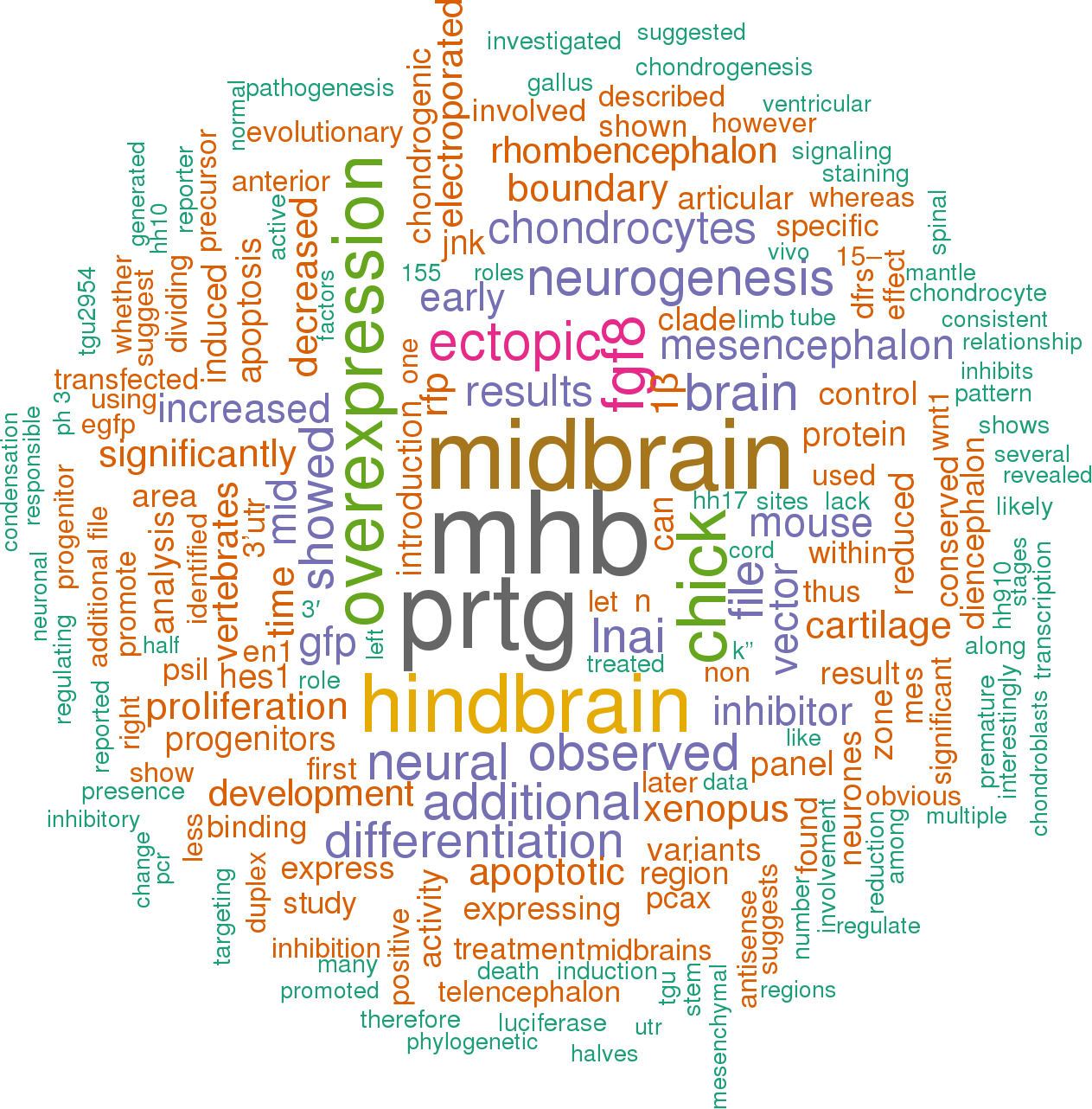
![]() 19 publications mentioning gga-mir-9-3Open access articles that are associated with the species Gallus gallus and mention the gene name mir-9-3. Click the [+] symbols to view sentences that include the gene name, or the word cloud on the right for a summary. |
|
1 |
![]()
Other miRNAs from this paper: gga-mir-15a, gga-mir-107, gga-mir-15b, gga-mir-9-2, gga-mir-9-1, gga-mir-15c, gga-mir-9-4, gga-mir-9b-1, gga-mir-9b-2
In zebrafish, Leucht et al. [15] described pea3, a downstream target of fgf8 as additional target of miR-9. Our target analysis scans did not suggest chick PEA3 as a direct target of miR-9 and we did not observe a visible downregulation of its expression in the MH 24 h after miR-9 overexpression.
[score:17]
The red, RFP expressing cells in (D) are those cells of (A) that do not express miR-9. Overexpression of miR-9 together with DFRS-S9 (B,E) showed that almost all cells in midbrain express only GFP (B) and no RFP (E) and therefore miR-9. With the DFRS-control sensor plasmid all transfected cells express GFP (C) and RFP (F).
[score:11]
Ectopic miR-9 expression did not change HES1 expression pattern visibly, nor did overexpression of miR-9-LNAi to block miR-9 or in vivo target protection.
[score:9]
Overexpression or reduction of miR-9 at HH9/10 (E1.5) in MH resulted in a down- or up-regulation of FGF8, respectively but no obvious changes in EN1 and EN2 expression.
[score:8]
Ectopic expression of an antisense oligonucleotide to the 3’UTR binding site for miR-9 of FGF8 enlarged FGF8 expression (j- l), as did the expression of miR-9-LNAi (m- p).
[score:7]
Ectopic expression and suppression of miR-9 identified FGF8 as in vivo target of miR-9. Ectopic miR-9 in the MHB resulted in premature neurogenesis in the IZ.
[score:7]
Nevertheless, since HES1 is a miR-9 target in other vertebrates we examined HES1 expression after miR-9 overexpression.
[score:7]
Interestingly, we did not find an obvious down regulation of EN1 expression along the MH area after overexpression of miR-9 at HH9/10 (Fig. 3j-l; n = 3/12).
[score:6]
Theoretical 3’UTR target sites of miR-9 were obtained from Target Scan (http://www.
[score:5]
Fig. 3 OTX2, GBX2, WNT1 and EN1 expressions were not disturbed by ectopic miR-9 expression.
[score:5]
Darnell et al. [53] described first miR-9 expression at HH22 with a strong expression in forebrain.
[score:5]
Ectopic miR-9 or miR-9-LNAi expression did not change the expression pattern of OTX2 and GBX2 (a), WNT1 (d, g), EN1 (j, m) or HES1 (s).
[score:5]
PEA3 expression is unchanged by miR-9 overexpression.
[score:5]
a- f Overexpression of miR-9 reduced FGF-8 expression.
[score:5]
Cells in the mantle zone however, and some cells on their way to the mantle zone expressed only EGFP, and therefore express miR-9 (Fig. 6 and Additional file 8: Movie S1).
[score:5]
At HH17 (j- k”) many cells in the mesencephalon were positive only for GFP (j- k”), whereas, cells in the MHB still expressed RFP (k- K″) and therefore no miR-9. : Di-diencephalon, Mes-mesencephalon, MHB-mid-hindbrain boundary, Rh-rhombencephalon, Tel-telencephalon Since the weak and diffuse expression at E2 and E3 (Fig. 1a, Additional file 3: Figure S3A-F) in the neural tube did not reveal an obvious lack of miR-9 at the MHB as has been described in zebrafish, mouse and Xenopus [15– 17], we employed another method to identify the activity of miR-9 at a cellular level.
[score:5]
We see observed first expression in telencephalon a stage later but general miR-9 expression already at HH13.
[score:5]
Interestingly, miR-9-LNAi overexpressing midbrains cells were also less likely to co-express pH 3 but showed in general a low amount of pH 3 positive cells.
[score:5]
Different studies have pointed to regional and species-specific differences in the response of neural progenitors to miR-9. In ovo and ex ovo electroporation was used to overexpress or reduce miR-9 followed by mRNA in situ hybridisation and immunofluorescent stainings to evaluate miR- expression and the effect of changed miR-9 expression.
[score:5]
These results show that FGF8 at the MHB is a direct target of miR-9 in chick a during early development.
[score:5]
We next addressed whether miR-9 misexpression influences PEA-3 expression as in zebrafish.
[score:5]
WNT1 expression was only reduced in very few cases after ectopic miR-9 duplex expression (Fig. 3d-i; n = 4/15).
[score:5]
Between E2 and E3 miR-9 was expressed in the chick brain from diencephalon to rhombencephalon (Fig. 1a-c; Additional file 3: Figure S3a-j; each stage n = 3 or more), and neither telencephalon nor spinal cord showed a strong expression (Fig. 1a).
[score:5]
At HH17 (j- k”) many cells in the mesencephalon were positive only for GFP (j- k”), whereas, cells in the MHB still expressed RFP (k- K″) and therefore no miR-9. : Di-diencephalon, Mes-mesencephalon, MHB-mid-hindbrain boundary, Rh-rhombencephalon, Tel-telencephalonSince the weak and diffuse expression at E2 and E3 (Fig. 1a, Additional file 3: Figure S3A-F) in the neural tube did not reveal an obvious lack of miR-9 at the MHB as has been described in zebrafish, mouse and Xenopus [15– 17], we employed another method to identify the activity of miR-9 at a cellular level.
[score:5]
Mir-9-5p overexpression at HH10 did not visibly reduce PEA-3 expression in midbrain at HH17 (n = 3; Additional file 4: Figure S4).
[score:4]
Our results showed that miR-9 is diffusely expressed in the brain early in development and becomes more focused later.
[score:4]
In midbrain expression of miR-9 reduced proliferation; but did not promote neurogenesis early in development.
[score:4]
MiR-9 overexpression at HH9–10 results in a reduction of FGF8 expression and premature neuronal differentiation in the mid-hindbrain boundary (MHB).
[score:4]
Although ectopic expression of miR-9 at HH9–10 did not result in ectopic neurogenesis within midbrain areas (Fig. 4), we detected that a broad overexpression of miR-9 reduced the size of the transfected midbrain compared to the uninjected control site Fig. 5a-c, n=26/42).
[score:4]
Early in development, miR-9 is diffusely expressed in the entire brain, bar the forebrain, and it becomes more restricted to specific areas of the CNS at later stages.
[score:4]
In the MHB and the hindbrain ectopic miR-9 can inhibit FGF8 signalling by down regulating FGF8 and promotes neurogenesis.
[score:4]
Our ectopic expression of miR-9 did not result in an obvious down regulation of HES1 (Fig. 3s-u, see above) within the MH area.
[score:4]
: Mes-mesencephalon, MHB-mid-hindbrain boundary, Rh-rhombencephalon To verify that miR-9 can affect Fgf8 expression at the MHB we knocked down miR-9 levels with miR-9-LNAi [18] at HH9/10.
[score:4]
: Mes-mesencephalon, MHB-mid-hindbrain boundary, Rh-rhombencephalonTo verify that miR-9 can affect Fgf8 expression at the MHB we knocked down miR-9 levels with miR-9-LNAi [18] at HH9/10.
[score:4]
All three constructs did result in miR-9 overexpression and down regulated RFP of the DFSR-S9 vector (Additional file 1: Figure S1B,E for miR-9-pSil, and data not shown).
[score:4]
This result suggests that cells producing ectopic miR-9 are able to down regulate FGF8 expression.
[score:4]
Interestingly, at later developmental stages more miR-9 expressing cells are observed in the mantle zone.
[score:4]
In zebrafish and Xenopus overexpression or down regulation of miR-9 began already at the 2-cell stage [15, 16].
[score:4]
The expression of miR-9 in neural progenitors in the CNS of chick, mouse, xenopus and zebrafish [15– 17, 20, 53, 75, 76] and the results of experimental over- and down regulations [15– 18, 60, 63] suggest that in most brain regions miR-9 promotes neuronal differentiation.
[score:4]
Transcription factors like Hes1 or TLX can inhibit miR-9 [18, 64, 79] and DNA binding proteins and FXFRqP genes are known to influence miR-9 regulation [80, 81].
[score:4]
This suggests that in midbrain miR-9 can inhibit proliferation but does not necessarily promote neurogenesis during early development.
[score:4]
Lateral view of the neural tube at MH level overexpressing miR-9 (a, b, d, e, j, k, m, n, s, t), miR-9-LNAi (g, h, m, n,) or pCAX-EGFP (p, q).
[score:3]
The time-limited sensibility for miR-9 influencing FGF8 is supported by the lesser effect of pre-miR-9 and miR-9-pSil on FGF8 expression we observed.
[score:3]
Reduced miR-9 activity resulted in an expansion of FGF8 expression (Fig. 2m- p; n = 5/6).
[score:3]
Broad overexpression of miR-9 along the DV axis of the midbrain result often in smaller midbrains (15/18).
[score:3]
We concluded that miR-9 is not influencing HAIRY-1 expression in the midbrain.
[score:3]
MiR-107 for example, inhibits dicer protein in zebrafish hindbrain, which normally activates miR-9 biogenesis and thus differentiation [65].
[score:3]
However, overexpression of miR-9 duplex at HH9–10 promoted ectopic neurones within the MHB (Fig. 4a, C- D’; n = 6/8) and in the anterior rhombencephalon (Additional file 5: Figure S5; n = 4/5) but not in the rest of the midbrain (Fig. 4a, c- E’; n = 19/20).
[score:3]
MiR-9 overexpression down regulated FGF8 at the MHB (Fig. 2a-f).
[score:3]
We showed that miR-9 overexpression in chick MH also generated ectopic neurones in the anterior hindbrain and the long lasting progenitor region of the MHB (IZ) but not in the midbrain.
[score:3]
Our results correlate with those of other vertebrates where one fundatmental function of miR-9 is to inhibit neural proliferation [15– 18, 60– 64].
[score:3]
We observed miR-9 expression first in posterior brain.
[score:3]
Ectopic expression of high concentration of miR-9 (25 μm) at HH8 resulted in an even more obvious reduction of midbrain size (Fig. 5d-f).
[score:3]
However, the number of phospho-histone-H3 positive cells (pH 3+) also expressing miR-9 (n=4), miR-9-LNAi (n=2) or pCAX-GFP (n=2) at E3 differed (Additional file 7: Figure S7).
[score:3]
Thus, our results agree with findings in mouse, Xenopus and zebrafish, which show that the MHB lacks miR-9 expression [15– 17, 19].
[score:3]
Fig. 6Dividing mesencephalic progenitor cells did not express miR-9. The right half of the mesencephalon was electroporated with miR-9 sensor DFRS-S9 ex ovo at HH10.
[score:3]
The presence of HES5 and HES6 in chick midbrain could be responsible for the lack of premature neurogenesis we observe after miR-9 overexpression.
[score:3]
Section of HH17 midbrains overexpressing pCAX-EGFP (A,A’,E,E’,E”’,I), miR-9-5p (B,B’F,F′,F″) or miR-9 LNAi (C,C’,G,G’,G”).
[score:3]
In zebrafish hindbrain and mouse telencephalon miR-9 is expressed in a range of progenitor cells with different commitments [18, 19].
[score:3]
In early chick midbrain miR-9 expressing cells might also belong to non-dividing progenitors.
[score:3]
Our results support the theory of a tissue specific regulation of miR-9 and multiple roles during neuronal development.
[score:3]
To promote differentiation versus proliferation the main target of miR-9 are Hes genes [15, 16, 18– 20, 66].
[score:3]
Our analysis reveals a closer relationship of chick miR-9 to mammalian miR-9 than to fish and a dynamic expression pattern in the chick neural tube.
[score:3]
We never observed any effect of miR-9 overexpression on the boundary between GBX2 and OTX2 (Fig. 3a-c; n = 3 for each, miR-9 duplex and miR-9-pSil).
[score:3]
In zebrafish, Xenopus and mouse primary targets for miR-9 are members of the Hes gene family [15, 16, 18– 20].
[score:3]
A locked nucleic acid (LNA) antisense inhibitor (miR-9-LNAi) (TCA TAC AGC TAG ATA ACC AAA G; Exiqon Product no.
[score:3]
In zebrafish hindbrain non-cycling progenitors and differentiating neurones expressed miR-9 [43].
[score:3]
Overexpression of miR-9 in the adjacent regions promoted neurogenesis only in hindbrain and not in midbrain.
[score:3]
microRNA Mid-hindbrain area Chick miR-9 Brain development The development of vertebrate midbrain and anterior hindbrain depends on coordinated signals from the organiser region at the mid-hindbrain boundary (MHB).
[score:3]
MiR-9 is downregulated in dividing cells.
[score:3]
Some of the miR-9-LNAi expressing cells in dorsal midbrain (f- F′) developed axons (arrows).
[score:3]
Overexpression of miR-9 in anterior hindbrain promotes neurogenesis.
[score:3]
Thus, our miR-9 misexpression experiments interfered with proteins after MHB has formed.
[score:3]
In zebrafish and Xenopus miR-9 overexpression promoted neural differentiation in the hindbrain and in zebrafish also in the MHB [15, 16, 19].
[score:3]
Overexpression of miR-9 did not promote ectopic neurogenesis within the midbrain (a, c- E’) but in the MHB (a, c).
[score:3]
In turn, Hes1 in mouse, and her6 in zebrafish suppress miR-9 transcription [18, 19].
[score:3]
The monomeric red fluorescence protein (mRFP) that is tagged by a 3’UTR complementary sequence of miR-9 indicates miR-9 activity: cells that express miR-9 silence mRFP.
[score:3]
b- f show a dividing cell that expressed both, GFP and RFP and thus no miR-9. : Di-diencephalon, Mes-Mesencephalon, NCC-neural crest cells, Rh-rhombencephalon; scale bar 100μm Additional file 8: Movie S1.
[score:3]
Whole mount staining of E4 (HH 23) heads showed miR-9 strongly expressed in diencephalon, weakly in the telencephalon and not at all in dorsal midbrain (Fig. 1b).
[score:3]
The change of miR-9 and its impact on the MHB in zebrafish and on neural development in zebrafish and Xenopus may therefore reflect an impact of miR-9 very early in development, whereby both sides of the brain are affected.
[score:3]
For long lasting effects we overexpressed miR-9 stem loop via the pSilencer U6.1 vector (miR-9-pSil).
[score:3]
Section of HH17 midbrains overexpressing pCAX-EGFP (A,B), miR-9-5p (C,D) or miR-9 LNAi (E,F).
[score:3]
Functional miR-9 overexpression.
[score:3]
Our theoretical target scan suggested a 3’UTR seed site of miR-9 for HES-1-B-like but not for HES1/HAIRY1A/HES4 in chick.
[score:3]
The DFRS-S9 expression pattern suggests that miR-9 becomes active in the midbrain from about HH14 onwards but not in the MHB and not yet in the hindbrain.
[score:3]
Evolutionary conservation of chick miR-9. Divergent expression of miR-9 in brain and spinal cord.
[score:3]
Our time-lapse experiments showed that dividing progenitors in the ventricular zone of the midbrain do not express miR-9 at HH12.
[score:3]
Expression of miR-9 in HH18 and E6 chick brains.
[score:3]
Overexpression of miR-9 at later stages (C; HH11) did not result in early neurogenesis in anterior hindbrain (white arrowheads, D).
[score:3]
The spatial and temporal miR-9 expression pattern we observed differs slightly from that described by Darnell et al. [53].
[score:3]
Similarly, ectopic miR-9-LNAi in the MH region caused no widening of EN1 expression (Fig. 3m-o; n = 6/6).
[score:3]
LNA-ISH against miR-9 at HH13 (a) revealed an initially weak expression from di- to rhombencephalon.
[score:3]
In zebrafish, Xenopus, and mouse miR-9 is expressed in regions adjacent to the MHB, but not in MHB [15– 17].
[score:3]
This result suggests a rather limited influence of miR-9 on EN1 expression or one, which is not visible by ISH.
[score:3]
The result is reminiscent of the indirect regulation of wnt1 in zebrafish [15] and agrees with the fact that there is no binding site for miR-9 in the 3’UTR of WNT1.
[score:3]
Overexpression of miR-9 leads to many committed neural cells since her6 is blocked and to many radila glia cells because elavl3 is reduced [65].
[score:3]
Horizontal sections revealed a general ubiquitous expression of miR-9 along the dorsoventral axis at E3 except for forebrain and spinal cord (HH18; Additional file 3: Figure S3A-F).
[score:3]
Our target analysis scan suggested 3’UTR seed sequences for miR-9 with FGF8, EN1 and EN2.
[score:3]
To confirm the early expression of miR-9 in the mid-hindbrain area, we used a miR-9 reporter sensor vector (DFRS-S9, [24]).
[score:3]
We observed smaller midbrain halves after overexpression of miR-9 in a very broad area 24 h after electroporation.
[score:3]
In midbrain we found that miR-9 is not expressed in dividing neural progenitor cells.
[score:3]
We never observed ectopic neurones in E3 (HH17–19) midbrains but at HH26 (E5) more cells transfected with pSil-miR-9 seemed to have located to the mantle zone than cells expressing pCAX-GFP (Additional file 7: Figure S7H,I).
[score:3]
The result suggests that miR-9-5p positive cells are around 60% less likely to express pH 3 (Additional file 7: Figure S7 K).
[score:3]
MiR-9 knockdown [43] leads to a disinhibition of her6 and elavl3, which results in more cycling progenitors and committed neurones.
[score:3]
MiR-9 (pSil-miR-9) was ectopically expressed in anterior hindbrain at HH9 (A,B) and HH11 (C,D) in left (A,B) or right (C,D) brain half.
[score:3]
To validate miR-9 endogenous expression on a cellular level, the dual fluorescence reporter sensor (DFRS) vector developed by De Pietri Tonelli et al. [23, 24] was used.
[score:3]
DFSR-S9 expression pattern revealed active miR-9 in the midbrain at around HH14 (E2) but not in the MHB and hindbrain (Fig. 1g-i; n = 8/9).
[score:3]
In zebrafish, mouse and Xenopus miR-9 expression begins in the telencephalon and spreads posteriorly to the rest of the brain and to the spinal cord in mouse and zebrafish.
[score:3]
MiR-9 influences FGF8 but not HAIRY1 expression within the MHBOur search for putative binding sites of miR-9 in the 3′ UTR of chick MHB core genes (Additional file 1: Figure S1G) yielded binding sites to the effector gene FGF8, the transcription factors ENGRAILED (EN) 1 and 2, and for HES-1-B-like, but none for the signalling protein WNT1 or HAIRY1a/HAIRY1b/HES4.
[score:3]
FGF8 expression was also not visibly changed by miR-9 transfections at later stages.
[score:3]
Note, miR-9 expression in the ventricular zone of HH26 diencephalon, mesencephalon and rhombencephalon.
[score:3]
MiR-9 inhibits proliferation in midbrain but not neurogenesis.
[score:2]
MiR-9 is a small non-coding RNA that is highly conserved between species and primarily expressed in the central nervous system (CNS).
[score:2]
We have investigated the expression and function of miR-9 during early development of the mid-hindbrain region (MH) in chick.
[score:2]
: Di-diencephalon, Mes-mesencephalon, MHB-mid-hindbrain boundary, Rh-rhombencephalonIn zebrafish, miR-9 down regulated her5 in the MHB [15] and thus influenced the stability of the MHB by destroying the non-differentiating IZ.
[score:2]
: Di-diencephalon, Mes-mesencephalon, MHB-mid-hindbrain boundary, Rh-rhombencephalon In zebrafish, miR-9 down regulated her5 in the MHB [15] and thus influenced the stability of the MHB by destroying the non-differentiating IZ.
[score:2]
MiR-9 influences FGF8 but not HAIRY1 expression within the MHB.
[score:2]
MiR-9 overexpression caused premature neurogenesis in MHB and hindbrain but not in midbrain.
[score:2]
427460–04) was employed to knock down mature miR-9 levels.
[score:2]
MiR-9 might not be sufficient to cause neural differentiation or specific factors, only present in midbrain, might regulate miR-9 or prevent premature differentiation.
[score:2]
MiR-9 expression promotes neurogenesis at the MHB and in anterior hindbrain.
[score:2]
To test whether any of these genes was regulated by miR-9 in vivo we performed miR-9 gain and loss of function experiments in ovo.
[score:2]
In early midbrain our results support a role of miR-9 in regulating proliferation and hence midbrain size.
[score:2]
An average of 15% of pH 3+ cells also expressed miR-9 compared to 51% GFP+/pH 3+ cells in control midbrains (Additional file 7: Figure S7 J,K).
[score:2]
MiR-9 misexpression and mitotic cells.
[score:2]
MiR-9-LNAi expressing cells in the MHB did not show long axons (arrowheads in F′).
[score:2]
The strongest down regulation was seen when the embryos were electroporated at HH9/10 with the miR-9 duplex (Fig. 2a-c; n = 7/9).
[score:2]
In the presence of miR-9, RFP, which contains a 3’UTR binding site for miR-9 is down regulated and only GFP is visible in the cell.
[score:2]
In most species both strands of miR-9 variants (guide- and passenger strand) participate in gene regulation, thus the number of miR-9s is even higher [43, 52].
[score:2]
MiR-9 is expressed differentially along the mid-hindbrain area.
[score:2]
Our findings indicate that miR-9 has regional specific effects in the developing mid-hindbrain region with a divergence of response of regional progenitors.
[score:1]
Several mechanisms could be responsible for the lack of ectopic neurogenesis in miR-9 transfected midbrains cells.
[score:1]
Our results suggest miR-9 helps to define the extent of the MHB in chick, as shown in zebrafish [15] and can promote neurogenesis in MHB and hindbrain but not in midbrain.
[score:1]
The consensus structure of mature sequences of miR-9 represents the degree of conservation in mature miR-9 sequences.
[score:1]
This method utilises a dual fluorescence vector (DFSR-S9; [24]) that reveals cells with active miR-9. In the absence of miR-9 the DFSR vector generates GFP and RFP.
[score:1]
The number of miR-9 genes is in part a reflection of the whole genome duplication (WGD) events that have occurred during vertebrate evolution, but also due to more restricted gene duplications.
[score:1]
Taken together, these results suggests in midbrain miR-9 is excluded from dividing neural progenitors cells.
[score:1]
Our findings further suggest that miR-9 has different roles in midbrain, MHB, and hindbrain.
[score:1]
B. Evolutionary relationship of pre-miR-9 family.
[score:1]
Accordingly, miR-9 did not interrupt the sharp boundary between OTX2 and GBX2, which is set up earlier.
[score:1]
The phylogenetic analysis of precursor sequences showed that miR-9 of vertebrates clusters into three clades (Additional file 2: Figure S2B): clade I consists of miR-9-1 and miR-9-2, clade II contains miR-9-3 and miR-9-4, while clade III consists of miR-9 sequences from Rattus norvegicus.
[score:1]
Next, we investigated the spatial and temporal expression of miR-9 in chick brain between embryonic day E2 (HH13) and E6 (HH29) (Fig. 1 and Additional file 3: Figure S3).
[score:1]
The loss and gain of miR-9 function resembled the control with ectopic EGFP-pCAX (Fig. 3p-r; n = 5).
[score:1]
The movie shows dividing neural progenitor cell lack miR-9. (AVI 2000 kb) In this study we have shown that chick miR-9 forms part of an evolutionary conserved family.
[score:1]
In both cases mature miR-9 has to be generated first whereas, mature miR-9-5p can be active immediately after electroporation.
[score:1]
At HH17 still a miR-9 free zone could be found in MHB in chick (Fig. 1j-K”, n = 3/3).
[score:1]
Both mature and precursor sequences of miR-9 from diverse vertebrates (Homo sapiens, Mus musculus, Rattus norvegicus, Gallus gallus, Taeniopygia guttata, Xenopus tropicalis, Fugu rubripes, Tetraodon nigroviridis, Danio rerio and Oryzias latipes) were downloaded from miRBase Release 21:June 2014 [34].
[score:1]
Sequence collection and phylogenetic analysis of miR-9. Computational analysis of miR-9 binding sites.
[score:1]
Interestingly, the miR-9-LNAi-GFP+ cells within the MHB did not sprout axons or label as neurones (white arrowhead in Fig. 4F’).
[score:1]
To introduce gga-miR-9 into the pSilencer U6.1 vector (Ambion), single stranded oligonucleotides containing a gga-miR-9 sense (5′-TCT TTG GTT ATC TAG CTG TAT GAT TCA AGA GAT CAT ACA GCT AGA TAA CCA AAG ATT TTTT-3′) and a miR-9 anti-sense (5′-AAT TAA AAA ATC TTT GGT TAT CTA GCT GTA TGA TCT CTT GAA TCA TAC AGC TAG ATA ACC AAA GAG GCC-3′) sequence were annealed and ligated into the ApaI/EcoRI digested pSilencer U6.1 vector (Ambion; miR-9-pSil).
[score:1]
The variants miR-9-3 and miR-9-4 of mammals and teleost fishes formed clade II.
[score:1]
Right brain halves transfected with miR-9-LNAi (m- p).
[score:1]
: FP-floor plate, Mes-mesencephalon, MHB-mid-hindbrain boundary, Rh-rhombencephalon, RP-roof plateWe next investigated whether miR-9 overexpression caused apoptosis or affected the number of cells undergoing mitosis.
[score:1]
LNA-ISH staining of whole-mount chick embryos (a- c) and miR-9 activity indicator DFRS-S9 (d- k”) showed that miR-9 is active from around HH13 in chick neural tube.
[score:1]
The movie shows dividing neural progenitor cell lack miR-9. (AVI 2000 kb) The miR-9 gene family has significantly expanded among vertebrates and consist of many variants.
[score:1]
Tunnel staining did not indicate additional apoptotic cells in midbrains independent of their transfection with miR-9-5p, miR-9-LNAi or pCAX-GFP (Additional file 6: Figure S6).
[score:1]
In mouse, zebrafish and Xenopus miR-9 affected either her5 (in zebrafish MHB) or Hes1 in the mouse telencephalon and Xenopus hindbrain [15, 16, 18, 20, 51].
[score:1]
Ectopic miR-9 in the left half of the MHB (a, c) induced ectopic neurones (white arrows), which are not found in the control right half (a, d).
[score:1]
C. CLUSTAL multiple sequence alignment of mature miR-9 sequence by MUSCLE (3.8).
[score:1]
The net effect of miR-9 is to keep cells in the ambivalent state of neural progenitors by accumulating in the cell.
[score:1]
A new player at the MHB is the microRNA-9 (miR-9).
[score:1]
Later, miR-9 localises to the ventricular zone like in other vertebrate brain regions [16, 17, 19].
[score:1]
The consensus structure of mature miR-9 was obtained using RNALogo [35].
[score:1]
In zebrafish hindbrain, miR-9 has been described to have a twofold effect.
[score:1]
Leucht et al., [15] showed that miR-9 is necessary to define the MHB.
[score:1]
The Neighbour-Joining phylogram shows the phylogenetic relationships of pre-miR-9 variants among vertebrates.
[score:1]
Chick miR-9 is evolutionary conserved.
[score:1]
MiR-9 is one of the ancient miR, which appeared with the bilateria, and the miR-9 subfamily is evolutionarily conserved among vertebrates [43, 44].
[score:1]
Brains electroporated with miR-9-LNAi at HH9–10 did not show an obvious loss of differentiated neurones in the dorsal midbrain, where the mesencephalic trigeminal nucleus (MTN) is generated (Fig. 4b, f, F’; n = 0/11).
[score:1]
GFP [+]/RFP [+] cells are devoid of endogenous miR-9 whereas, GFP [+]/RFP [−] cells produce miR-9. There are several other GFP [+]/RFP [+] visible, which very likely are mitotic cells.
[score:1]
Very likely, mature miR-9 accumulates in a cell and slowly reduces Hes1 [65] and hence contributes to the oscillation of Hes1 as Bonev et al. (2012) [18] have described in vitro.
[score:1]
Brain halves (C’, E’) or dorsal midbrain (F’) were electroporated at HH9–10 with miR-9 (a, c- E’) or miR-9-LNAi (f, F′).
[score:1]
E. The stem loop structure of Gallus gallus miR-9 variants.
[score:1]
In contrast to zebrafish, human, mouse and Xenopus En1 and En2 also possess a possible 3’UTR seed region for miR-9 (data not shown).
[score:1]
Left brain halves were electroporated at HH9/10 with miR-9 duplex (a- c), miR-9-pSil (d- f), pCAX-EGFP (g- i), and a single stranded antisense oligonucleotides against the 3’UTR FGF8 sequence (j— l).
[score:1]
This result suggests that within the MHB and the anterior hindbrain miR-9 can promote neurogenesis but not in midbrain.
[score:1]
Ectopic miR-9 in posterior midbrain (a, c, C’) or in more anterior midbrain regions (e, E’) did not promote early neurogenesis in midbrain.
[score:1]
At HH23 (b) miR-9 was strong in ventral hind- and midbrain and in diencephalon.
[score:1]
The colour denotes the Clades of miR-9 family in vertebrates (Clade I: red, Clade II: Rose and Clade III: Blue).
[score:1]
In mammals, Homo sapiens and Mus musculus have three variants (miR-9-1, miR-9-2, miR-9-3) whereas Rattus norvegicus displays six miR-9 genes.
[score:1]
Left brain half was electroporated with miR-9 duplex (A) at HH10.
[score:1]
We electroporated miR-9 as a duplex or a precursor form (pre-miR-9) for more immediate but shorter effects.
[score:1]
Whole-mount embryos were hybridized with digoxygenin labelled miR-9 LNA (20-40 nM) over night at 45°C in a Chaps buffer (50% Formamide, 1.3% SSC, 5 mM EDTA, 50 μg/μl tRNA, 0.1% Tween 20, 0.1% CHAPS, 100 μg/μl Heparin, 2% Blocking reagent); then washed in a less stringent solution (2xSSC, 0.1% CHAPS) followed by low salt washes (0.2% SSC, 0.1% CHAPS).
[score:1]
Sections of HH26 midbrain transfected with pCAX-EGFP (I) and pSil-miR-9 (D,D’,H).
[score:1]
Multiple sequence alignment of pre-miR-9 showed a conserved region localised to the stem region of the hairpin structures (Additional file 2: Figure S2A), which leads to mature sequences (Additional file 2: Figure S2C).
[score:1]
Our phylogenetic tree showed gga-miR-9-2 is more related to human miR-9 than to other vertebrates and gga-miR-9-1 is closely related with the dre-miR-9-4. This is different from the phylognetic relations presented in Yuva-Aydemir et al. [44], which is based on miR sequences from an earlier release of miRbase (release 16 versus 21).
[score:1]
The gga-miR-9-1 gives raise to functional mature miR-9 (miR-9-5p) and miR-9* (miR-9-3p), while gga-miR-9-2 only produce miR-9 (shown in colour).
[score:1]
In chick, both the variants (gga-miR-9-1 and gga-miR-9-2) produce the same mature product, miR-9-5p.
[score:1]
The miR-9 gene family has significantly expanded among vertebrates and consist of many variants.
[score:1]
MiR-9 in situ hybridisation was performed with a customised antisense dig-miR-9-LNA oligonucleotide (locked nucleic acid, Product No.
[score:1]
Variations in the sequences were observed in the flanking region of pre-miR-9. The evolutionary relationship of pre-miR-9 is shown in Additional file 2: Figure S2B.
[score:1]
Clade I has two subclades, namely miR-9-1 and miR-9-2. Clade II has miR-9-3 and miR-9-4. Clade III consists of miR-9 from Rattus norvegicus.
[score:1]
Therefore, we investigated whether overexpression of miR-9 has any influence on HAIRY-1/HES1 [50] in the MH area.
[score:1]
Only two variants (miR-9-1 and miR-9-2) of miR-9 are found in birds, such as Gallus gallus and Taeniopygia guttata.
[score:1]
Smaller midbrain halves and less mitotic miR-9+ cells suggest that although miR-9 does not promote neurogenesis in early midbrain, it can limit progenitor proliferation.
[score:1]
In the cortical ventricular zone miR-9 was high in cells with low Hes1 and vice versa and it was present in some of the differentiating neurones in the mantle zone.
[score:1]
A. CLUSTAL multiple sequence alignment of pre-miR-9 sequence by MUSCLE (3.8) shows that throughout vertebrate species miR-9 is conserved in the stem region of the hairpin (indicated by blue letters and asterix marks).
[score:1]
Phylogenetic analysis also showed that mature miR-9 sequences are highly conserved [43, 44].
[score:1]
Our results in chick suggested that miR-9 around the MHB might sharpen the FGF8 gradient in this area and thus very likely helps to sustain the MHB area and its size.
[score:1]
: FP-floor plate, Mes-mesencephalon, MHB-mid-hindbrain boundary, Rh-rhombencephalon, RP-roof plate We next investigated whether miR-9 overexpression caused apoptosis or affected the number of cells undergoing mitosis.
[score:1]
At HH10 no active miR-9 was observed, but three stages later the miR-9 sensor showed miR-9 positive cells in midbrain but not in MHB.
[score:1]
Horizontal sections through an E6 brain show miR-9 presence in the ventricular area of the neuroepithelium of posterior diencephalon, mesencephalon, rhombencephalon, and spinal cord (Additional file 3: Figure S3G-J), and in the mesenchyme surrounding the brain.
[score:1]
A control miR was generated using shuffled miR-9 sequence (Ambion; Leucht et al. [15]; sense - 5′-[UAU CAC UUC UAU AUG GUU UGG UG] [RNA], antisense: 5′-[CCA AAC CAU AUA GAA GUG AUA] [RNA] [TT] [DNA]).
[score:1]
To investigate in vivo whether progenitors or differentiating neurones, or both, normally express miR-9 we electroporated the miR-9 detector vector DFRS-S9 [24] into midbrain and established a time-lapse procedure for the chick brain (procedure will be published elsewhere).
[score:1]
Pre-miR-9 might also have had less effect since endogenous pre-mir-9 hairpin sequences not only generate mature miR-9 but also antisense i. e., miR-9* [56].
[score:1]
Mitotic cells were devoid of miR-9. The movie was generated from a time-lapse confocal microscopy study using Zen 2012 (Zeiss Inc).
[score:1]
The effect of the pre-miR-9 (5/6) and of the miR-9 pSilencer (Fig. 2d-f; n = 7/11) was less prominent.
[score:1]
The cladogram of mature miR-9 sequences revealed that chicken miR-9 is more closely related with the mammalian miR-9 (Additional file 2: Figure S2D).
[score:1]
The miR-9 sensor DFRS-S9 showed overlapping GFP (d) and RFP (e) positive cells in the mesencephalon at HH10 (c).
[score:1]
D. The cladogram shows the evolutionary relationship of mature miR-9 among different vertebrates.
[score:1]
The mature miR-9 obtained from both gga-miR-9-1 and gga-miR-9-2 has the same sequences and evolutionarily conserved.
[score:1]
Interestingly, both, decrease and increase of miR-9 showed this effect.
[score:1]
Within the midbrain miR-9 does not cause premature neuronal differentiation it rather reduces proliferation in the midbrain.
[score:1]
The miR-9 of fishes formed one cluster with amphibian.
[score:1]
We also investigated if miR-9 influences EN1 or WNT1 expression along the MH.
[score:1]
Taken together, in chick miR-9 is lacking in MHB.
[score:1]
Evolutionary relationship of miR-9 sequences was studied with the precursor sequences from vertebrates.
[score:1]
Our search for putative binding sites of miR-9 in the 3′ UTR of chick MHB core genes (Additional file 1: Figure S1G) yielded binding sites to the effector gene FGF8, the transcription factors ENGRAILED (EN) 1 and 2, and for HES-1-B-like, but none for the signalling protein WNT1 or HAIRY1a/HAIRY1b/HES4.
[score:1]
[1 to 20 of 218 sentences]
|
2 |
![]()
Other miRNAs from this paper: mmu-mir-9-2, mmu-mir-140, mmu-mir-146a, mmu-mir-10b, hsa-mir-196a-1, hsa-mir-10b, hsa-mir-34a, hsa-mir-196a-2, hsa-mir-221, hsa-mir-140, hsa-mir-9-1, hsa-mir-9-2, hsa-mir-9-3, hsa-mir-146a, mmu-mir-196a-1, mmu-mir-196a-2, mmu-mir-34a, mmu-mir-221, mmu-mir-9-1, mmu-mir-9-3, hsa-mir-196b, mmu-mir-196b, gga-mir-221, gga-mir-196-2, gga-mir-10b, gga-mir-140, gga-mir-146a, gga-mir-34a, gga-mir-196-1, gga-mir-196-3, gga-mir-9-2, gga-mir-9-1, gga-mir-196-4, gga-mir-196-5, ocu-mir-9-1, ocu-mir-9-2, ocu-mir-10b, ocu-mir-34a, ocu-mir-140, ocu-mir-146a, ocu-mir-196a-1, ocu-mir-196a-2, ocu-mir-196b, ocu-mir-221, gga-mir-9-4, mmu-mir-9b-2, mmu-mir-9b-1, mmu-mir-9b-3, gga-mir-9b-1, gga-mir-9b-2
Consistent with this, increased apoptosis of articular chondrocytes and PRTG level by DMM surgery was also inhibited with over -expression of miR-9 and stimulated with suppression of miR-9. From previously reported miRNA array data by inhibition of JNK signaling [11], we identified 14 up-regulated miRNAs and 12 down-regulated miRNAs whose expressions were altered during chondrogenesis (Additional file 1).
[score:17]
Here, for the first time, we found that PRTG exhibits chondro -inhibitory action in limb mesenchymal cells and that PRTG is a direct target of miR-9. From previously reported miRNA array data by inhibition of JNK signaling [11], we identified 14 up-regulated miRNAs and 12 down-regulated miRNAs whose expressions were altered during chondrogenesis (Additional file 1).
[score:16]
Second, the luciferase intensity of PRTG-UTR was specifically responsive to miR-9 over -expression suggesting that miR-9 may regulate PRTG protein expression by inducing translational suppression.
[score:10]
As well, inhibited precartilage condensation by JNK inhibition and PRTG over -expression was recovered by co-electroporation of PRTG-specific siRNA or co-introduction of miR-9 (Figure 3D) confirmed its efficiency with PRTG over-expressed cells (Figure 3C lower panel).
[score:9]
Target genes of miR-9 were predicted using miRNA target prediction algorithms, including TargetScan and miRDB and PRTG was identified as a potential target.
[score:9]
Consistent with this, increased apoptosis of articular chondrocytes and PRTG level by DMM surgery was also inhibited with over -expression of miR-9 and stimulated with suppression of miR-9. During development, most of our bones form through endochondral ossification in which bones are first laid down as cartilage precursor [1] and mitogen-activated protein kinase (MAPK) cascades are known to play essential roles in regulating mesenchymal cell chondrogenesis [2, 3].
[score:9]
Using these cells, we analyzed the changes in the expression of genes and proteins, tested the expression level of miR-9, and applied a target validation system.
[score:7]
And chondrocytes isolated from normal human articular cartilage expressed miR-9, and this expression was significantly reduced in OA chondrocytes, especially decreased its expression in parallel with the degree of cartilage degradation.
[score:7]
To validate the role of miR-9 in chondrocyte apoptosis during OA cartilage destruction in vivo, we overexpressed miR-9 in cartilage tissue by injecting miR-9 -expressing or si-miR-9 expressing lentiviruses into DMM mouse knee joints (Figure 6E).
[score:7]
Experimental evidence indicates that PRTG is a target of miR-9. First, the ability of miR-9 to regulate PRTG expression is likely direct, because it binds to the 3′UTR of PRTG mRNA.
[score:7]
And these inhibitory actions of PRTG on precartilage condensation and chondrogenic differentiation were recovered by co-introduction of miR-9. These data suggested that miR-9 suppresses sulfated proteoglycan accumulation and cartilage nodule formation for chondrogenic differentiation possibly by targeting PRTG.
[score:7]
To confirm that PRTG is a target for miR-9, we cloned the entire 3′ UTR of PRTG into a luciferase reporter vector, electroporated the vector into chondrogenic progenitors along with the precursor of miR-9 or a cognate non -targeting negative control, and assayed cell lysates for luciferase expression.
[score:6]
Figure 2 miR-9 targets PRTG and inhibits chondrogenic differentiation.
[score:5]
The RNA level of PRTG was also significantly decreased at 3, 6, and 9 days of culture i. e. at the time of proliferation and condensation with increased expression level of miR-9 and significantly increased at 12, 15, and 18 days of culture, i. e. at the time of hypertrophy and apoptosis with a decreased expression level of miR-9 (Figure 2F).
[score:5]
Apoptotic cell death, as assessed by FACS analysis (left panel) and by caspase-3 activity (right panel), was increased by the introduction of PRTG or treatment of JNK inhibitor and inhibited by co-induction of miR-9 (Figure 3C).
[score:5]
Apoptotic genes including ABL1, ATP6V1GNOL3, CASP1, 3, 7, CD40, CYLD, and FAS were induced with IL-1β treatments or PRTG over -expression whereas expression levels of those genes were decreased with miR-9 introduction.
[score:5]
In support of this prediction, we observed a significant induction in PRTG protein level in miR-9 inhibitor -treated or JNK inhibitor -treated chondroprogenitor cells.
[score:5]
Our results revealed that miR-9 inhibitor -induced apoptotic cell death may be responsible for JNK blockade -induced chondro -inhibitory action on precartilage condensation.
[score:5]
Among them, miR-9 was one of miRNA whose expression was substantially altered with inhibition of chondrogenic differentiation (determined using a P-value of 0.01 as a cutoff for significance).
[score:5]
Our study provides evidence for the mechanism through which miR-9 affects the survival/proliferation of chondrocytes and PRTG is one of the physiologic targets of miR-9 in the regulation of chondrocyte survival.
[score:4]
In addition, co-introduction of PRTG or inhibition of miR-9 significantly increased apoptosis in cells treated with TGF-β3 (Figure 4F), a known positive regulator of chondrocytes [27].
[score:4]
In order to examine the involvement of miR-9 during chondrogenesis, we exposed mesenchymal cells to 200 nM peptide nucleic acid -based antisense oligonucleotides (ASOs) against miR-9 (miR-9 inhibitor) whose knockdown efficiency was monitored by real time PCR (Figure 1C, upper panel).
[score:4]
Consistent with the results obtained with PRTG over -expression, knock-down of miR-9 promoted the apoptotic death of limb chondroblasts.
[score:4]
In sum, here, for the first time, we found that PRTG is regulated by miR-9, resulting in an inhibition of cell proliferation and survival in chondrogenic progenitors and articular chondrocytes.
[score:4]
This study shows that PRTG is regulated by miR-9, plays an inhibitory action on survival of chondroblasts and articular chondrocytes during chondrogenesis and OA pathogenesis.
[score:4]
MiR-9 is known as a growth inhibition factor and plays a role as in anti-proliferative activity in human gastric adenocarcinoma cells by negatively targeting NF-κB1 at the post-transcriptional level [35].
[score:4]
Down-regulation of miR-9 by blockade of JNK signaling was confirmed by quantitative RT-PCR (Figure 1B).
[score:4]
Most significant degeneration was observed in the combination of IL-1β and PRTG -treated cell or in the combination of IL-1β and miR-9 inhibitor -treated cell.
[score:3]
Change in expression level of miR-9 in was analyzed by real-time PCR.
[score:3]
Consisted with these observations, the protein level of PRTG was increased by co-treatment of miR-9 inhibitor (Figure 4B) and decreased by co-introduction of miR-9 (Figure 4C).
[score:3]
Figure 5 miR-9 and its target, PRTG is involved in chondrocyte apoptosis.
[score:3]
Treatment of cells with a miR-9 inhibitor caused a significant decrease in total cell numbers (Figure 1D) with significant increases in apoptotic cell death (Figure 1E) and caspase-3 activity (Figure 1F).
[score:3]
Human articular chondrocytes isolated from biopsy normal cartilage were electroporated with Prtg or miR-9 in the absence or presence of IL-1β and expression levels of apoptotic genes were examined and represented as heat-map.
[score:3]
To further investigate miR-9 involvement in limb formation, 18 HH stage chick embryos were treated with JNK inhibitor in the absence or presence of miR-9 inhibitors.
[score:3]
Studies have shown the roles of miR-9 and its validated target, protogenin (PRTG) in the differentiation of chondroblasts to chondrocyte and in the pathogenesis of osteoarthritis (OA).
[score:3]
And increased protein level of PRTG by JNK inhibitor treatment was significantly reduced with co-introduction of miR-9 (Figure 2A).
[score:3]
Most severe cartilage destruction was observed with the infection of si-miR-9 expression lentiviruses (MFC score of 3, MTP score of 3).
[score:3]
Seed sequences of putative targets for miR-9 (Figure 2B upper panel) were exchanged a purine for a pyrimidine and a pyrimidine to a purine.
[score:3]
This malformation was overcome by co-treatment of miR-9 inhibitor (Figure 3E).
[score:3]
The expressions of type II collagen (Col II), PRTG, and miR-9 were analyzed by real-time PCR (lower panel).
[score:3]
Furthermore, decreased in total cell number by JNK inhibitor or PRTG was reversed by co-introduction of PRTG siRNA or miR-9, respectively (Figure 3B, right panel).
[score:3]
We confirmed that IL-1β exposure to cells decreased the expression level of miR-9 (Figure 4A).
[score:3]
Our laboratory is currently undergoing study on the relationships between miR-9, PRTG, and MMP-13 to verify whether chondrocyte apoptosis by PRTG, a target for miR-9, is down-stream, up-stream, or independent of MMP-13 induction.
[score:3]
However, over -expression of miR-9 significantly reduced cartilage destruction (MFC score of 0, MTP score of 0.5).
[score:3]
Mice were killed 8 weeks after DMM surgery or 2 weeks after intraarticular injection (1 × 10 [9] plaque-forming units (PFU)) of miR-9 -expressing lentiviruses (lenti-miR-9) for histological and biochemical analyses.
[score:3]
A more significant degenerative phenotype and decreased level of type II collagen were observed in co-treatment of miR-9 inhibitor with IL-1β (Figure 4B) and IL-1β -induced degenerative changes were prevented by co-introduction of miR-9 (Figure 4C).
[score:3]
Here, we show that miR-9 targets PRTG, thus revealing a potential mechanism for apoptotic death of limb chondroblasts during endochondral ossification.
[score:3]
For further validation for apoptotic involvement of miR-9 and PRTG, normal chondrocytes were introduced with miR-9 in the absence or presence of IL-1β or PRTG and expression levels of genes involved in apoptosis was examined (Figure 5).
[score:3]
For miRNA target validation, chondroblasts were electroporated with 200 ng of a firefly luciferase reporter construct, 50 pmol of pre-miR-9 or pre-miR -negative (Ambion).
[score:3]
It has been shown that miR-9 is responsible for regulating viability of chondrocytes and reduction of miR-9 was observed in generative chondrocytes and this could be a reason for decreasing cell viability.
[score:2]
We found that cells transfected with the PRTG-3′ UTR vector plus miR-9 exhibited significantly less luciferase activity compared to cells that received the vector plus the non -targeting negative control (Figure 2B).
[score:2]
MiR-9 induces chondro -inhibitory action during chondrogenic differentiation of chick limb mesenchymal cells.
[score:2]
MiR-9 stimulated chondrogenic differentiation by regulating protogenin.
[score:1]
A more significant decrease was observed with co-treatment of miR-9 or PRTG (Figure 4D).
[score:1]
However, the co-treatment with the miR-9 precursor or PRTG-specific siRNA blocked this apoptotic signaling.
[score:1]
Figure 4 miR-9 is also involved in the degeneration of articular chondrocytes.
[score:1]
Furthermore, we suggested that miR-9 is one of important players in OA pathogenesis.
[score:1]
Reduction of miR-9 induction, which results in increased PRTG levels in OA pathogenesis, may be responsible for chondrocyte apoptosis, a typical hallmark of OA.
[score:1]
However, IL-1β -induced degeneration was significantly blocked by co-introduction of miR-9. We also observed that increased apoptotic cell death by IL-1β was blocked by co-introduction of miR-9 (Figure 4E right panel).
[score:1]
We hypothesized that miR-9 plays a distinct role in endochondral ossification and OA pathogenesis and the present study was undertaken to identify this role.
[score:1]
Induction of miR-9 successfully reduced PRTG protein level in myc-tagged PRTG/pCAGGS vector electroporated cells (Figure 2C).
[score:1]
We also performed functional study of miR-9 and PRTG.
[score:1]
The expression of mir-9 was measured with real-time PCR (upper panel) and Precartilage condensation was analyzed by PA staining at day 3 and Alcian blue staining at day 5 of culture (lower panel).
[score:1]
Here, we found another miRNA, miR-9 involved in JNK -induced chondrogenic differentiation.
[score:1]
Decreased intensities of PA at day 3 and Alcian blue staining at day 5 were observed with treatment of anti-miR-9 oligonucleotides (Figure 1C, lower panel).
[score:1]
In order to further study the role of miR-9 in survival of chondrocytes, dedifferentiation of articular chondrocytes was induced by IL-1β exposure.
[score:1]
Here, we also suggest the involvement of miR-9 in OA pathogenesis as well as chondrogenic differentiation of limb mesenchymal cells.
[score:1]
Here, we also found that cell viability was decreased in degenerated rabbit and human articular chondrocytes and miR-9: PRTG interplay is involved in the apoptotic process of IL-1β -induced degeneration.
[score:1]
Jones and colleagues (2009) suggest the involvement of miR-9 in OA bone and cartilage by mediating the IL-1β -induced production of TNF-α [36].
[score:1]
The protein and RNA levels of type II collagen and miR-9 were decreased whereas those levels of PRTG were increased as the progression of cartilage damage (Figure 6D).
[score:1]
For further investigation of involvement of miR-9 or PRTG, macroscopically normal human cartilage from 10 adult donors from both genders (mean age 37.4 years; age range 20–60 years), without history of joint disease was confirmed that the specimens were histological normal cartilage and used for isolating primary articular chondrocytes.
[score:1]
With the progression of chondrogenesis, decreased miR-9 level was observed at the time of numerous apoptotic cell deaths.
[score:1]
[1 to 20 of 72 sentences]
|
3 |
![]()
Other miRNAs from this paper: hsa-let-7a-1, hsa-let-7a-2, hsa-let-7a-3, hsa-let-7b, hsa-let-7c, hsa-let-7d, hsa-let-7e, hsa-let-7f-1, hsa-let-7f-2, hsa-mir-17, hsa-mir-24-1, hsa-mir-24-2, hsa-mir-25, mmu-let-7g, mmu-let-7i, mmu-mir-124-3, mmu-mir-9-2, mmu-mir-134, mmu-mir-137, mmu-mir-138-2, mmu-mir-145a, mmu-mir-24-1, hsa-mir-192, mmu-mir-194-1, mmu-mir-200b, hsa-mir-7-1, hsa-mir-7-2, hsa-mir-7-3, hsa-mir-215, hsa-mir-221, hsa-mir-200b, mmu-mir-296, mmu-let-7d, mmu-mir-106b, hsa-let-7g, hsa-let-7i, hsa-mir-124-1, hsa-mir-124-2, hsa-mir-124-3, hsa-mir-137, hsa-mir-138-2, hsa-mir-145, hsa-mir-9-1, hsa-mir-9-2, hsa-mir-9-3, hsa-mir-134, hsa-mir-138-1, hsa-mir-194-1, mmu-mir-192, mmu-mir-200a, mmu-let-7a-1, mmu-let-7a-2, mmu-let-7b, mmu-let-7c-1, mmu-let-7c-2, mmu-let-7e, mmu-let-7f-1, mmu-let-7f-2, mmu-mir-24-2, mmu-mir-346, hsa-mir-200c, mmu-mir-17, mmu-mir-25, mmu-mir-200c, mmu-mir-221, mmu-mir-124-1, mmu-mir-124-2, mmu-mir-9-1, mmu-mir-9-3, mmu-mir-138-1, mmu-mir-7a-1, mmu-mir-7a-2, mmu-mir-7b, hsa-mir-194-2, mmu-mir-194-2, hsa-mir-106b, hsa-mir-200a, hsa-mir-296, hsa-mir-369, hsa-mir-346, mmu-mir-215, gga-let-7i, gga-let-7a-3, gga-let-7b, gga-let-7c, gga-mir-221, gga-mir-17, gga-mir-138-1, gga-mir-124a, gga-mir-194, gga-mir-215, gga-mir-137, gga-mir-7-2, gga-mir-138-2, gga-let-7g, gga-let-7d, gga-let-7f, gga-let-7a-1, gga-mir-200a, gga-mir-200b, gga-mir-124b, gga-let-7a-2, gga-let-7j, gga-let-7k, gga-mir-7-3, gga-mir-7-1, gga-mir-24, gga-mir-7b, gga-mir-9-2, dre-mir-7b, dre-mir-7a-1, dre-mir-7a-2, dre-mir-192, dre-mir-221, dre-mir-430a-1, dre-mir-430b-1, dre-mir-430c-1, dre-let-7a-1, dre-let-7a-2, dre-let-7a-3, dre-let-7a-4, dre-let-7a-5, dre-let-7a-6, dre-let-7b, dre-let-7c-1, dre-let-7c-2, dre-let-7d-1, dre-let-7d-2, dre-let-7e, dre-let-7f, dre-let-7g-1, dre-let-7g-2, dre-let-7h, dre-let-7i, dre-mir-7a-3, dre-mir-9-1, dre-mir-9-2, dre-mir-9-4, dre-mir-9-3, dre-mir-9-5, dre-mir-9-6, dre-mir-9-7, dre-mir-17a-1, dre-mir-17a-2, dre-mir-24-4, dre-mir-24-2, dre-mir-24-3, dre-mir-24-1, dre-mir-25, dre-mir-92b, dre-mir-124-1, dre-mir-124-2, dre-mir-124-3, dre-mir-124-4, dre-mir-124-5, dre-mir-124-6, dre-mir-137-1, dre-mir-137-2, dre-mir-138-1, dre-mir-145, dre-mir-194a, dre-mir-194b, dre-mir-200a, dre-mir-200b, dre-mir-200c, dre-mir-430c-2, dre-mir-430c-3, dre-mir-430c-4, dre-mir-430c-5, dre-mir-430c-6, dre-mir-430c-7, dre-mir-430c-8, dre-mir-430c-9, dre-mir-430c-10, dre-mir-430c-11, dre-mir-430c-12, dre-mir-430c-13, dre-mir-430c-14, dre-mir-430c-15, dre-mir-430c-16, dre-mir-430c-17, dre-mir-430c-18, dre-mir-430a-2, dre-mir-430a-3, dre-mir-430a-4, dre-mir-430a-5, dre-mir-430a-6, dre-mir-430a-7, dre-mir-430a-8, dre-mir-430a-9, dre-mir-430a-10, dre-mir-430a-11, dre-mir-430a-12, dre-mir-430a-13, dre-mir-430a-14, dre-mir-430a-15, dre-mir-430a-16, dre-mir-430a-17, dre-mir-430a-18, dre-mir-430i-1, dre-mir-430i-2, dre-mir-430i-3, dre-mir-430b-2, dre-mir-430b-3, dre-mir-430b-4, dre-mir-430b-6, dre-mir-430b-7, dre-mir-430b-8, dre-mir-430b-9, dre-mir-430b-10, dre-mir-430b-11, dre-mir-430b-12, dre-mir-430b-13, dre-mir-430b-14, dre-mir-430b-15, dre-mir-430b-16, dre-mir-430b-17, dre-mir-430b-18, dre-mir-430b-5, dre-mir-430b-19, dre-mir-430b-20, mmu-mir-470, hsa-mir-485, hsa-mir-496, dre-let-7j, mmu-mir-485, mmu-mir-543, mmu-mir-369, hsa-mir-92b, gga-mir-9-1, hsa-mir-671, mmu-mir-671, mmu-mir-496a, mmu-mir-92b, hsa-mir-543, gga-mir-124a-2, mmu-mir-145b, mmu-let-7j, mmu-mir-496b, mmu-let-7k, gga-mir-124c, gga-mir-145, dre-mir-138-2, dre-mir-24b, gga-mir-9-4, mmu-mir-9b-2, mmu-mir-124b, mmu-mir-9b-1, mmu-mir-9b-3, gga-mir-9b-1, gga-let-7l-1, gga-let-7l-2, gga-mir-9b-2
microRNA-9 suppresses the proliferation, invasion and metastasis of gastric cancer cells through targeting cyclin D1 and Ets1.
[score:5]
MicroRNA-9 regulates neurogenesis in mouse telencephalon by targeting multiple transcription factors.
[score:3]
A feedback regulatory loop involving microRNA-9 and nuclear receptor TLX in neural stem cell fate determination.
[score:2]
MicroRNAd regulates the TLX/microRNA-9 cascade to control neural cell fate and neurogenesis.
[score:2]
MicroRNA-9: functional evolution of a conserved small regulatory RNA.
[score:1]
miR-9 is also a brain-enriched miRNA (Landgraf et al., 2007) and it is evolutionary conserved from flies to human (Yuva-Aydemir et al., 2011).
[score:1]
miR-9. Conclusion and perspectives.
[score:1]
MicroRNA-9 directs late organizer activity of the midbrain-hindbrain boundary.
[score:1]
[1 to 20 of 8 sentences]
|
4 |
![]()
Other miRNAs from this paper: gga-let-7i, gga-let-7a-3, gga-let-7b, gga-let-7c, gga-mir-155, gga-mir-16-1, gga-mir-26a, gga-mir-148a, gga-mir-133a-1, gga-mir-1a-2, gga-mir-30d, gga-mir-30a, gga-mir-133b, gga-mir-206, gga-mir-223, gga-mir-10b, gga-mir-199-2, gga-mir-16-2, gga-let-7g, gga-let-7d, gga-let-7f, gga-let-7a-1, gga-mir-146a, gga-mir-126, gga-mir-199-1, gga-mir-1a-1, gga-mir-133a-2, gga-mir-1b, gga-mir-133c, gga-mir-30e, gga-let-7a-2, gga-mir-34c, gga-let-7j, gga-let-7k, gga-mir-205a, gga-mir-31, gga-mir-122-1, gga-mir-122-2, gga-mir-142, gga-mir-9-2, gga-mir-9-1, gga-mir-146b, gga-mir-375, gga-mir-383, gga-mir-21, gga-mir-1329, gga-mir-22, gga-mir-1587, gga-mir-1c, gga-mir-199b, gga-mir-1736, gga-mir-1769, gga-mir-1773, gga-mir-122b, gga-mir-10a, gga-mir-146c, gga-mir-16c, gga-mir-222b, gga-mir-9-4, gga-mir-9b-1, gga-let-7l-1, gga-let-7l-2, gga-mir-122b-1, gga-mir-9b-2, gga-mir-122b-2
For instance, gga-miR-34c has six targets in the present network, including SOCS3, GCG, APP, THY1, COL11A1 and MYH7B; gga-miR-146b-3p was predicted to target GHR and AKT1; gga-miR-223 was predicted to target FOXO3 and ADAM17; and the predicted target gene of gga-miR-9-5p was TGFBR2.
[score:9]
TGFBR2 was regulated by miR-9-3p and interacted with TGFB and TGFBR1.
[score:2]
Su Y. H. Zhou Z. Yang K. P. Wang X. G. Zhu Y. Fa X. E. miR-142-5p and miR-9 may be involved in squamous lung cancer by regulating cell cycle related genes Eur.
[score:2]
[1 to 20 of 3 sentences]
|
5 |
![]()
Other miRNAs from this paper: hsa-let-7a-1, hsa-let-7a-2, hsa-let-7a-3, hsa-mir-19b-1, hsa-mir-19b-2, hsa-mir-98, hsa-mir-215, hsa-mir-217, hsa-let-7g, hsa-mir-133a-1, hsa-mir-133a-2, hsa-mir-9-1, hsa-mir-9-2, hsa-mir-9-3, hsa-mir-155, hsa-mir-29c, hsa-mir-375, gga-let-7a-3, gga-mir-155, gga-mir-19b, gga-mir-133a-1, gga-mir-217, gga-mir-215, gga-let-7g, gga-let-7a-1, gga-mir-133a-2, gga-let-7a-2, gga-mir-29c, gga-mir-9-2, hsa-mir-499a, gga-mir-9-1, gga-mir-375, hsa-mir-499b, gga-mir-9-4, gga-mir-9b-1, gga-mir-9b-2
Seven of the nine miRNAs, except for miR-155 and miR-9, had a relatively consistent expression in earlier developmental phases, increased significantly when puberty initiated and hold stably or showed further increment until laying the first egg.
[score:4]
Although the expression levels of miR-155 and miR-9 also increased significantly (P < 0.01) from 12 to 13 weeks, they then dropped significantly (P < 0.05, P < 0.01) and recovered to lower levels as early period.
[score:3]
Furthermore, miRNA-217, miRNA-155, miR-19b and miR-9 have target genes that are associated with puberty onset, such as FSHR, LEPR and circadian clock genes.
[score:3]
[1 to 20 of 3 sentences]
|
6 |
![]()
Other miRNAs from this paper: hsa-mir-9-1, hsa-mir-9-2, hsa-mir-9-3, gga-mir-9-2, gga-mir-9-1, gga-mir-9-4, gga-mir-9b-1, gga-mir-9b-2
Recently, mutations of TDP-43 in patient iPSC-derived MNs associated with reduced levels of micro -RNA 9 (miR-9) and its precursor pre-miR-9-2, suggesting miR-9 downregulation to be a potential common event in ALS and FTLD (Zhang et al., 2013).
[score:5]
Downregulation of microRNA-9 in iPSC-derived neurons of FTD/ALS patients with TDP-43 mutations.
[score:5]
[1 to 20 of 2 sentences]
|
7 |
![]()
Other miRNAs from this paper: gga-mir-155, gga-mir-221, gga-mir-92-1, gga-mir-19b, gga-mir-20a, gga-mir-19a, gga-mir-18a, gga-mir-17, gga-mir-16-1, gga-mir-26a, gga-mir-181a-1, gga-mir-16-2, gga-mir-7-2, gga-mir-146a, gga-mir-301a, gga-mir-181a-2, gga-mir-200b, gga-mir-7-3, gga-mir-101-1, gga-mir-7-1, gga-mir-7b, gga-mir-9-2, gga-mir-9-1, gga-mir-146b, gga-mir-375, gga-mir-21, gga-mir-101-2, gga-mir-146c, gga-mir-16c, gga-mir-19b-2, gga-mir-9-4, gga-mir-9b-1, gga-mir-9b-2
miR-9 inhibits ovarian and gastric cancer cell growth through modulation of the NF-κB signaling pathway (Guo et al., 2009; Wan et al., 2010; Wang et al., 2010).
[score:3]
MicroRNA-9 inhibits ovarian cancer cell growth through regulation of NF-kB1.
[score:3]
Induction and regulatory function of miR-9 in human monocytes and neutrophils exposed to proinflammatory signals.
[score:2]
Regulation of the transcription factor NF-kB1 by microRNA-9 in human gastric adenocarcinoma.
[score:2]
[1 to 20 of 4 sentences]
|
8 |
![]()
Other miRNAs from this paper: gga-mir-16-1, gga-mir-15a, gga-mir-206, gga-mir-107, gga-mir-16-2, gga-mir-9-2, gga-mir-9-1, gga-mir-21, gga-mir-144, gga-mir-16c, gga-mir-210a, gga-mir-9-4, gga-mir-9b-1, gga-mir-210b, gga-mir-9b-2
HIF-1 and microRNAs build a network of hypoxia response, HIF-1 was reported as regulator of miR-9, it induced upregulation of miR-9 in PASMCs.
[score:5]
As a phenotypic switch, knockdown of miR-9 was followed by hypoxia exposure attenuation in PASMCs proliferation [62].
[score:2]
[1 to 20 of 2 sentences]
|
9 |
![]()
Other miRNAs from this paper: gga-mir-29a, gga-mir-29b-1, gga-let-7i, gga-let-7a-3, gga-let-7b, gga-let-7c, gga-mir-125b-2, gga-mir-155, gga-mir-221, gga-mir-92-1, gga-mir-26a, gga-mir-30d, gga-mir-30b, gga-mir-30a, gga-mir-30c-2, gga-mir-103-2, gga-mir-137, gga-let-7g, gga-let-7d, gga-let-7f, gga-let-7a-1, gga-mir-146a, gga-mir-103-1, gga-mir-130b, gga-mir-126, gga-mir-34a, gga-mir-30e, gga-mir-30c-1, gga-let-7a-2, gga-let-7j, gga-let-7k, gga-mir-29c, gga-mir-29b-2, gga-mir-101-1, gga-mir-27b, gga-mir-9-2, gga-mir-9-1, gga-mir-146b, gga-mir-455, gga-mir-429, gga-mir-1416, gga-mir-1306, gga-mir-1677, gga-mir-101-2, gga-mir-146c, gga-mir-7460, gga-mir-26a-2, gga-mir-92-2, gga-mir-125b-1, gga-mir-143, gga-mir-9-4, gga-mir-9b-1, gga-let-7l-1, gga-let-7l-2, gga-mir-9b-2
For example, CXCR4 is involved in cytokine-cytokine receptor interaction and was identified as a potential target of gga-miR-155 and gga-miR-9-3p.
[score:3]
The present study of splenic miRNA and mRNA profiles from chickens after Salmonella challenge has identified differential expression of several miRNAs linked to immune responses, including miR-155, miR-9, miR-30 which have been reported previously and several miRNAs, such as miR-101-3p and miR-130b-3p, which were shown here to be associated with the immune response to infection with SE.
[score:3]
Several miRNAs previously reported to be involved in immune responses such as miR-155, miR-9, miR-30, miR-126, and miR-29 families were identified.
[score:1]
[1 to 20 of 3 sentences]
|
10 |
![]()
Other miRNAs from this paper: gga-mir-29b-1, gga-let-7i, gga-mir-33-1, gga-let-7a-3, gga-let-7b, gga-let-7c, gga-mir-222a, gga-mir-221, gga-mir-92-1, gga-mir-19a, gga-mir-17, gga-mir-15a, gga-mir-148a, gga-mir-32, gga-mir-1a-2, gga-mir-206, gga-mir-218-1, gga-mir-103-2, gga-mir-181b-1, gga-let-7g, gga-let-7d, gga-let-7f, gga-let-7a-1, gga-mir-146a, gga-mir-103-1, gga-mir-218-2, gga-mir-130a, gga-mir-181b-2, gga-mir-1a-1, gga-mir-200a, gga-mir-200b, gga-mir-1b, gga-mir-100, gga-let-7a-2, gga-let-7j, gga-let-7k, gga-mir-29b-2, gga-mir-101-1, gga-mir-27b, gga-mir-31, gga-mir-142, gga-mir-9-2, gga-mir-9-1, gga-mir-429, gga-mir-21, gga-mir-451, gga-mir-460a, gga-mir-1416, gga-mir-22, gga-mir-454, gga-mir-1434, gga-mir-1306, gga-mir-1551, gga-mir-1563, gga-mir-1653, gga-mir-1684a, gga-mir-1805, gga-mir-101-2, gga-mir-10a, gga-mir-146c, gga-mir-458a, gga-mir-2188, gga-mir-3535, gga-mir-222b, gga-mir-1684b, gga-mir-458b, gga-mir-33-2, gga-mir-92-2, gga-mir-143, gga-mir-9-4, gga-mir-9b-1, gga-let-7l-1, gga-let-7l-2, gga-mir-9b-2
miR-22 can regulate the PTEN/AKT pathway and target HDAC6 [66– 68]; miR-206 and miR-1a can suppress hepatic lipogenesis [69]; miR-29b and miR-9 are involved in insulin sensitivity and diabetes [70, 71]; miR-31 and miR-32 participate in differentiation of stem cells into adipocyte and lipid metabolism in oligodendrocytes [72, 73].
[score:6]
[1 to 20 of 1 sentences]
|
11 |
![]()
Other miRNAs from this paper: hsa-let-7a-1, hsa-let-7a-2, hsa-let-7a-3, hsa-let-7f-1, hsa-let-7f-2, hsa-mir-25, hsa-mir-29a, hsa-mir-92a-1, hsa-mir-92a-2, hsa-mir-192, hsa-mir-129-1, hsa-mir-219a-1, hsa-mir-124-1, hsa-mir-124-2, hsa-mir-124-3, hsa-mir-132, hsa-mir-9-1, hsa-mir-9-2, hsa-mir-9-3, hsa-mir-129-2, hsa-mir-134, hsa-mir-29c, hsa-mir-219a-2, gga-mir-29a, gga-let-7a-3, gga-mir-92-1, gga-mir-124a, gga-let-7f, gga-let-7a-1, gga-mir-219a, gga-mir-124b, gga-let-7a-2, gga-mir-29c, gga-mir-9-2, hsa-mir-92b, gga-mir-9-1, gga-mir-124a-2, gga-mir-2954, tgu-mir-219b, tgu-mir-2954, tgu-mir-2976-1, tgu-mir-2976-3, tgu-mir-2976-2, tgu-mir-2985-1, tgu-mir-2985-2, tgu-mir-129, tgu-mir-9-2, tgu-mir-9-1, tgu-mir-124-1, tgu-mir-124-2, tgu-mir-124-3, tgu-let-7a-1, tgu-let-7a-3, tgu-let-7a-2, tgu-let-7a-4, tgu-let-7f, tgu-mir-29a-1, tgu-mir-29a-2, tgu-mir-219a, tgu-mir-92-1, tgu-mir-92-2, gga-mir-219b, tgu-mir-25, tgu-mir-192, tgu-mir-132, hsa-mir-219b, gga-mir-124c, gga-mir-92-2, gga-mir-9-4, gga-mir-2985-1, gga-mir-9b-1, gga-mir-132a, gga-mir-132b, gga-mir-132c, gga-mir-2985-2, gga-mir-129-1, gga-mir-129-2, gga-mir-129-3, gga-mir-9b-2
Probe sequences used for each target miRNA are given in Table 4. Table 4 Probes used for Taqman analysis of specific miRNA sequences miRBase name Company name Sequence detected tgu-let-7a let-7a 5'-UGAGGUAGUAGGUUGUAUAGUU-3' tgu-let-7f let-7f 5'-UGAGGUAGUAGAUUGUAUAGUU-3' tgu-miR-124 miR-124 5'-UAAGGCACGCGGUGAAUGCC-3' tgu-miR-9 miR-9 5'-UCUUUGGUUAUCUAGCUGUAUGA-3' tgu-miR-129-5p miR-129-5p 5'-CUUUUUGCGGUCUGGGCUUGC-3' tgu-miR-129-3p miR-129-3p 5'-AAGCCCUUACCCCAAAAAGCAU-3' tgu-miR-29a miR-29c 5'-UAGCACCAUUUGAAAUCGGU-3' tgu-miR-92 miR-92a 5'-UAUUGCACUUGUCCCGGCCUGU-3' tgu-miR-25 miR-25 5'-CAUUGCACUUGUCUCGGUCUGA-3' RNU6B RNU6B 5'-CGCAAGGAUGACACGCAAAUUCGUGAAGCGUUCCAUAUUUUU-3' tgu-miR-2954-5p novel51F-5p 5'-GCUGAGAGGGCUUGGGGAGAGGA-3' tgu-miR-2954-3p novel51F-3p 5'-CAUCCCCAUUCCACUCCUAGCA-3' (Northern validated) tgu-miR-2954R-5p novel51R-5p 5'-UGCUAGGAGUGGAAUGGGGAUG-3' tgu-miR-2954R-3p novel51R-3p 5'-UCCUCUCCCCAAGCCCUCUCAGC-3' Northern blotting to confirm novel miRNA tgu-miR-2954-3p was performed by modifying the protocol of [97].
[score:3]
[1 to 20 of 2 sentences]
|
12 |
![]()
Other miRNAs from this paper: gga-let-7i, gga-let-7a-3, gga-let-7b, gga-let-7c, gga-mir-125b-2, gga-mir-20a, gga-mir-18a, gga-mir-26a, gga-mir-32, gga-mir-1a-2, gga-mir-124a, gga-mir-223, gga-mir-18b, gga-mir-10b, gga-mir-190a, gga-mir-7-2, gga-let-7g, gga-let-7d, gga-let-7f, gga-let-7a-1, gga-mir-1a-1, gga-mir-124b, gga-mir-1b, gga-let-7a-2, gga-let-7j, gga-let-7k, gga-mir-7-3, gga-mir-7-1, gga-mir-7b, gga-mir-9-2, gga-mir-20b, gga-mir-9-1, gga-mir-22, gga-mir-454, gga-mir-1c, gga-mir-10a, gga-mir-124a-2, gga-mir-124c, gga-mir-26a-2, gga-mir-125b-1, gga-mir-190b, gga-mir-9-4, gga-mir-9b-1, gga-mir-10c, gga-let-7l-1, gga-let-7l-2, gga-mir-9b-2
In addition, the similar phenomenon was also observed in S. mansoni (miR-4, miR-6, miR-9, miR-32, miR-125, miR-3, and miR-5 were expressed in adult worms only, and miR-20, miR-18, miR-22, miR-26, and bantam were expressed in schistosomula only) (Simoes et al., 2011).
[score:5]
[1 to 20 of 1 sentences]
|
13 |
![]()
Other miRNAs from this paper: gga-mir-155, gga-mir-196-2, gga-mir-133a-1, gga-mir-1a-2, gga-mir-124a, gga-mir-133b, gga-mir-206, gga-mir-140, gga-mir-1a-1, gga-mir-133a-2, gga-mir-124b, gga-mir-1b, gga-mir-133c, gga-mir-196-1, gga-mir-196-3, gga-mir-9-2, gga-mir-9-1, gga-mir-1c, gga-mir-124a-2, gga-mir-124c, gga-mir-196-4, gga-mir-196-5, gga-mir-9-4, gga-mir-9b-1, gga-mir-9b-2
Interestingly, miRNA -mediated exchange of BAF variants was described during neuronal differentiation, where BAF53a is downregulated by miR-9* and miR-124 (Yoo et al., 2009).
[score:4]
[1 to 20 of 1 sentences]
|
14 |
![]()
Other miRNAs from this paper: gga-mir-29a, gga-let-7i, gga-mir-135a-2, gga-let-7a-3, gga-let-7b, gga-mir-99a, gga-let-7c, gga-mir-125b-2, gga-mir-92-1, gga-mir-16-1, gga-mir-15a, gga-mir-26a, gga-mir-153, gga-mir-148a, gga-mir-138-1, gga-mir-1a-2, gga-mir-30d, gga-mir-16-2, gga-mir-7-2, gga-mir-138-2, gga-let-7g, gga-mir-135a-1, gga-let-7d, gga-let-7f, gga-let-7a-1, gga-mir-130a, gga-mir-126, gga-mir-1a-1, gga-mir-34a, gga-mir-1b, gga-let-7a-2, gga-mir-34b, gga-mir-34c, gga-let-7j, gga-let-7k, gga-mir-135a-3, gga-mir-7-3, gga-mir-7-1, gga-mir-122-1, gga-mir-7b, gga-mir-122-2, gga-mir-9-2, gga-mir-9-1, gga-mir-490, gga-mir-1434, gga-mir-1c, gga-mir-135b, gga-mir-1682, gga-mir-1683, gga-mir-1684a, gga-mir-1744, gga-mir-122b, gga-mir-130c, gga-mir-146c, gga-mir-15c, gga-mir-16c, gga-mir-6549, gga-mir-6516, gga-mir-7471, gga-mir-92-2, gga-mir-125b-1, gga-mir-9-4, gga-mir-9b-1, gga-let-7l-1, gga-let-7l-2, gga-mir-122b-1, gga-mir-9b-2, gga-mir-122b-2
For example, miR-138 [75– 77], miR-29a [78– 81], miR-490-3p [82, 83], miR-9-3p [84– 86], and miR-135 [87– 90] have pivotal roles in tumorigenesis and tumor progression by acting as tumor suppressors.
[score:3]
[1 to 20 of 1 sentences]
|
15 |
![]()
Other miRNAs from this paper: mmu-mir-9-2, mmu-mir-182, mmu-mir-183, mmu-mir-96, mmu-mir-9-1, mmu-mir-9-3, gga-mir-183, gga-mir-9-2, gga-mir-9-1, gga-mir-96, gga-mir-182, gga-mir-9-4, mmu-mir-9b-2, mmu-mir-9b-1, mmu-mir-9b-3, gga-mir-9b-1, gga-mir-9b-2
However, no statistically significant change in luminescence is seen in cells co -transfected with the reporter and a comparable expression vector for miR-9 (pGFP-9) compared to the control.
[score:2]
Controls for reporter specificity were conducted using an unrelated miRNA, miR-9 housed in the pGFP backbone (pGFP-9), and the miR-96 luciferase reporter.
[score:1]
[1 to 20 of 2 sentences]
|
16 |
![]()
Other miRNAs from this paper: gga-let-7a-3, gga-mir-155, gga-mir-19a, gga-mir-217, gga-let-7a-1, gga-let-7a-2, gga-mir-23b, gga-mir-9-2, gga-mir-9-1, gga-mir-9-4, gga-mir-9b-1, gga-mir-9b-2
Analysis also showed that the enrichment of the targets of other miRNAs such as gga-miR-9*, gga-miR-217, gga-miR-19a and gga-miR-23b was also significant.
[score:3]
[1 to 20 of 1 sentences]
|
17 |
![]()
Other miRNAs from this paper: hsa-let-7a-1, hsa-let-7a-2, hsa-let-7a-3, hsa-let-7b, hsa-let-7c, hsa-let-7d, hsa-let-7e, hsa-let-7f-1, hsa-let-7f-2, hsa-mir-16-1, hsa-mir-21, hsa-mir-16-2, mmu-let-7g, mmu-let-7i, mmu-mir-9-2, mmu-mir-151, mmu-mir-10b, hsa-mir-192, mmu-mir-194-1, mmu-mir-199a-1, hsa-mir-199a-1, mmu-mir-122, hsa-mir-10a, hsa-mir-10b, hsa-mir-199a-2, hsa-mir-199b, hsa-mir-210, hsa-mir-214, mmu-let-7d, hsa-let-7g, hsa-let-7i, hsa-mir-122, hsa-mir-9-1, hsa-mir-9-2, hsa-mir-9-3, hsa-mir-194-1, mmu-mir-192, mmu-let-7a-1, mmu-let-7a-2, mmu-let-7b, mmu-let-7c-1, mmu-let-7c-2, mmu-let-7e, mmu-let-7f-1, mmu-let-7f-2, mmu-mir-16-1, mmu-mir-16-2, mmu-mir-21a, mmu-mir-10a, mmu-mir-210, mmu-mir-214, mmu-mir-199a-2, mmu-mir-199b, mmu-mir-9-1, mmu-mir-9-3, hsa-mir-194-2, mmu-mir-194-2, hsa-mir-365a, mmu-mir-365-1, hsa-mir-365b, hsa-mir-151a, gga-let-7i, gga-let-7a-3, gga-let-7b, gga-let-7c, gga-mir-16-1, gga-mir-194, gga-mir-10b, gga-mir-199-2, gga-mir-16-2, gga-let-7g, gga-let-7d, gga-let-7f, gga-let-7a-1, gga-mir-199-1, gga-let-7a-2, gga-let-7j, gga-let-7k, gga-mir-122-1, gga-mir-122-2, gga-mir-9-2, mmu-mir-365-2, gga-mir-9-1, gga-mir-365-1, gga-mir-365-2, hsa-mir-151b, mmu-mir-744, gga-mir-21, hsa-mir-744, gga-mir-199b, gga-mir-122b, gga-mir-10a, gga-mir-16c, gga-mir-214, sma-let-7, sma-mir-71a, sma-bantam, sma-mir-10, sma-mir-2a, sma-mir-3479, sma-mir-71b, mmu-mir-21b, mmu-let-7j, mmu-mir-21c, mmu-let-7k, gga-mir-365b, sma-mir-8437, sma-mir-2162, gga-mir-210a, gga-mir-9-4, mmu-mir-9b-2, mmu-mir-9b-1, mmu-mir-9b-3, gga-mir-9b-1, gga-mir-10c, gga-mir-210b, gga-let-7l-1, gga-let-7l-2, gga-mir-122b-1, gga-mir-9b-2, gga-mir-122b-2
Consistent with the array results, there was an increase in miR-199-5p, miR-199-3p, miR-214, miR-21, miR-210, and a reduction of miR-192, miR-194, miR-365, miR-122 and miR-151 in the liver tissue of S. mansoni infected mice as compared to naïve mice; miR-9 and miR-744 did not display differential expression and were not analysed further (Table 1).
[score:2]
[1 to 20 of 1 sentences]
|
18 |
![]()
Other miRNAs from this paper: gga-mir-135a-2, gga-mir-107, gga-mir-135a-1, gga-mir-135a-3, gga-mir-9-2, gga-mir-9-1, gga-mir-135b, gga-mir-9-4, gga-mir-9b-1, gga-mir-9b-2
Our group reported that the HBx -elevated HULC promoted hepatoma cell proliferation via decreased p18 [10] and increased abnormal lipid metabolism in hepatoma cells through a miRNA-9 mediated RXRA signaling pathway [11].
[score:1]
[1 to 20 of 1 sentences]
|
19 |
![]()
Other miRNAs from this paper: gga-mir-133a-1, gga-mir-1a-2, gga-mir-133b, gga-mir-206, gga-mir-1a-1, gga-mir-133a-2, gga-mir-1b, gga-mir-133c, gga-mir-9-2, gga-mir-9-1, gga-mir-1c, gga-mir-9-4, gga-mir-9b-1, gga-mir-9b-2
miR-9 was recognized as the exhibit hallmarks of spinal muscular atrophy [8].
[score:1]
[1 to 20 of 1 sentences]
|