sort by

45 publications mentioning mmu-mir-497b

Open access articles that are associated with the species Mus musculus and mention the gene name mir-497b. Click the [+] symbols to view sentences that include the gene name, or the word cloud on the right for a summary.
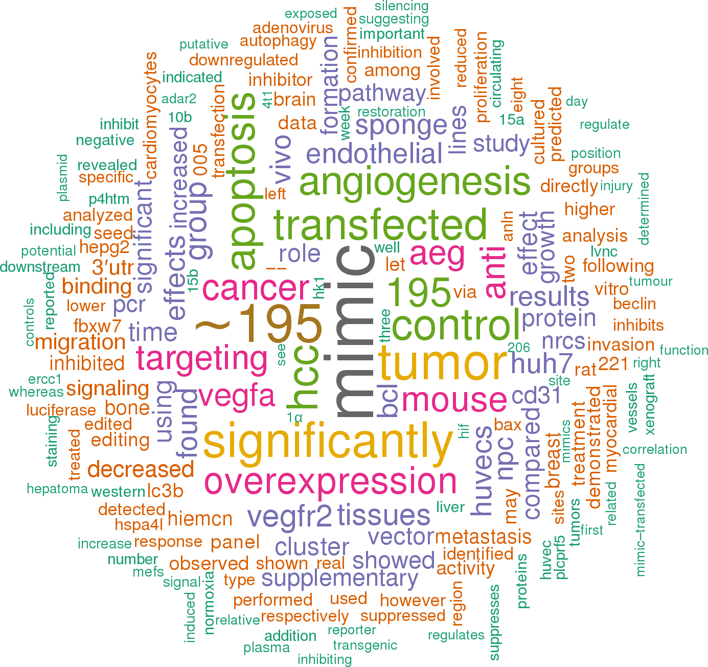
1
[+] score: 442
These data indicated that miR-497 may negatively regulate VEGFA expression at the translational level by directly targeting its 3′UTR. [score:9]
Consistently, the overexpression of VEGFA in miR-497 transfectants obviously abrogated the inhibitory effects of miR-497 on HUVEC migration and capillary tube formation in Huh7 cells (Figure 5), while down-regulation of VEGFA in anti-miR-497 transfectants obviously abrogated the pro-angiogenic effects of anti-miR-497 on HepG2 cells (Supplementary Figure S2). [score:8]
Subsequent experiments confirmed that miR-497 exerted its anti-angiogenic and anti-metastatic effects by directly inhibiting VEGFA and AEG-1. We previously identified a specific miRNA expression profiling in liver cancer and the microarray data were deposited in NCBI's Gene Expression Omnibus (GEO) public database (http://www. [score:8]
Together, the above results demonstrated that miR-497 suppresses HCC metastasis by directly targeting AEG-1. Figure 6 A. miR-497 and its putative binding sequences in the 3′UTR of AEG-1. Mutations were generated in the complementary sites that bind to the seed region of miR-497. [score:7]
In conclusion, our findings indicate that miR-497 suppresses angiogenesis and metastasis of HCC cells in vitro and in vivo by inhibiting the expression of VEGF and AEG-1, and highlight the therapeutic potential of miR-497 in HCC. [score:7]
miR-497 inhibits HCC angiogenesis by directly targeting VEGFA. [score:6]
These data indicated that miR-497 suppressed VEGFA expression and the negative regulation of VEGFA by miR-497 might contribute partially to anti-angiogenesis effects of miR-497 involved in HCC. [score:6]
These results suggested that miR-497 suppressed AEG-1 expression, and the negative regulation of AEG-1 by miR-497 might contribute partially to anti-metastasis effects of miR-497 involved in HCC. [score:6]
MiR-497 inhibits VEGFA expression by targeting its 3′UTR. [score:6]
We also demonstrated for the first time that miR-497 suppressed AEG-1 expression by directly binding to the 3′-UTR of AEG-1. Moreover, restoration of AEG-1 could partially reverse the anti-invasion and anti-metastasis effects induced by miR-497. [score:6]
Previously, we have also identified a group of differentially expressed miRNAs between cancerous hepatocytes and normal primary human hepatocytes through human microRNA arrays, and found that miR-497 was one of the significantly down-regulated miRNAs [13]. [score:6]
Furthermore, we also demonstrated that miR-497 suppressed VEGFA expression by binding directly to the 3′-UTR of VEGFA. [score:6]
To unravel the mechanism underlying miR-497 disrupted angiogenesis, we searched for positive regulators of angiogenesis using miRNA target prediction software (TargetScan and microRNA. [score:6]
Since miR-497 was significantly downregulated in HCC, we successfully constructed a recombinant lentiviral vector named LV-miR-497 (and LV-miR-NC as control) to transduce Huh7 cells (Figure 9A), and increase the expression of miR-497 (Figure 9B). [score:6]
Suppression of VEGFA in HCC cells displayed a significantly reduced capacity to promote HUVEC migration and capillary tube formation, which phenocopied the effects of miR-497 overexpression (Supplementary Figure S1). [score:5]
Furthermore, western blot analysis also showed that the restoration expression of miR-497 in Huh7 cells reduced the expression of VEGFA at the protein level (Figure 4E). [score:5]
These results indicated that overexpression of miR-497 in HCC cells could inhibit pro-angiogenic activity of HCC cells in vitro. [score:5]
The inhibition of migration ability of HUVEC cells by miR-497 overexpression was antagonized by introduction of VEGFA (Olympus DP70, magnification ×200). [score:5]
A. Forty-eight hours after co -transfected with the following RNA duplex/expression plasmid combinations: miR-NC/empty vector pcDNA3.1(+) (panel 1), miR-NC/ AEG-1 (panel 2), miR-497/ pcDNA3.1 (panel 3) or miR-497/ AEG-1 (panel 4), expression of AEG-1 protein in Huh7 cells were analyzed by immunoblotting. [score:5]
For miR-497, it was reported to inhibit ovarian cancer cell migration and invasion through targeting of SMAD specific E3 ubiquitin protein ligase and modulate gastric cancer cell invasion by repressing eIF4E [25, 26]. [score:5]
MiR-497 down-regulation was correlated with the overexpression of AEG-1 in human HCC specimens (Figure 1C and 1D). [score:5]
The migration ability of HUVEC cells were significantly inhibited by miR-497 overexpression in both Huh7 and PLC/PRF/5 cells. [score:5]
D. and E. Overexpression of AEG-1 rescue the anti-invasion effect of miR-497 ectopic expression (Olympus DP70, magnification ×200). [score:5]
Figure 3MiR-497 suppresses HCC migration and invasion in vitro A. Restoration of miR-497 inhibited HCC cell migration. [score:5]
Restoration of miR-497 expression reduced AEG-1, CD34 and VEGFA expression in xenograft tumors established by Huh7 cells. [score:5]
In HCC, previous studies have shown that miR-497 blocked cell cycle at G1 phase by suppressing the expression of CCNE1, CDC25A, CCND3, CDK4, BTRC, and Checkpoint kinase 1 [11, 12]. [score:5]
Consistently, the overexpression of AEG-1 in miR-497 transfectants obviously abrogated the inhibitory effects of miR-497 on cell migration and invasion (Figure 7). [score:5]
Besides, the expression of VEGFA, another potential target gene of miR-497, was significantly higher in HCC tissues than that in matched adjacent non-tumor tissues (Figure 1F). [score:5]
To clarify the cellular mechanisms underlying miR-497 mediated tumor suppression, resected tissues from those subcutaneous xenograft tumors were analyzed to verify AEG-1, CD34 and VEGFA expression. [score:5]
Among the eight HCC cell lines, the expression of miR-497 in Huh7, PLC/PRF/5, SMMC-7721, MHCC-97H, MHCC-97L and Hep3B was much lower than its expression in HepG2 and SK-HEP-1 (Figure 1A). [score:5]
The average miRNA expression in anti-miR-497 group was designated as 1. B. Down-regulation of miR-497 increased the protein levels of cellular VEGFA and AEG-1. HepG2 cells that were transfected with anti-miR-NC (lane 1) or anti-miR-497 (lane 2) for 48 h were analyzed by western blot assay. [score:5]
Wang W et al. have demonstrated that miR-497 suppresses angiogenesis by targeting VEGFA in ovarian cancer, which showed similar results to our findings [9]. [score:5]
Figure 7 A. Forty-eight hours after co -transfected with the following RNA duplex/expression plasmid combinations: miR-NC/empty vector pcDNA3.1(+) (panel 1), miR-NC/ AEG-1 (panel 2), miR-497/ pcDNA3.1 (panel 3) or miR-497/ AEG-1 (panel 4), expression of AEG-1 protein in Huh7 cells were analyzed by immunoblotting. [score:5]
revealed that miR-497 directly suppressed the activity of luciferase reporter with wild-type 3′UTR of AEG-1 (Figure 6B). [score:4]
The migration ability of HUVEC cells were significantly promoted by miR-497 down-regulation in HepG2 cells (Olympus DP70, magnification ×200). [score:4]
F. Down-regulation of miR-497 promotes HepG2 cell migration. [score:4]
As shown in Figure 8, down-regulation of miR-497 significantly increased the protein levels of cellular VEGFA and AEG-1 as well as the secretory levels of VEGFA in CM. [score:4]
Using transwell assays, we observed that both the migratory (Figure 3A) and invasive (Figure 3B) activities of HCC cells were suppressed by miR-497 overexpression. [score:4]
E. VEGFA protein was downregulated in Huh7 cells transfected with miR-497 mimic. [score:4]
Additionally, we found that the expression of miR-497 was also generally suppressed in 8 hepatoma cell lines compared with 2 normal hepatic cell lines. [score:4]
C. AEG-1 mRNA was downregulated in Huh7 cells transfected with miR-497 mimic. [score:4]
Down-regulation of miR-497 promotes angiogenesis, migration and invasion of HepG2 cells. [score:4]
miR-497 represses HCC metastasis by negatively regulating AEG-1 expression. [score:4]
The endothelial recruitment assay, performed in 24-transwell chamber with 8 μm pore insert, revealed that the restoration of miR-497 expression significantly suppressed the ability of HCC cells to promote human umbilical vein endothelial cell (HUVEC) migration (Figure 2B and 2C). [score:4]
Furthermore, in vivo analysis indicated that the induction of miR-497 expression markedly decreased the pulmonary metastasis of circulating HCC cells. [score:3]
C. The amount of secreted VEGFA was increased by inhibition of miR-497. [score:3]
E. Inhibition of miR-497 facilitated the HCC cell–promoted HUVEC tube formation (Olympus DP70, magnification × 100). [score:3]
Here, we disclosed that miR-497 could suppress HCC metastasis and invasion in vitro. [score:3]
Luciferase activity in pLUC-wt-VEGFA group displayed a significant decrease following ectopic expression of miR-497. [score:3]
These data provide strong evidence that miR-497 could inhibit HCC angiogenesis and metastasis in vivo. [score:3]
miR-497 impairs the growth of hepatoma xenografts and suppresses metastasis in vivo. [score:3]
A. Relative expression of miR-497 was detected by SYBR Green qRT–PCR in two hepatoma cell lines (Huh7 and PLC/PRF/5) transfected with miR-NC or miR-497 mimic. [score:3]
revealed that co-transfection of miR-497 significantly inhibited the activity of luciferase reporter with wild-type 3′UTR of VEGFA, whereas this effect was abrogated when the predicted 3′UTR binding site was mutated (Figure 4B). [score:3]
In accordance with the microarray results, the expression of miR-497 was lower in HCC cells than in normal hepatic cell lines (Figure 1A). [score:3]
A. Relative expression of miR-497 detected by qRT–PCR in HepG2 cell lines transfected with anti-miR-497 or anti-miR-NC. [score:3]
Reduced expression of miR-497 was found in multiple cancers including HCC [11]. [score:3]
miR-497 mimics or inhibitors (anti-miR-497) and their matched negative controls (miR-NC or anti-miR-NC) were purchased from Guangzhou Ribobio Co. [score:3]
Next, we examined miR-497 expression levels in 36 pairs of HCC tissues and the corresponding adjacent noncancerous tissues. [score:3]
Among the predicted targets of miR-497, we focused on AEG-1 (Figure 6A) because of its carcinogenicity in several cancers. [score:3]
Figure 8 A. Relative expression of miR-497 detected by qRT–PCR in HepG2 cell lines transfected with anti-miR-497 or anti-miR-NC. [score:3]
Figure 2MiR-497 exerts antiangiogenic activity in vitro A. Relative expression of miR-497 was detected by SYBR Green qRT–PCR in two hepatoma cell lines (Huh7 and PLC/PRF/5) transfected with miR-NC or miR-497 mimic. [score:3]
Figure 5 A. Forty-eight hours after co -transfected with the following RNA duplex/expression plasmid combinations: miR-NC/empty vector pcDNA3.1(+) (panel 1), miR-NC/VEGFA (panel 2), miR-497/ pcDNA3.1 (panel 3) or miR-497/VEGFA (panel 4), Huh7 cells were analyzed by immunoblotting. [score:3]
To further confirm the anti-angiogenic and anti-metastatic function of miR-497 in HCC cells, loss-of-function studies using anti-miR-497 were performed in HepG2 cells which express high levels of miR-497. [score:3]
Luciferase activity in pLUC-wt-AEG-1 group displayed a significant decrease following ectopic expression of miR-497. [score:3]
Recently, the expression level of miR-497 was reported to be reduced in multiple types of cancers including renal cancer [8], ovarian cancer [9], pancreatic cancer [10] as well as HCC [11]. [score:3]
The average miRNA expression in LV-miR-NC group was designated as 1. C. Huh7 cells transfected with lentivirus LV-miR-497 or LV-miR-NC were injected subcutaneously into nude mice. [score:3]
result indicated that enhancing miR-497 expression had no obviously effects on the mRNA level of VEGFA (Figure 4C). [score:3]
A. Forty-eight hours after co -transfected with the following RNA duplex/expression plasmid combinations: miR-NC/empty vector pcDNA3.1(+) (panel 1), miR-NC/VEGFA (panel 2), miR-497/ pcDNA3.1 (panel 3) or miR-497/VEGFA (panel 4), Huh7 cells were analyzed by immunoblotting. [score:3]
Over -expression of VEGFA attenuates the anti-angiogenic effect of miR-497. [score:3]
Our data indicated that the expression of miR-497 in HCC tissues was significantly lower than that in matched adjacent non-tumor tissues, which is in agreement with a previous report [11]. [score:3]
miR-497 suppresses pro-angiogenic and metastasis activity of HCC cells. [score:3]
Overexpression of AEG-1 attenuated the anti-metastatic effect of miR-497. [score:3]
MiR-497 is down-regulated in HCC cells and tissues. [score:3]
Reduced expression of miR-497 in liver cancer cell lines and tissues. [score:3]
Interestingly, the abundance of miR-497 was inversely correlated with that of AEG-1 (Figures 1C and 1D), a potential target of miR-497. [score:3]
Interestingly, predicated target genes of miR-497 including vascular endothelial growth factor A (VEGFA) and astrocyte elevated gene-1 (AEG-1), which play pivotal roles in angiogenesis and metastasis in HCC respectively [14– 16]. [score:3]
In our study, inverse correlation was observed between miR-497 and AEG-1 expression in human HCC tissues, which had not been shown before. [score:3]
B. Restoration of miR-497 inhibited HCC cell invasion. [score:3]
As shown in Figure 2A, the significant increasing of miR-497 expression could be verified by in hepatoma cell lines transfected with 50nM miR-497 mimics. [score:3]
G. Inhibition of miR-497 promotes HepG2 cell invasion. [score:3]
In this study, we examined the expression levels of miR-497 in eight hepatoma cell lines (HepG2, Huh7, PLC/PRF/5, SMMC-7721, SK-HEP-1, MHCC97-H, MHCC97-L and Hep3B) and two normal hepatic cell lines (Chang liver and L02) by to assess the microarray data. [score:3]
Next, the mechanism by which miR-497 inhibited tumor metastasis was elucidated. [score:3]
Both in vitro and in vivo data indicated that miR-497 was capable of suppressing HCC angiogenesis. [score:3]
Consistently, miR-497 was also significantly suppressed in HCC tissues (Figure 1B). [score:3]
A. Restoration of miR-497 inhibited HCC cell migration. [score:3]
These results suggested that miR-497 repressed tumor angiogenesis by inhibiting VEGFA in HCC cells. [score:3]
Lentivirus -based miR-497 overexpression. [score:3]
D. Restoration of miR-497 suppressed the HCC cell–promoted HUVEC tube formation. [score:3]
MiR-497 directly targets AEG-1 in hepatocarcinoma. [score:3]
Restoration of miR-497 in HCC cells significantly suppressed tumor angiogenesis and metastasis in vitro and in vivo. [score:3]
As shown in Figure 10, the LV-miR-497 group displayed reduced AEG-1, CD34 and VEGFA expression in the tumor tissues. [score:3]
D. AEG-1 was inversely correlated with miR-497 expression in the human HCC tissues. [score:3]
D. Expression of miR-497 reduced the protein levels of cellular AEG-1. Huh7 cells that were transfected with miR-NC or miR-497 for 48 h were analyzed by western blot assay. [score:2]
Consistently, LV-miR-497 group showed a status of proliferation inhibition and apoptosis enhancement compared with LV-miR-NC group (Figure 11). [score:2]
However, whether the dysregulation of miR-497 contributes to HCC angiogenesis or metastasis remains unclear. [score:2]
MiR-497 inhibits the tumor angiogenesis and metastasis of hepatoma xenografts in nude mice. [score:2]
The expression levels of miR-497 were analyzed by Bulge-LoopTM miRNA qRT-PCR Primer (RiboBio, China) and normalized to U6. [score:2]
In addition, by using capillary tube formation assays, we observed that the morphological differentiation of HUVEC cells was affected by miR-497 overexpression in HCC cells (Figure 2D and 2E). [score:2]
Mutations were generated in the complementary site that binds to the seed region of miR-497. [score:2]
Moreover, the expression levels of AEG-1 mRNA and protein were both significantly decreased in Huh7 cells transfected with miR-497 compared with controls (Figure 6C and 6D). [score:2]
A. miR-497 and its putative binding sequences in the 3′UTR of AEG-1. Mutations were generated in the complementary sites that bind to the seed region of miR-497. [score:2]
MiR-497 suppresses HCC migration and invasion in vitro. [score:2]
Figure 4 A. Putative binding sequence between the 3′UTR of VEGFA and miR-497. [score:1]
In order to elucidate the role of miR-497 in vivo, we constructed a recombinant lentivirus termed LV-miR-497 to generate stable gain-of-function of miR-497 in hepatoma cells. [score:1]
Effects of miR-497 on the proliferation and apoptosis in vivo. [score:1]
The recombinant lentivirus LV-miR-497 and its control LV-miR-NC were prepared as previously described [30]. [score:1]
Then, LV-miR-497 or LV-miR-NC transfected Huh7 cells were injected in the flanks of athymic nude mice to establish subcutaneous HCC xenograft. [score:1]
A. Effect of miR-497 on the proliferation of xenograft tumors established by Huh7 cells. [score:1]
HepG2 cells that were transfected with anti-miR-NC or anti-miR-497 were added to transwell chambers with Matrigel coatings and incubated for 72 h, followed by staining with crystal violet (Olympus DP70, magnification ×200). [score:1]
In 96-well plates, Hela cells were cotransfected with 0.1 mg of pLUC-wt-gene or pLUC-mut-gene, 0.01 mg of pMIR-REPORT β-galactosidase plasmid served as an internal transfection efficiency control, and miR-NC or miR-497 mimic with a 50 nM final concentration. [score:1]
But the role of miR-497 in HCC metastasis has not been explored. [score:1]
Considering the important roles of miR-497 in HCC, we next used HCC xenograft mo dels to further confirm the above findings in vivo. [score:1]
B. and C. Introduction of AEG-1 antagonized the anti-migration effect of miR-497 (Olympus DP70, magnification ×200). [score:1]
During the further characterization of miR-497 in HCC, we found that miR-497 functioned as an angiogenesis and metastasis suppressor. [score:1]
Huh7 (upper) and PLC/PRF/5 (lower) cells that were transfected with miR-NC (left) or miR-497 (right) mimic were added to transwell chambers with Matrigel coatings and incubated for 24 hours (Huh7) or 36 h (PLC/PRF/5), followed by staining with crystal violet. [score:1]
A. The expressions of miR-497 in eight human liver cancer cell lines (HepG2, Huh7, PLC/PRF/5, SMMC-7721, SK-HEP-1, MHCC97-H, MHCC97-L and Hep3B) and two human normal hepatic cell lines (Chang liver and L02) were measured by. [score:1]
Tumor sizes in LV-miR-497 group were much smaller than that in LV-miR-NC group (Figure 9C, 9D and 9E; tumor incidence for LV-miR-497 versus LV-miR-NC groups: 6/6 versus 6/6). [score:1]
B. Effect of miR-497 on the apoptosis of xenograft tumors established by Huh7 cells. [score:1]
B. pLUC-wt-AEG-1 or pLUC-mut-AEG-1 vector was cotransfected with miR-NC or miR-497 mimics. [score:1]
Huh7 (upper) and PLC/PRF/5 (lower) cells that were transfected with miR-NC (left) or miR-497 (right) mimic were added to transwell chambers and incubated for 24 hours (Huh7) or 36 h (PLC/PRF/5), followed by staining with crystal violet. [score:1]
Huh7 cells transfected with lentivirus miR-497 or miR-NC were injected into the tail vein of nude mice. [score:1]
HepG2 cells that were transfected with anti-miR-NC or anti-miR-497 were added to transwell chambers and incubated for 72 h, followed by staining with crystal violet (Olympus DP70, magnification × 200). [score:1]
Figure 11Effects of miR-497 on the proliferation and apoptosis in vivo A. Effect of miR-497 on the proliferation of xenograft tumors established by Huh7 cells. [score:1]
B. miR-497 (measured by) was down-regulated in HCC tissues compared with the matched adjacent non-tumorous liver tissues. [score:1]
HUVEC cells formed incomplete and fluffy tubular structures in the presence of CM obtained from miR-497 transfected HCC cells. [score:1]
Additionally, LV-miR-NC or LV-miR-497 transfected Huh7 cells were injected into the tail vein of nude mice to assess the metastatic activity. [score:1]
In this study, we have found the possibly effects of miR-497 in HCC angiogenesis for the first time. [score:1]
B. pLUC-wt-VEGFA or pLUC-mut-VEGFA vectors were cotransfected with miR-497 mimic or miR-NC. [score:1]
A. Putative binding sequence between the 3′UTR of VEGFA and miR-497. [score:1]
D. The amount of secreted VEGFA was decreased by restoration of miR-497. [score:1]
The LV-miR-497 or LV-miR-NC transfected Huh7 cells were harvested from tissue culture flasks using trypsin and washed three times with PBS. [score:1]
These results afford reference for the option of cell mo del for further research of miR-497 in HCC. [score:1]
Conditional medium from Huh7 cells that were transfected with miR-NC or miR-497 mimic were analyzed by ELISA. [score:1]
B. SYBR Green qRT–PCR was used to evaluate relative expression of miR-497 in Huh7 cells transfected with lentivirus LV-miR-497 or LV-miR-NC. [score:1]
Moreover, anti-miR-497 could not only promote the migration and tube formation ability of HUVEC cells, but also stimulate the migration and invasion ability of HepG2 cells (Figure 8). [score:1]
D. Introduction of VEGFA antagonized the anti-tube formation effect of miR-497. [score:1]
Subsequently, to clarify the role of miR-497 in HCC metastasis, we analyzed the effects of miR-497 on the migration and invasion ability of HCC cells. [score:1]
HCC cells were placed in the lower compartments and transfected with miR-497 mimics, anti-miR-497 or si-VEGFA or their matched negtive control for 36 h and refreshed with 600 μl serum-free medium before the recruitment experiments. [score:1]
Conditional medium from HepG2 cells that were transfected with anti-miR-NC or anti-miR-497 were analyzed by ELISA. [score:1]
Double-stranded oligonucleotides corresponding to the wild-type (wt) or mutant (mut) miR-497 binding site in the 3′-UTR of VEGFA and AEG-1 were synthesized and subcloned into the pMIR-REPORT system (Applied Biosystems). [score:1]
Figure 1 A. The expressions of miR-497 in eight human liver cancer cell lines (HepG2, Huh7, PLC/PRF/5, SMMC-7721, SK-HEP-1, MHCC97-H, MHCC97-L and Hep3B) and two human normal hepatic cell lines (Chang liver and L02) were measured by. [score:1]
[1 to 20 of 142 sentences]
2
[+] score: 357
And overexpression of miR-497 played an important role in suppressing tumor angiogenesis and inhibiting tumor growth with down-regulation of VEGFR2. [score:10]
The results in this study demonstrated that up-regulation of miR-497 could markedly induce HUVECs apoptosis through inhibiting PI3K/Akt signaling pathway by targeting upstream regulator VEGFR2. [score:9]
And overexpression of miRNA-497 inhibited the survival of 4T1 breast cancer cells via targeting VEGFR2 and its downstream signal pathway proteins Bcl-2 and Bax expression (see Supplementary material online, Fig. S4B–D), which markedly increased 4T1 breast cancer cells apoptosis (see Supplementary material online, Fig. S4E–F). [score:9]
Our current study presented the first evidence that up-regulation of miR-497 greatly induced HUVECs apoptosis and inhibited HUVECs growth via targeting VEGFR2 and its downstream signaling pathway proteins, which provided the direct evidence that miR-497 was able to modulate tumor angiogenesis (summarized in Fig. 7). [score:9]
Firstly, we demonstrated, for the first time, that VEGFR2 was the target gene of miR-497, which could be down-regulated by overexpression of miR-497 in HUVECs and 4T1 breast cancer cells. [score:8]
The results showed that transfection of the miR-497 mimic led to a significant decrease in the expression of Raf, p-MEK, and p-ERK proteins in HUVECs (P < 0.05) (Fig. 2B,D,F), accompanied by parallel down-regulation of VEGFR2 expression. [score:8]
Taken together, these results demonstrated that up-regulation of miR-497 was capable of inhibiting the proliferation of HUVECs via targeting VEGFR2/Raf/MEK/ERK signal pathway. [score:8]
In vitro overexpression of miR-497 is able to induce HUVECs apoptosis and inhibit cell proliferation via targeting VEGFR2/PI3K/AKT and VEGFR2/Raf/MEK/ERK signaling pathways respectively, which play important role in regulating miR-497 -mediated anti-angiogenesis role. [score:8]
The results showed that HUVECs transfected with miR-497 mimic, but not miR-497 inhibitor, proliferated at a lower rate as compared with control group (P < 0.05) (Fig. 2A), indicating that up-regulation of miR-497 could suppress the growth of HUVECs. [score:7]
How to cite this article: Tu, Y. et al. Overexpression of miRNA-497 inhibits tumor angiogenesis by targeting VEGFR2. [score:7]
Zhang et al. identified down-regulation of miR-497 as an important mechanism of up-regulation of IGF1-R in colorectal cancer cells, which contributed to malignancy of colorectal cancer 29. [score:7]
Western blotting analysis displayed that overexpression of miR-497 could down-regulate the endogenous levels of Raf, p-ERK and p-MEK in vivo (P > 0.05) (Fig. 6), meanwhile the expression of total ERK and MEK did not change compared to control (P > 0.05) (Fig. 6). [score:7]
Concomitantly, the expression of VEGFR2 could be successfully suppressed by miR-497 overexpression in vivo, which was consistent with the in vitro results. [score:7]
Overexpression of miR-497 inhibits angiogenesis in 4T1 tumor bearing mice using BLI in vivoFor real-time visualization of the anti-angiogenesis effects of miR-497 via targeting VEGFR2, we used VEGFR2-luc transgenic mice to monitor tumor growth and angiogenesis in vivo by using BLI. [score:7]
These data demonstrate that overexpression of miRNA-497 inhibit the survival of HUVECs via targeting VEGFR2 and its downstream signal pathway. [score:7]
The western blotting results showed that treatment of HUVECs with miR-497 mimic significantly decreased VEGFR2 and its downstream proteins p-AKT, Bcl-2 expression, while increased Bax expression, demonstrating that miR-497 mimic significantly inhibited the angiogenesis responses induced by HUVECs (Fig. 1B–F). [score:7]
In the same way, the HUVECs were transfected with miR-497 inhibitor or inhibitor control at final oligonucleotide concentration of 50 nM to knockdown miR-497. [score:6]
Up-regulation of miR-497 inhibits tumor growth by inducing cell apoptosis. [score:6]
Secondly, as shown in Fig. 7, up-regulation of miR-497 level was able to induce HUVECs apoptosis via targeting VEGFR2/PI3K/AKT signaling pathway and trigger anti-proliferative action through VEGFR2/Raf/ERK/MEK signaling pathway in HUVECs. [score:6]
These data suggest that miR-497 may inhibit translation of VEGFR2 by directly acting on response elements specific for miR-497 in the VEGFR2 3′-UTR region in HUVECs. [score:6]
In order to further demonstrate that miR-497 may inhibit VEGFR2 expression through direct interaction with predicted binding sites located in the 3′-UTR region of VEGFR2 in HUVECs, we cloned a reporter vector in which luciferase cDNA was followed by a fragment of the 3′-UTR from VEGFR2 mRNA containing the putative miR-497 binding sequences. [score:6]
Taken together, the up-regulation of miR-497 inhibits tumor growth by inducing cell apoptosis. [score:6]
HUVECs and 4T1 breast cancer cells were transfected with 100 nmol/L of miR-497 mimic, miR-497 mimic-NC, miR-497 inhibitor, or miR-497 inhibitor-NC for 24 h. miR-497 expression was evaluated by real-time quantitative PCR after transfection. [score:5]
Overexpression of miR-497 inhibits tumor angiogenesis in vivo. [score:5]
Overexpression of miR-497 inhibits tumor angiogenesis in vivoIn this study, immunofluorescent staining with CD31 and CD61 was applied to testify MVD in the tumor sections. [score:5]
Briefly, HUVECs (50 ~ 60% density) transfected with 100 nM miR-497 mimic, miR-497 mimic-NC, miR-497 inhibitor or inhibitor-NC were seeded in 96-well plates and incubated at 37 °C for 48 h. Cells transfected nothing were used as control. [score:5]
One of the predicted targets of miR-497 is VEGFR2 by using the specific program TargetScan (http://www. [score:5]
In addition, miR-497 may induce endothelial cells apoptosis by directly down -regulating anti-apoptotic factor Bcl-2 expression. [score:5]
Overexpression of miR-497 inhibits angiogenesis in 4T1 tumor bearing mice using BLI in vivo. [score:5]
And in vivo BLI data show that miR-497 inhibits tumor angiogenesis via targeting VEGFR2 and its downstream signaling pathways, suggesting that miR-497 may be a promising intervention in the management of tumor angiogenesis and a potential drug candidate for cancer therapy in future. [score:5]
Recently, Zhao et al. found that ectopic expression of miR-497 in vivo significantly inhibited tumor growth and tumor angiogenesis in severe combined immune deficiency (SCID) mouse xenograft mo del of non-small cell lung cancer 35. [score:5]
More importantly, the negative correlation between miR-497 expression and MVD of tumor tissues was found in this study, which suggested that the miR-497 played a vital role in suppressing tumor angiogenesis. [score:5]
In addition, we also demonstrated that overexpression of miR-497 could induce HUVECs apoptosis by targeting Bcl-2 (see Supplementary material online, Fig. S3), which was in line with previous studies 27. [score:5]
In accord with previous study 30, we also found that overexpression of miR-497 led to the inhibition of Raf/MEK/ERK signaling pathway, suggesting miR-497 was involved in modulating proliferation in HUVECs in vitro. [score:5]
Overexpression of miR-497 induced apoptosis of HUVECs and 4T1 cells via targeting VEGFR2. [score:5]
No statistical significance of miR-497 expression was found among the control, miR-497 mimic-NC and miR-497 inhibitor-NC groups (P > 0.05) (Fig. 1A). [score:5]
In line with what we expected, overexpression of miR-497 could significantly inhibit breast tumor growth and produced ant-angiogenesis effect in vivo. [score:5]
The results showed that miR-479 mimic increased while miR-497 inhibitor decreased the expression of miR-497 in HUVECs compared with the control group (P < 0.05). [score:4]
To evaluate the pro-apoptosis role of miR-497 via targeting VEGFR2 and its downstream proteins in vivo, western blotting was performed to detect the expression of VEGFR2, Bax and Bcl-2. The data showed that compared with control, VEGFR2 and Bcl-2 expression in miR-497 mimic group was significant lower, while pro-apoptosis protein Bax was higher (P < 0.05) (Fig. 4B–D). [score:4]
Next, in order to elucidate the anti-angiogenesis effects of miR-497 mediated by down-regulation of VEGFR2/Raf/ERK/MEK pathway in breast tumor, we investigated the expression of angiogenesis-related signal pathway proteins in tumor lysates from these three groups. [score:4]
For the miR-497 up-regulation, the cells were transfected with miR-497 mimic or mimic control (GenePharma Co. [score:4]
Data from TUNEL assay showed that the percentage of apoptosis of each group was as follows: control (6.3 ± 0.5%), miR-497 mimic (35.6 ± 1.2%), mimic-NC (7.2 ± 0.8%), inhibitor (5.2 ± 0.3%), and inhibitor-NC (6.6 ± 0.7%) (see Supplementary material online, Fig. S2). [score:4]
Up-regulation of miR-497 mitigated VEGFR2 level and increased the levels of apoptosis related proteins in human umbilical vein endothelial cells (HUVECs). [score:4]
Effects of miR-497 overexpression on apoptosis in mouse breast tunmor mo del. [score:3]
Overexpression of miR-497 influenced the proliferation of cultured HUVECs. [score:3]
We then tested the influence of miR-497 on the expression of VEGFR2 and its downstream proteins Akt, Bcl-2 and Bax in HUVECs, which were related to apoptosis. [score:3]
In brief, these results not only help us to comprehend the mechanisms underlying the anti-angiogenesis effects of miR-497 but also improve our view of miRNAs that may serve as potential therapeutic and drug targets in future. [score:3]
Thirdly, overexpression of miR-497 produced anti-tumor and anti-angiogenesis effects in breast tumor-bearing mice, and VEGFR2-luc mouse was a useful tool in monitoring the therapeutic anti-angiogenesis effect of miR-497 in vivo. [score:3]
However, the decreased degree of miR-497 in HUVECs after miR-497 inhibitor treatment was not obvious, only down to 50% relative to control group. [score:3]
However, the direct role of miR-497 in regulating tumor angiogenesis has not yet been fully disclosed. [score:3]
miR-497 modulates HUVECs proliferation via targeting VEGFR2 and its downstream Raf/MEK/ERK signal pathway. [score:3]
VEGFR2 is the target gene of miR-497 in HUVECs. [score:3]
For real-time visualization of the anti-angiogenesis effects of miR-497 via targeting VEGFR2, we used VEGFR2-luc transgenic mice to monitor tumor growth and angiogenesis in vivo by using BLI. [score:3]
received intratumoral injections of miR-497 mimic or miR-497 mimic-NC respectively for 1 dose (50 mg/kg mimic or inhibitor dissolved in 100 μL mixed solution) at day 0. The same volume of saline was locally injected into the tumor mass as control. [score:3]
The above results confirmed that miR-497 could markedly inhibit breast tumor angiogenesis in vivo. [score:3]
Moreover, the previous studies provided information that miR-497 had effects on inhibiting cell viability and inducing apoptosis in several tumor cell lines. [score:3]
Effects of miR-497 overexpression on Raf/MEK/ERK signaling pathway in mouse breast tumor mo del. [score:3]
In concordance with our expected, miR-497 mimic had no effect on total MEK and ERK proteins expression (P > 0.05) (Fig. 2C,E). [score:3]
For instance, Liu and co-workers found that has-miR-497 could play a role in both gastric and lung cancer cell lines, at least in part modulating apoptosis via targeting Bcl-2 28. [score:3]
Transfection of miR-497 mimic or inhibitor into cultured HUVECs. [score:3]
In addition, the immunostaining analysis further demonstrated that miR-497 was involved in inhibiting tumor angiogenesis in mouse tumor mo del. [score:3]
To further confirm the role of miR-497 in the anti-proliferative action in HUVECs, we performed gain- and loss-of-function studies on the expression of miR-497 using in vitro cells transfection. [score:3]
Schematic illustrations explain the possible targeting and signalling mechanisms by which miR-497 produces anti-angiogenesis effects in in vitro and in vivo mo del. [score:3]
Creevey et al. verified that overexpression of miR-497 reduced cell viability and increased apoptosis in MNA cells at the same time, which could be used as a potential therapeutic agent for high-risk neuroblastoma 34. [score:3]
Increased miR-497 represses angiogenesis by targeting VEGFR2, which lead to decreases in the activation of both PI3K/AKT and Raf/MEK/ERK signaling pathways. [score:3]
From the above data, we concluded that up-regulation of miR-497 markedly increased HUVECs apoptosis compared to control group (P < 0.05) (see Supplementary material online, Fig. S2). [score:3]
In conclusion, our study indicates that VEGFR2, which is required for inducing angiogenesis, is a key downstream target of miR-497. [score:3]
Then real-time PCR was used to quantify the expression of miR-497 with specific Taqman assays (Applied Biosystems, USA) and Taqman universal master mix (Applied Biosystems, USA). [score:2]
The mice were sacrificed and tumor tissues were removed after in vivo bioluminescent imaging on 14 d. The miR-497 expression level of tumor tissues was confirmed by real-time PCR assay. [score:2]
Compared with the control, the expression level of miR-497 significantly increased in miR-497 mimic group (P < 0.05) (Fig. 4A). [score:2]
In addition, we also found that miR-479 mimic increased the expression of miR-497 in 4T1 breast cancer cells compared with the control group (P < 0.05) (see Supplementary material online, Fig. S4A). [score:2]
However, miR-497 mimic-NC could not play the same role (P > 0.05) (Fig. 2A). [score:1]
We then transfected this luciferase reporter vector with either wild-type or mutant miR-497 binding sequences into HUVECs. [score:1]
Shen et al. reported that miR-497 induced apoptosis of breast cancer cells by acting on Bcl-w 31. [score:1]
In order to disclose the relationship between miR-497 and tumor angiogenesis, VEGFR2-luc transgenic mice were applied to repetitively and non-invasively monitor tumor growth and angiogenesis by using BLI. [score:1]
The tumor sample from miR-497 mimic group had less capillary blood vessels than the control group (35 ± 1.2 vs. [score:1]
However, the miR-497 mimic has no significant effect on the reporter vector with mutated miR-497 binding sequences (see Supplementary material online, Fig. S1C). [score:1]
As shown in Fig. 1B, the miR-497 mimic administered at a concentration of 50 nM decreases luciferase activity of the reporter vector containing miR-497 binding sequences (see Supplementary material online, Fig. S1B). [score:1]
After tumor cells injection for seven days, the VEGFR2-luc transgenic mice with tumor were randomized into three groups with the similar mean tumor volume: control group, miR-497 mimic group, miR-497 mimic-NC group. [score:1]
The following day, cells were co -transfected with 80 ng of pMIR-REPORT Luciferase vector, including the 3′UTR of VEGFR2 (with either wild-type or mutant miR-497 binding sites), pRL-TK control vector (encoding Renilla luciferase, 8 ng), and miR-497 mimic or mimic control at a final concentration of 50 nm by using Lipofectamine 2000 (Invitrogen) according to the manufacturer’s instructions. [score:1]
Furthermore, we synthesized another luciferase reporter fused to the VEGFR2 3′-UTRs, but with a mutant miR-497 binding sequence. [score:1]
However, there was no statistical difference observed between control and miR-497 mimic-NC from 0 d to 14 d (P > 0.05) (Fig. 3A,B). [score:1]
We defined the miR-497 mimic treatment day as 0 d, and as shown in Fig. 3A, there was no significant difference of tumor volume and bioluminescence signal intensity among three groups at 0 d (P > 0.05). [score:1]
Taken together, these results suggested that miR-497 could act as an apoptosis inducer in HUVECs and 4T1 breast cancer cells in vitro. [score:1]
Mice were randomized into three groups with the very similar mean tumor volume: control group, miR-497 mimic group, miR-497 mimic-NC group (n = 5 in each group). [score:1]
The results demonstrated that the mean MVD of control, miR-497 mimic and miR-497 mimic-NC groups was 76 ± 1.6, 35 ± 1.2 and 82 ± 3.5, respectively. [score:1]
Real-time PCR was performed using the SYBR Green PCR Master Mix Kit (Applied Biosystems, USA) on a 7500 Fast Real-Time PCR System (Applied Biosystems, USA) to quantify the level of miR-497. [score:1]
However, tumor sizes of miR-497 mimic treatment group were found to be smaller than those in control at 5 d (P < 0.05) (Fig. 3C), which was much more apparent on 14 d (Fig. 3C). [score:1]
[1 to 20 of 88 sentences]
3
[+] score: 285
In cultured NRCs, showed that miR-497 mimic significantly down-regulated protein expressions of Bcl2, LC3B-II and beclin-1, and up-regulated Bax expression (Figure 4a and 4b). [score:11]
The downregulation of miR-497 in response to myocardial ischemia or IR may work as an important adaptive mechanism to upregulate the expression levels of Bcl2 and LC3B. [score:9]
In non-cardiomyocytes, miR-497 has been demonstrated to promote ischemic neuronal death by downregulating Bcl-2 and Bcl-w [21], and inhibit tumorigenesis as a tumor suppressor [22, 23]. [score:8]
Although the downregulation of miR-497 in response to AR would expect an increase of Bcl-2 in cardiomyocytes, other proapoptotic factors may antagonize this effect and eventually lead to a net effect of Bcl-2 downregulation. [score:7]
Roy et al reported that only miR-15b but no other family members was upregulated in mouse heart subjected to IR for 2 or 7 days [25], while in rats with cerebral IR for 24 h or 48 h, only miR-497 in brain tissue was significantly upregulated [26]. [score:7]
Because a single microRNA can regulate a number of different mRNAs, and the same mRNA can be silenced by multiple miRs, it is therefore important to further explore the target network of miR-497 in ischemic diseases. [score:6]
In mice with MI, myocardial miR-497 expression was dynamically down-regulated from 1 day to 4 weeks (Figure 1b). [score:6]
By using adenovirus carrying short hairpin RNA for becelin-1 (sh-beclin-1) and lysosomal inhibitor Bafilomycin A1 (Baf), we further investigated whether the increase of LC3B-II by miR-497 sponge is affected by beclin 1. As shown in Figure 4f, the upregulation of LC3B-II was enhanced by Baf and antagonized by Ad-sh-beclin-1 and autophagic inhibitor wortmannin. [score:6]
Although Bax is not a predicted target gene of miR-497, its expression can be regulated by miR-497, which is likely secondary to the change of Bcl-2, a similar phenomenon was also reported elsewhere [12]. [score:6]
Myocardial miR-497 expression is consistently downregulated in response to MI or AR. [score:6]
Because complementary sites on the lncRNA (long noncoding RNA) allow them to recognize and bind to miRNAs and act as highly specific sensors for their regulation, identifying the lncRNA that regulates miR-497 would also be helpful for searching new therapeutic targets of myocardial IR. [score:5]
Addition of Baf to miR-497 mimic or sponge increased Bax expression, suggesting that inhibition of autophagic flux promotes apoptosis (Figure 5a–5d). [score:5]
MiR-497 directly targets Bcl2 and LC3-B. MiR-497 mimic induces cardiomyocyte apoptosis and inhibits autophagy. [score:5]
Effects of miR-497 expression on protein expression related to autophagy and apoptosis. [score:5]
Inhibiting lysosomal degradation with Baf increased beclin-1 and LC3B-II expression levels in the presence of miR-497 mimic or sponge (Figure 5a–5d). [score:5]
Furthermore, overexpression of miR-497 increased cardiomyocyte apoptosis and inhibited autophagic flux, whereas silencing of miR-497 reduced cardiomyocyte apoptosis and enhanced autophagic flux. [score:5]
In this study, we clearly demonstrated that overexpression or knockdown of miR-497 alone decreased and increased Bcl-2 protein, respectively. [score:4]
Real-time quantitative PCR showed that miR-497 was expressed in the tissues of heart, lung, kidney, liver and brain, and the expression levels were significantly higher in heart when compared with other tissues (Figure 1a). [score:4]
In the present study, we identified miR-497 as a critical regulator of cardiomyocyte apoptosis and autophagy via targeting Bcl-2 and LC3B genes. [score:4]
To elucidate the in vivo effects of miR-497 inhibition on IR injury, we directly injected Ad-miR-497-sponge or Ad-miR-497 into left ventricular myocardium of mice 3 days before the onset of IR procedure. [score:4]
Overexpression and silencing of miR-497 and becline-1 knockdown. [score:4]
In this study, we found downregulated miR-497 could moderately increase autophagy and reduce myocardial damage during IR (45 min/24 h) and AR (3 h/2 h), which indicate that a moderate enhancement of autophagy would attenuate myocardial injury induced by IR. [score:4]
We found that miR-497 was enriched in cardiac tissue and was dramatically downregulated in response to IR or AR insult. [score:4]
Thus, miR-497 and its downstream targets may serve as valuable therapeutic entry points and thereby improve cardiac performance after IR injury. [score:3]
Our data suggest that miR-497 is a new therapeutic target for myocardial IR injury. [score:3]
In cultured NRCs exposed to AR, loss and gain function of miR-497 also exerted significant influence on protein expression of LC3B-II, beclin-1, Bcl-2 and Bax. [score:3]
Effect of miR-497 mimic and sponge on the expression of Bcl2 and LC3B protein in neonatal rat cardiomyocytes (NRCs). [score:3]
In both gastric and lung cancer cell lines, miR-497 modulates apoptosis by targeting Bcl2 [30]. [score:3]
Intriguingly, miR-497 expression was dramatically decreased in a time -dependent manner when NRCs were exposed to 2 h of reoxygenation following 3–24 h of anoxia (Figure 1c). [score:3]
To inhibit the activity of miR-497, a U6 sponge was used. [score:3]
As a result, miR-497 mimics significantly inhibited autophagosome formation (Figure 2d) in cardiomyocytes during AR. [score:3]
The miR-15 family members including miR-15a, miR-15b, miR-16, miR-195, miR-424, and miR-497, show 5′-end sequence similarity and many common targets [16, 17]. [score:3]
Figure 1 a. Real-time PCR analysis indicates that the miR-497 expression was higher in adult mouse heart than in other tissues (including lung, kidney, liver and brain). [score:3]
Similar to the results in cultured NRCs, treatment with Ad-miR-497-sponge enhanced cardiomyocyte autophagy and inhibited apoptosis in IR mice, while Ad-miR-497 exerted opposite effects (Figure 7a–7e). [score:3]
Either specific mimic or anti -RNA was transfected to overexpress or to silence miR-497 in the cultured cells. [score:3]
Silencing of endogenous miR-497 provides protection against IR -induced cardiomyocyte death and apoptosis by targeting Bcl-2 and LC3B. [score:3]
Western blot shows the protein expression of Bcl-2, Bax and LC3B-II c. beclin-1 and P62 d. which were significantly changed in miR-497 sponge -treated cells e, f. Western blotting of LC3B-II in NRCs treated with/without miR-497 sponge (S), vector (V), Ad-sh-beclin-1 (sh-b), Ad-sh-control (sh-c), bafilomycin A1 (Baf), wortmannin (W). [score:3]
In mouse N2A cells after oxygen-glucose deprivation, miR-497 was found to promote apoptosis by targeting Bcl-2 and Bcl-w [21]. [score:3]
a. Real-time PCR analysis indicates that the miR-497 expression was higher in adult mouse heart than in other tissues (including lung, kidney, liver and brain). [score:3]
The Ad-miR-497-sponge or miR-497 mimic or Ad-sh-beclin-1 was directly transfected to the cultured NRCs (multiplicity of infection (MOI) = 10), while in vivo infection of Ad-miR-497-sponge or Ad-miR-497 in heart was reached by direct myocardial injection through a left thoracotomy in mice anesthetized with a mixture of xylazine (5 mg/kg, intraperitoneal) and ketamine (100 mg/kg, intraperitoneal) as reported elsewhere [35]. [score:3]
In cultured neonatal rat cardiomyocytes (NRCs) exposed to various time periods of anoxia, miR-497 expression was not changed for 3 or 6 h of anoxia but significantly decreased 24 h later (P < 0.001; Figure 1c). [score:3]
50 nM of miR-497 mimic was identified to increase the expression of miR-497 by more than 2 folds (Figure 2a). [score:3]
Using bioinformatics algorithms, anti-apoptosis gene Bcl2, and autophagy gene LC3B, were predicted as putative targets of miR-497 (Figure 1d). [score:3]
Similar to our findings on miR-497, Hullinger et al has demonstrated that miR-15b, also a member of miR-15 family, aggravates myocardial IR injury by targeting Bcl-2 [15]. [score:3]
Overexpression of miR-497 enhanced apoptosis and autophagy in cultured neonatal rat cardiomyocytes (NRCs). [score:3]
b. Levels of myocardial miR-497 expression determined by real-time-PCR in different groups. [score:3]
MiR-497 sponge exerted opposite effects and increased autophagic influx as determined by P62 downregulation (Figure 4c–4e). [score:3]
In human neuroblastoma cells, overexpression of miR-497 was shown to increase reactive oxygen species formation, disrupt mitochondrial membrane potential, and induce cytochrome C release [24]. [score:3]
Western blot shows the effects of miR-497 overexpression on Bcl-2, Bax, LC3B-II and beclin-1. b. Quantitation for panel A. NRCs were transfected with miR-497 sponge or vector adenovirus. [score:3]
Effects of miR-497 knockdown in cultured neonatal rat cardiomyocytes (NRCs) exposed to anoxia/reoxygenation (A/R). [score:2]
Briefly, Ad-miR-497-sponge or Ad-miR-497 or the corresponding control virus particles (1 × 10¹¹ viral genomes/ml) were administered by direct injection in the left ventricular free wall (3 sites, 20 μl/site) using a syringe with a 30-gauge needle, and 3 days later, sham or IR surgery was performed. [score:2]
MiR-497 and its target proteins were changed in response to myocardial infarction (MI) or anoxia/reoxygenation. [score:2]
The mir-497 clones were sequenced completely to confirm the absence of cloning artifacts and mutation. [score:2]
MTT (3-(4,5-dimethyl thiazol-2-yl)-2,5-diphenyl tetrazolium bromide) cell viability assay shows that inhibition of miR-497 increased cell survival in the context of AR (Figure 5e). [score:2]
In cultured cardiomyocytes under normoxia, change of miR-497 expression levels exerted influence on autophagic signal but did not lead to detectable change of vesicle formation as measured by non-selective MDC staining or EM, while under AR, miR-497 significantly regulated both autophagic flux and vesicle formation. [score:2]
Accumulating evidence shows that miR-497 is involved in the regulation of apoptosis. [score:2]
Adenovirus carrying miR-497-sponge or miR-497 were directly injected into the myocardium. [score:2]
MiR-497 is predicted to target anti-apoptosis gene Bcl-2 and autophagy gene microtubule -associated protein 1 light chain 3 B (LC3B). [score:2]
We noted that miR-497 mimics decreased Bcl-2, LC3B-II and beclin-1 and increased Bax in cardiomyocytes in the presence of AR (Figure 5a–5b), while opposite effects were noted by treatment with miR-497 sponge (Figure 5c–5d). [score:1]
a. NRCs were transfected with miR-497 mimic or negative control (NC). [score:1]
The pEntr-U6-miR-497-sponge (Ad-miR-497-spong) was transfected to 293A cells using lipofectamine 2000. [score:1]
Figure 5 a. Examples of western blotting showing effects of miR-497 sponge and Baf on LC3B-II, beclin-1, Bcl-2 and Bax. [score:1]
Two-way ANOVA analysis was performed, F = 29.254 for treatment with miR-497 sponge (S) or vector (V), P < 0.001; F = 403.582 for treatment with normoxia or A 3 h/R 2 h, P < 0.0001. [score:1]
Replication deficient adenoviruses for miR-497 sponge and null virus were prepared using the Getaway System (Invitrogen) according to the manufacturing's instructions. [score:1]
c. Hoechst staining shows that miR-497 sponge -treated cardiomyocytes were resistant to A/R -induced apoptosis. [score:1]
Consistently, miR-497 levels were significantly decreased when the mice were subjected to ischemia for 45 min and reperfusion for 24 h. Meanwhile, miR-497 levels were further reduced in Ad-miR-497-sponge -treated group but significantly increased in Ad-miR-497 -treated group (Figure 6b). [score:1]
Transfections of miR-497 mimic were performed with a Lipofectamine RNAiMAX kit (Invitrogen) according to the manufacturer's instructions. [score:1]
c. Effect of miR-497 mimic on cell apoptosis determined by TUNEL in NRCs exposed to normoxia or anoxia/reoxygenation. [score:1]
Figure 3 a. Infective efficiency and silencing effect of adenovirus-miR-497 sponge and vector in NRCs after 24 h adenoviral infection. [score:1]
Two-way ANOVA test was performed, F = 43.090 for treatment with miR-497 mimic (M) or NC (N); F = 106.406 for treatment with normoxia or anoxia/ reoxygenation (A 3 h/R 2 h), n = 9 for each group. [score:1]
Preparation of adenovirus-miR-497. [score:1]
Myocardial infarct size determined with triphenyltetrazolium chloride (TTC) staining was significantly smaller in mice treated with Ad-miR-497-sponge than in mice receiving scramble vector infection, while myocardial infarct size was larger in Ad-miR-497 -treated mice than in control vector -treated group (Figure 6c and 6d). [score:1]
Figure 2 a. The levels of miR-497 determined by real time-PCR in response to 24 h transfection of miR-497 mimic or negative control (NC). [score:1]
Sponge indicates miR-497 sponge, Mimic indicates miR-497 mimic. [score:1]
The cardioprotective effect of anti-miR-497 was further confirmed in mice with IR insult. [score:1]
c. In neonatal rat cardiomyocytes exposed to anoxia or anoxia/reoxygenation, miR497 was also changed in a time -dependent manner. [score:1]
Figure 4 a. NRCs were transfected with miR-497 mimic or negative control (NC). [score:1]
b. Effect of miR-497 mimic on cell apoptosis determined by Hoechst staining in NRCs exposed to normoxia or anoxia/reoxygenation (A/R). [score:1]
a. The levels of miR-497 determined by real time-PCR in response to 24 h transfection of miR-497 mimic or negative control (NC). [score:1]
d. Quantitation of myocardial infarct size in 4 IR groups treated with scramble, miR-497 sponge, vector and miR-497 mimic, respectively. [score:1]
These results indicate that silencing of miR-497 attenuates AR injury in cardiomyocytes. [score:1]
Briefly, the oligonucleotides for miR-497 were synthesized by Shanghai General Biotech with the following sequences: 5′-gtacaaaccacagtgtgctgctgcgtatacaaaccacagtgtgctgctggcatg-3′ (forward) and 5′-ccagcagcacactgtggtttgtatacgcagcagcacactgtggtttgtactgca-3′ (reverse), which contained two perfect miR-497 binding sites. [score:1]
Effects of Ad-miR-497 sponge and Ad-miR-497 on myocardial autophagy and apoptosis in mice subjected to ischemia/reperfusion (IR). [score:1]
To further examine the contribution of miR-497 to AR injury, NRCs were transfected with the Ad-miR-497-sponge. [score:1]
Effect of miR-497 on proteins related to autophagy and apoptosis in neonatal rat cardiomyocytes (NRCs) exposed to anoxia/reoxygenation (A/R). [score:1]
d. Effect of miR-497 mimic on formation of autophagosome (indicated by arrow) which was determined by monodansylcadaverine (MDC) staining in NRCs exposed to normoxia or anoxia/reoxygenation (A/R). [score:1]
Double-stranded miR-497 mimic and the miRNA negative control (NC) (GenePharma, Shanghai, China) at a final concentration of 50 nM were introduced into the NRCs. [score:1]
The Hoechst and TUNEL staining analyses showed that miR-497-sponge exerted no influence on cardiomyocytes under normoxia, but significantly decreased apoptosis in cardiomyocytes with AR insult (P < 0.05, Figure 3c and 3d). [score:1]
Two-way ANOVA F = 8.944 for treatment with miR-497 mimic or NC, P < 0.05, while F = 63.246 for treatment with normoxia or anoxia/ reoxygenation, P < 0.001. [score:1]
Preparation of adnovirus-miR-497 sponge. [score:1]
Autophagy detected by MDC was significantly enhanced in miR-497 sponge -treated NRCs than in vector -treated cells (Figure 3f and 3g). [score:1]
These results indicate a pro-apoptosis and an anti-autophagic role of miR-497 in cardiomyocytes. [score:1]
To characterize miR-497 function, we overexpressed synthetic mature miR-497 (miR-497 mimic) in NRCs. [score:1]
a. Examples of western blotting showing effects of miR-497 sponge and Baf on LC3B-II, beclin-1, Bcl-2 and Bax. [score:1]
MiR-497 regulates autophagy and apoptosis related signal proteins in the context of AR. [score:1]
In response to AR, the apoptosis was significantly enhanced by miR-497 mimic (P < 0.01; Figure 2b and 2c). [score:1]
Two-way ANOVA analysis was performed, F = 6.182 for treatment with miR-497 sponge or vector or control, P < 0.01; F = 199.523 for treatment with normoxia or A 3 h/R 2 h, P < 0.001. [score:1]
As shown in Figure 3a, a satisfactory infection efficiency of miR-497 sponge and silencing effect were obtained. [score:1]
Note the complementarity at the 5’ and 3’ ends of miR-497, where the crucial seed regions are located. [score:1]
Myocardial miR-497 levels were decreased in a time -dependent manner in response to MI for 1 day to 4 weeks in mice. [score:1]
These findings indicate that miR-497 is involved in myocardial ischemia and preferentially in IR injury, suggesting that miR-497 is an IR-related microRNA in the murine heart. [score:1]
Two-way ANOVA test was performed, F = 69.131 for treatment with miR-497 mimic or NC; F = 138.498 for treatment with normoxia or anoxia/ reoxygenation, n = 5. Experiments were repeated 3 times. [score:1]
Briefly, pDC316-mCMV-EGFP-miR-497 and virus backbone plasmid pBHGlox delIE13cre were co -transfected into cultured HEK293 cells by using polyfectamine, then the recombinant adenovirus were collected and amplified in HEK293 cells. [score:1]
d. Sequence alignment of the 3′UTR of Bcl-2 and LC3B from various species with the seed sequence of miR-497. [score:1]
a. Infective efficiency and silencing effect of adenovirus-miR-497 sponge and vector in NRCs after 24 h adenoviral infection. [score:1]
The AdMAXTM system was used for the generation of recombinant adenovirus carrying miR-497 or empty vector (pEGFP). [score:1]
Mouse miR-497 (synthetized by Invitrogen) was inserted into vector pDC316-mCMV-EGFP by using restriction enzymes NheI and HindIII. [score:1]
Sponge, adenovirus-miR-497 sponge; miR-497, adenovirus-miR-497. [score:1]
Immediately after transfection of miR-497 mimic or sponge or vector, NRCs were placed in either a low oxygen atmosphere or normoxia conditions. [score:1]
monodansylcadaverine (MDC) is the specific marker for autolysosomes, thus we examined the effect of miR-497 mimic on the incorporation of MDC into NRCs in response to AR. [score:1]
Sponge = adenovirus-miR-497 sponge; miR-497 = adenovirus-miR-497. [score:1]
Two-way ANOVA analysis was performed, F = 3.456 for treatment with miR-497 sponge or vector or control, P < 0.05; F = 131.769 for treatment with normoxia or anoxia/ reoxygenation (A 3 h/R 2 h), P < 0.001. [score:1]
Silencing of miR-497 improves cell survival. [score:1]
However the exact function of miR-497 in cardiomyocytes or heart remains completely unknown. [score:1]
Construction of plasmid pDC316-mCMV-EGFP-CMV-mir-497 and adenovirus packaging of the recombinant plasmid were completed by a professional company (Biowit Technologies, Shenzhen, China). [score:1]
They were synthesized by GenePharma Company with the following sequences: rno-miR-497 mimics (5′-cagcagcacacugugguuugua-3′) and miRNA NC (5′-uucuccgaacgugucacgutt-3′) which shares minimal sequence identity in mammals. [score:1]
b. Quantitation of panel A. c. Examples of western blotting showing effects of miR-497 mimic and Baf on LC3B-II, beclin-1, Bcl-2 and Bax. [score:1]
[1 to 20 of 117 sentences]
4
[+] score: 185
We confirmed that six of the eight selected down-regulated hsa-miRNAs (miR-145, miR-497, miR-150, miR-342-5p, miR-34b* and miR-100) were significantly down-regulated in NPC tissues, whereas miR-195 and miR-143 exhibited no significant difference between the two groups of subjects (Fig. 1D). [score:7]
Thus, on the basis of our results as well as recent reports, we suggest that down-regulation of miR-497 in NPC plays a causative role in NPC via up-regulation of ANLN and HSPA4L. [score:7]
Exogenous miR-497 significantly down-regulated the mRNA levels of ANLN (Fig. 5A, left) and HSPA4L (Fig. 5A, right), indicating that miR-497 can regulate the expression of these genes in NPC cells. [score:7]
We confirmed the up-regulation of ANLN and HSPA4L in NPC primary tumors, and found that exogenous miR-497 indeed caused down-regulation of ANLN and HSPA4L in NPC cells. [score:7]
Down-regulation of miR-497 may contribute to tumor growth and angiogenesis by targeting HDGF (hepatoma-derived growth factor) in non-small cell lung cancer [34]. [score:6]
To search for target genes of miR-497, we used TargetScan Human (Release 6.2: June 2012, http://www. [score:5]
Zheng et al. [37] showed that IL-1–mediated IL6 expression was significantly repressed by miR-497 via the MAPK/ERK pathway, suggesting that miR-497 may play a suppressive role in inflammation-related cancers such as NPC [38]. [score:5]
F. Pearson correlation of miR-497 expression (expressed on the ΔCt scale) between tissues and plasma. [score:5]
Our findings suggest that miR-497 acts as a tumor suppressor by targeting ANLN and HSPA4L in NPC. [score:5]
Using the TargetScan algorithm, we identified two genes, ANLN (anillin, actin -binding protein) and HSPA4L (heat shock 70 kDa protein 4–like) as potential targets of miR-497. [score:5]
Target gene expression levels in NPC cells following transfection with miR-497 mimic. [score:5]
In addition, miR-497 plays a tumor-suppressive role in human cancer cell lines by targeting BCL2, thereby inducing apoptosis [36]. [score:5]
Among the dysregulated human miRNAs detected in tissues, miR-497 was concordantly down-regulated in plasma; therefore, miR-497 is a promising novel biomarker for diagnosis of NPC. [score:5]
MiR-497 inhibited tumor growth in vivoTo examine the role of miR-497 in NPC development, we performed a xenograft study in which miR-497 mimic– or control mimic–transfected HK1 and HONE1 cells were transplanted into the flanks of BALB/c athymic nu/nu mice. [score:4]
Compared to the negative control, miR-497 mimic significantly inhibited growth of HK1/EBV, HK1, and CNE1 cells (Fig. 2A, left, middle, right, respectively), suggesting that miR-497 has a tumor-suppressive function. [score:4]
The results of these analyses revealed concordant down-regulation of miR-497 in tissues and plasma. [score:4]
miR-497 is down-regulated in human tumors, such as prostate cancer [22], hepatocellular carcinoma [23], neuroblastoma [24], cervical cancer [25], breast cancer [26], colorectal cancer [27], and gastric cancer [28]. [score:4]
Therefore, epigenetic silencing by DNA methylation may be one of the mechanisms underlying down-regulation of miR-497 in EBV-related NPC. [score:4]
Significant down-regulation of ANLN and HSPA4L in NPC cells by exogenous miR-497. [score:4]
Thus, miR-497 was concordantly down-regulated in tissues and plasma, suggesting that it would could be useful as a biomarker for NPC. [score:4]
Circulating miR-497 is significantly down-regulated in plasma of NPC. [score:4]
The mechanism responsible for miR-497 down-regulation remains unknown. [score:4]
Prior to our in vitro functional analyses, we confirmed by quantitative RT-PCR that miR-497 was down-regulated in NPC cell lines, as it is in NPC tissues relative to NNE tissues (Supplementary Fig. 2A). [score:4]
In conclusion, the concordant down-regulation of miR-497 in tissues and plasma make this miRNA potentially very useful as a diagnostic biomarker for NPC. [score:4]
Tumor growth in vivo was also suppressed by exogenous miR-497. [score:3]
In addition, we confirmed that miR-497 was expressed at significantly higher levels in the miR-497 mimic tumors at day 13 (Supplementary Fig. 2E). [score:3]
Expression of miR-497 was significantly correlated between tissues and plasma (Fig. 1F, Pearson correlation coefficient; r = 0.490, P = 0.007), whereas the other miRNAs exhibited no significant correlation (Table S1). [score:3]
The tumor xenograft experiment indicated that miR-497 plays a role in suppressing NPC tumorigenicity. [score:3]
Specifically, we transfected miR-497 mimic and negative control into NPC cell lines, and confirmed the expression level at the indicated time points (Supplementary Fig. 2B-2D). [score:3]
A. Expression levels of ANLN (left) and HSPA4L (right) in HK1 cells (n = 4) after transfection with miR-497 mimic. [score:3]
Our functional studies using miR-497 mimic demonstrated that this miRNA efficiently suppresses cell proliferation and migration, and significantly induces apoptosis. [score:3]
Figure 5 A. Expression levels of ANLN (left) and HSPA4L (right) in HK1 cells (n = 4) after transfection with miR-497 mimic. [score:3]
Notably, we demonstrated in this study that ANLN and HSPA4L are potential targets of miR-497. [score:3]
E. miR-497 expression levels were determined by quantitative RT-PCR in plasma of the same patients mentioned above. [score:3]
Exogenous miR-497 suppresses cell proliferation and migration, and induces apoptosis, in NPC cell lines. [score:3]
Expression levels of miR-497 were significantly higher in NPC cells treated with miR-497 mimic for 72 h than in cells treated with the control mimic, although the levels of miR-497 gradually decreased after mimic transfection (Supplementary Fig. 2B–2D). [score:3]
Transfection of a miR-497 mimic or small interfering RNAs (siRNAs) against ANLN and HSPA4L inhibited cancer phenotypes in NPC cells. [score:3]
miR-497 inhibits NPC tumor growth in vivo. [score:3]
To examine the role of miR-497 in NPC development, we performed a xenograft study in which miR-497 mimic– or control mimic–transfected HK1 and HONE1 cells were transplanted into the flanks of BALB/c athymic nu/nu mice. [score:2]
This study demonstrated for the first time that miR-497 is a regulator of ANLN and HSPA4L. [score:2]
One of the the 13 dysregulated miRNAs detected in tissues, miR-497, was present at significantly lower levels in NPC plasma than in noncancerous control plasma (P < 0.01, Fig. 1E), whereas the levels of other miRNAs were not significantly altered (Table S1). [score:2]
MiR-497 inhibited tumor growth in vivo. [score:2]
miR-497 mimic induced activation of caspase-3 protein, which exhibited cytoplasmic localization (an arrow and the enlarged). [score:1]
C. Cell migration in NPC cells transfected with miR-497 mimic. [score:1]
To investigate the biological function of miR-497 down-regulation in NPC, we assessed the effect of exogenously administered miRNA on cell proliferation using the MTT assay. [score:1]
Subcutaneous tumor growth of miR-497 mimic–transfected cells was slower than that of control mimic–transfected cells, and tumor volumes were significantly smaller until day 14 (HK1: Fig. 3A inset, HONE1: Fig. 3D). [score:1]
Additionally, immunocytochemistry (ICC) analyses revealed that protein levels of ANLN (Fig. 5B, left) and HSPA4L (Fig. 5B, right) were significantly decreased by transfection with miR-497 mimic. [score:1]
Although reduced expression of miR-497 was documented in a previous report [33], the biological function of miR-497 has not been characterized. [score:1]
HK1/EBV (left), HK1 (middle), and CNE1 (right) cells were transfected with control or miR-497 mimic, and then cultured for 72 h. After transfection, harvested cells were seeded in a migration chamber and cultured for 24 h. Migratory cells were stained, photographed under a microscope, and quantitatively analyzed (n = 3). [score:1]
HK1 and CNE1 cells were transfected with control or miR-497 mimic, and then stained with annexin V-FITC and PI and subjected to flow cytometry (n = 3). [score:1]
Because tumor growth of HK1 cells was relatively slow, we continued the observation after day 14, but thereafter there was no significant difference in the volumes of HK1 xenografts (Fig. 3A) or tumor weight at sacrifice (day 26: miR-497 mimic, 30.0 ± 19.0 mg vs. [score:1]
Caspase-3, which is activated in apoptotic cells, was observed in the cytoplasm of HK1/EBV cells transfected with miR-497 mimic, but not in cells transfected with control mimic. [score:1]
Subcutaneous xenografts were established by inoculating 2 × 10 [6] miR-497 mimic–transfected cells into the left flank, or an equal number of control mimic–transfected cells into the right flank. [score:1]
Furthermore, the percentage of cells with activated caspase-3 was significantly higher in miR-497 mimic–transfected cells (Fig. 2B, left). [score:1]
In HK1 (Fig. 2B, middle) and CNE1 cells (Fig. 2B, right) transfected with miR-497 mimic, the apoptosis rate was significantly higher than in cells transfected with control mimic, suggesting the involvement of miR-497 in inducing apoptosis in NPC cells. [score:1]
Therefore, we focused on miR-497 in our subsequent exploration of miRNA functions in NPC. [score:1]
Migratory cells were observed less frequently among NPC cells transfected with miR-497 mimic than among those transfected with control mimic (Fig. 2C). [score:1]
The tumor weight of HONE1 xenografts was significantly lower in the miR-497 mimic tumors than in the control mimic tumors (322.8 ± 94.5 mg vs. [score:1]
C. Image of subcutaneous xenografts in the mouse flanks (the arrows indicate the presence of tumors in mouse injected with control mimic–transfected cells (R) and miR-497 mimic–transfected cells (L)) and excised tumors. [score:1]
[1 to 20 of 59 sentences]
5
[+] score: 142
Overexpression of miR-497∼195 promoted the CD31 and endomucin mRNA expression (Fig. 6c,d), whereas inhibition of miR-497∼195 attenuated the CD31 and endomucin mRNA expression in BMECs (Fig. 6c,d). [score:9]
Endothelial-cell-specific overexpression of the Notch1 intracellular domain (NICD) increased the number of type H vessel 6. We found that HIF-1α and NICD protein levels were up-regulated by overexpression of miR-497∼195 (Fig. 6e–g), however the mRNA level of HIF-1α and Notch1 were not changed (Supplementary Fig. 6a,b). [score:8]
To predict the targets of miR-497∼195, we employed TargetScan. [score:5]
Our study showed that EC-specific miR-497∼195 activation post transcriptionally inhibited Fbxw7 and P4HTM expression, maintained Notch activity and HIF-1α protein stability in endothelial cells and promoted type H vessel formation. [score:5]
Our results demonstrated that overexpression or inhibition of miR-497∼195 cluster altered endogenous levels of Fbxw7 and P4HTM protein without changing mRNA level (Fig. 6e,h,i and Supplementary Fig. 6c,d). [score:5]
We observed moderate elevation of miR-15a, miR-15b and miR-16 expression levels, and great increase in miR-497∼195 expression in CD31 [hi]Emcn [hi] endothelial cells compared to CD31 [lo]Emcn [lo] endothelial cells (Supplementary Fig. 1a). [score:4]
Grünhagen et al. 26 showed that miR-497∼195 cluster regulates osteoblast differentiation by targeting BMP signalling. [score:4]
MiR-497∼195 cluster was strongly expressed in CD31 [hi]Emcn [hi] endothelial cellsTo determine the role of miRNAs in type H vessel generation, CD31 [hi]Emcn [hi] and CD31 [lo]Emcn [lo] endothelial cells were sorted by fluorescence-activated cell sorting (FACS) from bone marrow cells of 1-month-old C57BL/6 mice to identify dysregulated miRNAs by performing miRNA microarray analysis. [score:4]
MiR-497∼195 cluster transgenic mice showed increased CD31 [hi]Emcn [hi] vessels and bone formationWe then generated transgenic mice overexpressing miR-497∼195 specifically in endothelial cells to investigate whether overexpression of miR-497∼195 in vivo would contribute to CD31 [hi]Emcn [hi] vessels growth and bone formation. [score:3]
These results demonstrate that miR-497∼195 maintains Notch and HIF-1α activity by targeting Fbxw7 and P4HTM. [score:3]
Among the predicted genes, Fbxw7 (F-box and WD-40 domain protein 7) and P4HTM (Prolyl 4-hydroxylase, transmembrane) genes were the novel targets of miR-497∼195. [score:3]
Conversely, mice transgenically overexpressing miR-497∼195 in endothelial cells showed significantly higher bone mass and more CD31 [hi]Emcn [hi] vessels. [score:3]
MiR-497 and miR-195 belong to miR-15 family, thus we also tested the expression of other members of the miR-15 family in bone marrow endothelial cells (BMECs). [score:3]
Taken together, the results suggest that overexpression of miR-497∼195 in endothelial cells promotes CD31 [hi]Emcn [hi] vessels formation and osteogenesis in mice. [score:3]
BMECs were sorted by FACS and transfected with the agomiR-497∼195 or antagomiR-497∼195 to overexpress or silence miR-497∼195 cluster, respectively (Fig. 6a,b). [score:3]
We then generated transgenic mice overexpressing miR-497∼195 specifically in endothelial cells to investigate whether overexpression of miR-497∼195 in vivo would contribute to CD31 [hi]Emcn [hi] vessels growth and bone formation. [score:3]
The expression of miR-15a, miR-15b and miR-16 showed no significant difference between miR-497∼195 [−/−] and miR-497∼195 [lox/lox] controls indicating that genetic manipulation of miR-497∼195 in ECs has little effect on other members of the miR-15 family (Supplementary Fig. 2b). [score:3]
However, our results show that the miR-497∼195 cluster is strongly expressed in CD31 [hi]Emcn [hi] endothelial cells that promote angiogenesis and osteogenesis by maintaining endothelial Notch and HIF-1α activity in murine long bone. [score:3]
The higher level of miR-497∼195 cluster expression in CD31 [hi]Emcn [hi] endothelial cells was further confirmed by quantitative real-time PCR (qRT-PCR; Fig. 1b). [score:3]
One line with a highest (sixfold) overexpression of miR-497∼195 in endothelial cells was selected from five transgenic founders and bred in C57BL/6 strain for six generations to obtain offsprings with a defined genetic background. [score:3]
To clarify whether miR-497∼195 cluster can directly regulate Fbxw7, luciferase reporter constructs containing the unaltered or mutated predicted miRNA -binding site of Fbxw7 (WT-pGL3-Fbxw7 and MUT-pGL3-Fbxw7, respectively) were generated. [score:3]
qRT-PCR confirmed that the expression level of miR-497∼195 cluster in miR-497∼195 [−/−] mice was one third of which in miR-497∼195 [lox/lox] controls (Supplementary Fig. 2a). [score:3]
In the present study, we found that the miR-497∼195 cluster, which is strongly expressed in CD31 [hi]Emcn [hi] endothelial cells and gradually decreased during ageing, plays an important part in type H vessel formation. [score:3]
By contrast, inhibition of miR-497∼195 attenuated NICD and HIF-1α protein in BMES without influencing the mRNA level of Notch1 and HIF-1α. [score:3]
Among them, the expression of miR-497 and miR-195, which are found clustered at the same locus, is 3 times higher in CD31 [hi]Emcn [hi] than in CD31 [lo]Emcn [lo] endothelial cells (Fig. 1a). [score:3]
We used qRT-PCR to detect the expression of miR-497 and miR-195. [score:3]
The fraction of EdU [+] ECs in bone marrow was significantly increased in Tg mice relative to their WT control indicating a positive regulatory role for miR-497∼195 in bone ECs proliferation. [score:2]
qRT-PCR revealed five folds higher of miR-497∼195 expression in Tg mice as compared with that in controls (Supplementary Fig. 4a) and no alterations in the transcription of miR-15a, miR-15b and miR-16 (Supplementary Fig. 4b). [score:2]
The Fbxw7 and P4HTM mutants for the miR-497∼195 seed regions were prepared using the QuikChange Site-Directed Mutagenesis Kit (Stratagene) to get mouse MUT-pGL3-Fbxw7 or MUT-pGL3-P4HTM. [score:2]
To generate endothelium specific miR-497∼195 knockout mice, mice carrying loxP-flanked miR-497∼195 alleles (miR-497∼195 [lox/lox]) and Cdh5-Cre transgenics were interbred. [score:2]
MiR-497∼195 cluster targets Fbxw7 and P4HTM. [score:2]
For endothelium specific miR-497∼195 knockout experiment, five to six male mice were used for each group at each observed time point (1, 3 and 12 months) for each independent experiment. [score:2]
MiR-497∼195 cluster targets Fbxw7 and P4HTM to maintain endothelial Notch and HIF-α activity. [score:2]
MiR-497∼195 cluster was strongly expressed in CD31 [hi]Emcn [hi] endothelial cells. [score:2]
These data suggest that miR-497∼195 may play an important role in regulating type H vessel formation and degeneration along with ageing in mouse and human. [score:2]
How to cite this article: Yang, M. et al. MiR-497∼195 cluster regulates angiogenesis during coupling with osteogenesis by maintaining endothelial Notch and HIF-1α activity. [score:1]
Flow cytometric analysis showed a significant decrease of CD31 [hi]Emcn [hi] endothelial cells number in bone marrow of miR-497∼195 [−/−] mice as well as decreased fraction of EdU [+] bone ECs (Fig. 3c,d and Supplementary Fig. 2c,d). [score:1]
Notably, the miR-497∼195 levels in human and mice endothelial cells sorted from bone marrow cells were negatively correlated with age (Fig. 1c–f). [score:1]
MiR-497∼195 cluster knockout mice showed impaired CD31 [hi]Emcn [hi] vessels and bone formationTo investigate the role of miR-497∼195 in endothelial cells in vivo, we generated endothelial-cell-specific miR-497∼195 cluster knockout (miR-497∼195 [−/−]) mice by combining loxP-flanked miR-497∼195 alleles (miR-497∼195 [lox/lox]) and Cdh5 (PAC)-Cre transgene. [score:1]
Calcein double labelling confirmed that miR-497∼195 [−/−] mice had decreased bone formation rates (BFRs; Fig. 4f–h). [score:1]
First, we revealed the role of the miR-497∼195 cluster in the formation of type H vessel. [score:1]
We transfected WT-pGL3-Fbxw7 or MUT-pGL3-Fbxw7 with agomiR-497∼195 or agomiR-NC into BMECs and measured the effects of miR-497∼195 on luciferase translation by the level of luciferase enzyme activity. [score:1]
017968) and loxP-flanked miR-497∼195 mice (Stock No. [score:1]
Endothelial-cell-specific MiR-497∼195 cluster knockout mice showed less CD31 [hi]Emcn [hi] vessels formation. [score:1]
Taken together, these results suggest that miR-497∼195 [−/−] mice have reduced CD31 [hi]Emcn [hi] vessels and bone formation. [score:1]
As the animals aged, the decreasing of the miR-497∼195 cluster in endothelial cells was correlated with the pronounced reduction of CD31 [hi]Emcn [hi] vessels and remarkable decrease of Osterix [+] osteoprogenitors (Fig. 2a–d) as well as the loss of bone mass (Fig. 2e–i). [score:1]
Endothelial-cell-specific MiR-497∼195 cluster knockout mice showed less bone formation. [score:1]
The miR-497∼195 [lox/lox] littermates were used as controls. [score:1]
MiR-497∼195 cluster knockout mice showed impaired CD31 [hi]Emcn [hi] vessels and bone formation. [score:1]
Effects of miR-497∼195 on the reporter constructs were determined at 48 h after transfection. [score:1]
For functional analysis of miR-497∼195, the segments of the mouse Fbxw7 and P4HTM 3′-UTR, including the predicted miR-497∼195–binding site, were PCR-amplified. [score:1]
To generate endothelial-cell-specific miR-497∼195 transgenic (Tg) mice, we first constructed Cdh5 pre-miR-497∼195 vector by subcloned the mouse pre-miR-497∼195 cDNA (synthesized by Genscript Co. ) [score:1]
The fragments of the Cdh5 pre-miR-497∼195 were purified and microinjected into C57BL/6 F2 mouse oocytes, and the oocytes were then surgically transferred into pseudopregnant C57BL/6 dams. [score:1]
These data show that miR-497∼195 bind specifically to the 3′-UTR of Fbxw7 or P4HTM. [score:1]
Sequence analysis revealed conserved binding site for miR-497∼195 cluster in the 3′-UTR of the Fbxw7 and P4HTM gene (Supplementary Fig. 6g). [score:1]
miR-497∼195 [−/−] mice also showed significantly decreased number of Osterix [+] osteoprogenitors and bone surface osteoblasts relative to their control littermates (Fig. 3a,b and Supplementary Fig. 3a,b), however, the number and surface of osteoclasts in bone surface were unchanged (Supplementary Fig. 3c–e). [score:1]
We investigated the target genes by miR-497∼195 in endothelial cells. [score:1]
However the total endothelial cell number and the branch number of α-SMA [+] artery were not significantly changed in miR-497∼195 [−/−] mice (Fig. 3e and Supplementary Fig. 2e,f). [score:1]
These results suggested that miR-497∼195 promote the formation of CD31 [hi]Emcn [hi] endothelial cells by maintaining endothelial Notch and HIF-1α activity. [score:1]
Then the plasmid (Cdh5 pre-miR-497∼195) was transfected into ECs using Lipofectamine 2000 (Invitrogen). [score:1]
For endothelial-cell-specific miR-497∼195 transgene experiment, five to six male mice were used for each group at each observed time point (1, 3 and 12 months) for each independent experiment. [score:1]
[1 to 20 of 61 sentences]
6
[+] score: 55
Consistently, we find SerbinB1 mRNA to be downregulated by ∼2-fold in the absence of ADAR2 supporting the idea that this mRNA is targeted by unedited miR-497 (Table 3). [score:6]
Another highly edited miRNA, miR-497, that is edited in the seed region and the star sequence to 52% and 73%, respectively, is not only redirected but also downregulated by more than 2.5-fold, upon loss of editing. [score:5]
Interestingly, miR-497 has been found to positively regulate neuronal death in mouse brain and target genes like Bcl-2, Bcl-w, cyclin D2 and SerpinB1 therefore underlining the importance for a tight regulation of this miRNA (52, 53). [score:5]
Confirming this hypothesis, we found miR-497* as the strongest downregulated miRNA (−2.6-fold) in ADAR2 -deficient brain (see Tables 1 and 2). [score:4]
This finding is even more astonishing when considering that maturation of miR-497 is downregulated upon lack of editing. [score:4]
For example miR-497 is exclusively targeted by ADAR2, whereas miR-376b is edited by ADAR1, as it is even more efficiently edited in the absence of ADAR2. [score:3]
In the absence of ADAR2, we found decreased SerpinB1 mRNA levels which is predicted to be targeted by the native but not the edited miR-497 underscoring the impact of RNA editing in the seed region of this miRNA. [score:3]
Yin K. -J. Deng Z. Huang H. Hamblin M. Xie C. Zhang J. Chen Y. E. miR-497 regulates neuronal death in mouse brain after transient focal cerebral ischemiaNeurobiol. [score:2]
Moreover, as it was shown that miR-497 regulates neuronal death in mouse brain (52, 53), editing of this miRNA may play an important role in fine tuning the effect of miR-497 on apoptosis in neuronal cells. [score:2]
The change from A to G at editing site in mmu-miR-497*, position 20 was accomplished by PCR site-directed mutagenesis. [score:2]
To demonstrate the role of editing on pri-mir-497 processing more directly we analyzed the efficiency of the DROSHA cleavage step using Hek293 cells. [score:2]
Yadav S. Pandey A. Shukla A. Talwelkar S. S. Kumar A. Pant A. B. Parmar D. miR-497 and miR-302b regulate ethanol -induced neuronal cell death through BCL2 protein and cyclin D2J. [score:2]
For detection of pri- and pre-miR-497, the following probe: 5′-TTTGGACGTGGCCACAGTGCCG-3′ (complementary to the loop region of the miRNA) was end-labeled using PNK (Thermo Scientific) and γ-ATP[32] according to manufacturer's protocol and hybridized over night at 37°C. [score:1]
Figure 4. Editing of pri-mir-497 enhances its processing efficiency. [score:1]
pri-miR-497 is edited to 73% in the brain (Table 2). [score:1]
The editing sites, position 20 in miR-497* and position 2 in miR-497, are opposite each other in the double-stranded pre-miRNA structure (Figure 4A), suggesting that ADAR2 can edit both sides of the stem loop. [score:1]
Also mmu-miR-497 is found highly edited (52%) in wild type and almost unedited in Adar2 [−/−] (1%). [score:1]
Mfold energy predictions of unedited and singly edited pri-miR-497* are indicated. [score:1]
The second most edited miRNA was mmu-miR-497* (mmu-miR-497-3p), which is edited to 73% at position 20 in wild type. [score:1]
edu) for unedited (top) and edited pri-miR-497 (editing at position 74 = position 20 in miR-497*; bottom). [score:1]
Mature miR-497 is edited to 52% at position 2 within its seed region. [score:1]
This demonstrates that editing of pri-mir-497 by ADAR2 stimulates processing of the primary transcript. [score:1]
The highest change in abundance was 2.76-fold up for miR-335-5p and −2.59-fold down for miR-497* (Table 1). [score:1]
Wild-type Hek293 cells that show negligible ADAR2 activity were transfected with either unedited or pre-edited (A to G change at position 20 in mmu-miR-497*) primary miRNA and processing efficiencies from pri- to pre-miRNA were analyzed by northern blots (Figure 4B). [score:1]
The northern blot shows a 3-fold (+/−20%) more efficient processing of edited over unedited pri-mir-497 (Figure 4C). [score:1]
Thus, editing of position 20 of miR-497* changes the structure around the DROSHA cleavage site and thereby might enhance DROSHA cleavage efficiency. [score:1]
For miR-497, we could show that a pre-edited version is processed more efficiently by DROSHA. [score:1]
[1 to 20 of 27 sentences]
7
[+] score: 47
Relatively robust expression of miR-195 and miR-221 was detected in the cultured cells, while miR-497 expression was significantly lower. [score:5]
Similar finding have also been reported in a recent study examining miR-195 and miR-497 in both primary human breast tumours and various breast cancer cell lines, highlighting their inhibitory role in breast cancer [22]. [score:3]
Summary statistics and graphical techniques were used to summarise and compare the change in mean log expression level for each response variable (i. e. miR-497, miR-195 and miR-221) between the groups (i. e. Control, MFP and SC) and across time (i. e. Weeks 1, 3 and 6). [score:3]
The expression of a panel of breast cancer associated miRNAs (miR-10b, miR-221, miR-195 and miR-497) was examined on the basis of their reported relevance [29], [30]. [score:3]
RNA was extracted from cultured MDA-MB-231 cells, reverse transcribed and RQ-PCR carried out targeting miR-195, miR-497, miR-221, and miR-10b. [score:3]
Furthermore there was no significant relationship between the expression of circulating miR-221, miR-195 and miR-497 at week 6 and final tumour volume detected. [score:3]
MiR-221, miR-195, and miR-497 were detected in all samples and expression was found to remain unchanged in healthy controls throughout the 6 week duration of the study. [score:3]
MiR-195 and miR-497 expression was observed to increase again at week 6, however this was not significant. [score:3]
There was evidence of a significant difference in mean log miR-497 expression across time where the level was significantly lower at Week 3 compared to Week 1 (p = 0.049) and Week 6 (p = 0.05, Figure 4). [score:2]
Although no direct relationship between circulating miRNAs levels and tumour burden was observed, a significant positive correlation was observed between miR-497 and miR-195 within tissue. [score:2]
MiR-195 and miR-497 were both detected in circulation of all animals however were not significantly altered in expression in tumour bearing animals compared to controls at termination of the study. [score:2]
The expression levels of miR-195 and miR-497 in MFP and SC cancer tissues were significantly decreased when compared to healthy tissue (p<0.05, p<0.001, Figure 2D, E) respectively. [score:2]
At 6 weeks following tumour induction there was no significant difference in miR-221 (Figure 3A), miR-195 (Figure 3B), or miR-497 (Figure 3C) expression at a circulating level in tumour bearing animals (n = 15) compared to healthy controls (n = 5). [score:2]
There was no evidence of a significant difference in mean log miR-497 between the three groups. [score:1]
MiR-195 and miR-497 have been shown to both originate from the miR-16 super family [54]. [score:1]
Further significant correlations were detected between miR-221 and miR-497 (r = 0.4, p<0.001, Figure 5) and miR-221 and miR-195 in the circulation (r = 0.4, p<0.05, Figure 5). [score:1]
0050459.g005 Figure 5Further significant positive correlations were observed between circulating miR-195 and miR-497, circulating miR-195 and miR-221, and between miR-497 and miR-221. [score:1]
Prior to in vivo inoculation, expression of miR-10, miR-221, miR-195 and miR-497 was investigated in the cultured MDA-MB-231 cell line. [score:1]
Across all blood samples at each time point (total n = 60), a significant positive correlation was detected between miR-195 & miR-497 in the circulation (r = 0.56, p<0.001, Figure 5). [score:1]
A potential relationship between miR-497 and tumour burden was also observed in this study although it failed to reach significance (p = 0.07). [score:1]
There was a significant positive correlation between miR-497 and miR-195 detected in all tissues examined of the murine mo del (r = 0.61, p<0.001, Figure 2F). [score:1]
Further significant positive correlations were observed between circulating miR-195 and miR-497, circulating miR-195 and miR-221, and between miR-497 and miR-221. [score:1]
When analysing miRNA release over the 6 week study it was observed that both miR-195 and miR-497 were significantly decreased at week 3 following tumour induction. [score:1]
Circulating miR-195 and miR-497 was significantly decreased at week 3 following tumour induction (B, C). [score:1]
[1 to 20 of 24 sentences]
8
[+] score: 25
Of the ten miRNAs downregulated in Ercc1 [−/−] MEFs, eight (miR-449a, miR-455*, miR-128, miR-497, miR-543, miR-450b-3p, miR-872 and miR-10b) were also down-regulated in both the progeroid and old WT mouse livers compared to the WT young (20 week) control mouse livers (Figure 1). [score:6]
MiR-455*, miR-497 and miR-543 were significantly downregulated in P7 Ercc1 [−/−] MEFs compared to P7 WT MEFs (Table 1) and P7 versus P3 Ercc1 [−/−] MEFs (Table 3)grown at 3% O [2], suggesting that these miRNAs may be dysregulated as a result of deficient DNA repair and/or sequential passaging. [score:4]
Eight miRNAs (miR-449a, miR-455*, miR-128, miR-497, miR-543, miR-450b-3p, miR-872 and miR-10b) are significantly downregulated in the livers of progeroid Ercc1 [−/Δ] and naturally aged mice compared to young adult mice (Figure 1). [score:3]
Additionally, we identified six downregulated miRNAs (miR-301a, miR-326, miR-455*, miR-497, miR-543 and miR-872) in late-passage P7 Ercc1 [−/−] MEFs compared to the WT MEFs grown in 3% O [2] (Table 1). [score:3]
Previously confirmed gene targets of the miRNAs identified in this study that are linked to cellular senescence and aging (miR-449a, miR-455*, miR-128, miR-497, miR-543, miR-450b-3p, miR-872 and miR-10b) are listed in Supplemental Table S1. [score:3]
Additionally, we demonstrate that several miRNAs differentially expressed in the Ercc1 [−/−] MEFs (miR-449a, miR-455*, miR-128, miR-497, miR-543, miR-450b-3p, miR-872 and miR-10b) were also dysregulated in liver tissues of both progeroid Ercc1 [−/Δ] and old WT mice compared to young WT mice. [score:3]
In summary, we identified several miRNAs that are similarly dysregulated in senescent primary MEFs and senescent tissues of progeroid and naturally aged mice (miR-449a, miR-455*, miR-128, miR-497, miR-543, miR-450b-3p, miR-872 and miR-10b). [score:2]
We analyzed the levels of 13 miRNAs confirmed to be dysregulated in P7 Ercc1 [−/−] MEFs compared to P3 Ercc1 [−/−] MEFs (miR-680, miR-320, miR-22, miR-449a, miR-455*, miR-675-3p, miR-128, miR-497, miR-543, miR-450b-3p, miR-872, miR-369-5p and miR-10b) in RNA samples prepared from the livers of WT young (20 weeks), the progeroid Ercc1 [−/Δ] mice, and WT old mice (30 months). [score:1]
[1 to 20 of 8 sentences]
9
[+] score: 19
These results indicate that miR-497, miR-351 and miR-31 may serve as mediators of neutrophilic inflammation by targeting genes that regulate neutrophil recruitment to the airways; predicted targets of each miRNA are listed in Additional file 9: Table S6. [score:6]
In the second approach, we constructed putative miRNA/mRNA regulatory networks and identified three miRNAs (miR-497, miR-351 and miR-31) as candidate master regulators of genes associated with neutrophil recruitment. [score:3]
The trans-eQTL locus shared by miR-322 and miR-503 was also weakly associated with the expression of miR-351 and miR-497 (p [adjusted] < 0.1). [score:3]
The bioinformatic analysis we conducted to identify miRNAs that may act as key regulators of genes involved in granulocyte (eosinophil and neutrophil) recruitment pointed to three miRNAs of interest for neutrophils, namely miR-497, miR-351 and miR-31. [score:2]
We identified three miRNAs, miR-497, miR-351 and miR-31, that are candidate master regulators of genes associated with neutrophil recruitment. [score:2]
For the set of genes that were positively correlated with neutrophils (n = 674 at FDR < 0.1), we identified miR-497, miR-351 and miR-31 as candidate regulatory hubs (Fig.   7). [score:2]
Pairwise Pearson Correlation Values Among miR-322, miR-252, miR-497, and miR-503. [score:1]
[1 to 20 of 7 sentences]
10
[+] score: 14
For example, we found a significant downregulation of miR-27a, miR-411 and miR-497 in bladder cancer patient B09 and a significant upregulation of miR-379, miR-381 and miR-411 in kidney cancer patient K44 (Figure  5B). [score:7]
However, the editing sites in miR-379 and miR-497 appear to be targets of ADARB1 [12, 13]. [score:3]
In addition, our remapping method proved to be more sensitive and was able to detect editing in additional species, for example, of miR-379 in human, and of miR-497* in mouse and opossum (Figure  3; Table S2 in Additional file 2). [score:1]
Several editing events within the seed were found to be much more ancient than previously recognized: editing of miR-27a was found in placental mammals and platypus, thus presumably dating back at least 220 million years, while editing of miR-187*, miR-497 and miR-1251 was shared between placental mammals and marsupials, whose last common ancestor lived 180 million years ago [20]. [score:1]
Editing of miR-376b, miR-376c, miR-379, miR-381, miR-411 and miR-497 was significantly correlated with age in both species, demonstrating that the age-related increase of editing frequencies at specific sites is conserved between species (Figure  4B). [score:1]
Human Macaque Mouse Opossum Platypus Chicken Human SNP Opossum SNP Known ValidatedmiR-27a6>1%>1%  >1% No-Human [e]---miR-99b*2 >1%>1%   No-Human [c]-Mouse [c]Mouse [c]miR-140*16   >5% >5%NoNo----miR-187*5  >1%>1%  NoNo----miR-301a20 >1% >5% >5%NoNo----miR-376a-13>1%>1%>1%   No-Human [b,c,d]-Mouse [b,c,d,e,f]Mouse [b,c]miR-376b6>5% >5%   No-Human [b,c,d]-Mouse [b,c,d,f,g]Mouse [b,c]miR-376c6>5% >5%   No-Human [e]-Mouse [b,d,e,f]Mouse [b]miR-3795 >5%>5%   No-Human [a,c,e]-Mouse [c,d,e,f]Mouse [c]miR-3814>5%>5%>5%   No-Human [e]Human [e]Mouse [d,f,g]-miR-4115>5%>5%>5%   No-Human [b,d]-Mouse [c,d,e]Mouse [c]miR-45517 >1%   >1%No-Human [e]Human [e]--miR-4972>5%>5%>5%>5%  NoNoHuman [e]Human [e]Mouse [d,e]-miR-497*20>5%>5%    NoNoMouse [d]---miR-12516 >5%>5%>1%  NoNoMouse [d,e]-                     - -Summary of the output from the miRNA editing detection pipeline, run with a 5% or 1% frequency cutoff. [score:1]
[1 to 20 of 6 sentences]
11
[+] score: 14
According to previous studies in cancer (MCF-7) cells EGCG up-regulates the expression of miR-16, a member of the miR-15b family (family of miR-16/miR-15a/miR-497/miR-322/miR-195) and consequently, EGCG down-regulates Bcl-2 expression level and thus counteracts cancer progression [25]. [score:11]
A. Cartoon showing the murine mmu-miR-15b (family of miR-16/miR-15a/miR-497/miR-322/miR-195) with STIM2 3’-untranslated region (3’-UTR) with seed sequence. [score:3]
[1 to 20 of 2 sentences]
12
[+] score: 14
Other miRNAs from this paper: hsa-let-7a-1, hsa-let-7a-2, hsa-let-7a-3, hsa-let-7b, hsa-let-7c, hsa-let-7d, hsa-let-7e, hsa-let-7f-1, hsa-let-7f-2, hsa-mir-17, hsa-mir-18a, hsa-mir-19b-1, hsa-mir-19b-2, hsa-mir-21, hsa-mir-23a, hsa-mir-31, hsa-mir-92a-1, hsa-mir-92a-2, hsa-mir-96, hsa-mir-98, hsa-mir-99a, hsa-mir-106a, mmu-let-7g, mmu-let-7i, mmu-mir-23b, mmu-mir-99a, mmu-mir-127, mmu-mir-128-1, mmu-mir-136, mmu-mir-142a, mmu-mir-145a, mmu-mir-10b, mmu-mir-182, mmu-mir-183, mmu-mir-187, mmu-mir-193a, mmu-mir-195a, mmu-mir-200b, mmu-mir-206, mmu-mir-143, hsa-mir-139, hsa-mir-10b, hsa-mir-182, hsa-mir-183, hsa-mir-187, hsa-mir-210, hsa-mir-216a, hsa-mir-217, hsa-mir-219a-1, hsa-mir-221, hsa-mir-222, hsa-mir-224, hsa-mir-200b, mmu-mir-302a, mmu-let-7d, mmu-mir-106a, hsa-let-7g, hsa-let-7i, hsa-mir-23b, hsa-mir-128-1, hsa-mir-142, hsa-mir-143, hsa-mir-145, hsa-mir-127, hsa-mir-136, hsa-mir-193a, hsa-mir-195, hsa-mir-206, mmu-mir-19b-2, mmu-mir-200a, mmu-let-7a-1, mmu-let-7a-2, mmu-let-7b, mmu-let-7c-1, mmu-let-7c-2, mmu-let-7e, mmu-let-7f-1, mmu-let-7f-2, mmu-mir-18a, mmu-mir-21a, mmu-mir-23a, mmu-mir-31, mmu-mir-92a-2, mmu-mir-96, mmu-mir-98, hsa-mir-200c, mmu-mir-17, mmu-mir-139, mmu-mir-200c, mmu-mir-210, mmu-mir-216a, mmu-mir-219a-1, mmu-mir-221, mmu-mir-222, mmu-mir-224, mmu-mir-19b-1, mmu-mir-92a-1, mmu-mir-128-2, hsa-mir-128-2, mmu-mir-217, hsa-mir-200a, hsa-mir-302a, hsa-mir-219a-2, mmu-mir-219a-2, hsa-mir-363, mmu-mir-363, hsa-mir-302b, hsa-mir-302c, hsa-mir-302d, hsa-mir-371a, hsa-mir-18b, hsa-mir-20b, hsa-mir-452, mmu-mir-452, ssc-mir-106a, ssc-mir-145, ssc-mir-216-1, ssc-mir-217-1, ssc-mir-224, ssc-mir-23a, ssc-mir-183, ssc-let-7c, ssc-let-7f-1, ssc-let-7i, ssc-mir-128-1, ssc-mir-136, ssc-mir-139, ssc-mir-18a, ssc-mir-21, hsa-mir-146b, hsa-mir-493, hsa-mir-495, hsa-mir-497, hsa-mir-505, mmu-mir-20b, hsa-mir-92b, mmu-mir-302b, mmu-mir-302c, mmu-mir-302d, hsa-mir-671, mmu-mir-216b, mmu-mir-671, mmu-mir-497a, mmu-mir-495, mmu-mir-146b, mmu-mir-708, mmu-mir-505, mmu-mir-18b, mmu-mir-493, mmu-mir-92b, hsa-mir-708, hsa-mir-216b, hsa-mir-935, hsa-mir-302e, hsa-mir-302f, ssc-mir-17, ssc-mir-210, ssc-mir-221, mmu-mir-1839, ssc-mir-146b, ssc-mir-206, ssc-let-7a-1, ssc-let-7e, ssc-let-7g, ssc-mir-128-2, ssc-mir-143, ssc-mir-10b, ssc-mir-23b, ssc-mir-193a, ssc-mir-99a, ssc-mir-98, ssc-mir-92a-2, ssc-mir-92a-1, ssc-mir-92b, ssc-mir-142, ssc-mir-497, ssc-mir-195, ssc-mir-127, ssc-mir-222, ssc-mir-708, ssc-mir-935, ssc-mir-19b-2, ssc-mir-19b-1, ssc-mir-1839, ssc-mir-505, ssc-mir-363-1, hsa-mir-219b, hsa-mir-371b, ssc-let-7a-2, ssc-mir-18b, ssc-mir-187, ssc-mir-218b, ssc-mir-219a, mmu-mir-195b, mmu-mir-145b, mmu-mir-21b, mmu-let-7j, mmu-mir-21c, ssc-let-7d, ssc-let-7f-2, ssc-mir-20b-1, ssc-mir-20b-2, ssc-mir-31, ssc-mir-182, ssc-mir-216-2, ssc-mir-217-2, ssc-mir-363-2, ssc-mir-452, ssc-mir-493, ssc-mir-671, mmu-let-7k, ssc-mir-7138, mmu-mir-219b, mmu-mir-216c, mmu-mir-142b, mmu-mir-935, ssc-mir-9843, ssc-mir-371, ssc-mir-219b, ssc-mir-96, ssc-mir-200b
Cell cycle and Neurotrophin signaling pathway were regulated by ssc-miR-20b, ssc-miR-128, ssc-miR-497, ssc-miR-195 and ssc-miR-371-5p through corresponding putative target genes. [score:4]
P53 signaling pathway was regulated by ssc-miR-20b, ssc-miR-497 and ssc-miR-195 through targeting CCNG2, CDKN1A, CASP8, GADD45G, CHEK1, SESN1 and CCNE1. [score:4]
Ssc-miR-106a, ssc-miR-363, ssc-miR-195, ssc-miR-497, ssc-miR-146b, ssc-miR-92b-5p, ssc-miR-20b and ssc-miR-935 were highly expressed in hpiPSCs than that in mpiPSCs (Fig 3A). [score:3]
Ssc-miR-195 and ssc-miR-497 were highly expressed in hpiPSCs and they were also located in the same genome loci in chromosome 12. [score:3]
[1 to 20 of 4 sentences]
13
[+] score: 14
Our data provides a detailed map of miRNA brain expression in rats and shows that there are some differences in the expression in the cerebellum of a subset of the detectable transcripts, which are either highly enriched (miR-206 and miR-497) or nearly depleted (miR-132, miR-212, miR-221 and miR-222). [score:5]
Notably, we found reciprocal expression profiles for a subset of the miRNAs predominantly found (> ten times) in either the cerebellum (miR-206 and miR-497) or the forebrain regions (miR-132, miR-212, miR-221 and miR-222). [score:3]
Accumulations of miR-497 in the cerebellum, of miR-7 in the hypothalamus, and of miR-221 and miR-222 in the hippocampus have also been described in mice [39] and zebrafish (larval and adult brain), where miR-222 is expressed in specific groups of differentiating cells in the rostral parts of the brain [42]. [score:3]
Inversely, miR-206 and miR-497 were relatively more abundant (>10 times) in the cerebellum compared with the various forebrain regions, whereas the hypothalamus clustered separately from the other regions due to the expression of miR-489, which was nearly absent in the cerebellum and the hippocampus. [score:2]
The within-region variability of miR-132 (sd = 0.38 and 0.37), miR-320 (sd = 0.38 and 0.44), miR-497 (sd = 0.47 and 0.27) and let-7a (sd = 0.40 and 0.55) did not differ from the regional average of the hippocampus (sd = 0.39) and the hypothalamus (sd = 0.43) respectively (P-values from 0.13 to 0.95). [score:1]
[1 to 20 of 5 sentences]
14
[+] score: 11
In conclusion, we have shown that miR-29 and the miR-15 family members miR-195 and miR-497 are differentially regulated between the newborn and the six-week old murine aorta, and using in vitro assays we have demonstrated that they regulate elastin by means of multiple MREs in both the 3′ UTR and the CDS. [score:2]
Computational analysis revealed eleven additional 7–8mer binding sites for miR-29 in the coding sequence (CDS) of elastin (Fig. 4A, dashed lines), as well as eight MREs with perfect complementarity to the seed sequence of the miR-15 family members miR-195/miR-497 (Fig. 4A, arrows). [score:1]
According to the presence of MREs the short construct was repressed only by miR-497 mimics, but the long construct was repressed by both mimics. [score:1]
Therefore, some genes have a lower number of miR-497 MREs in comparison to the number of miR-195 MREs. [score:1]
miR-29 and the miR-15 family members miR-195/miR-497 form three intergenic clusters in mouse chromosomes 1, 6, and 11.. [score:1]
revealed eleven additional 7–8mer binding sites for miR-29 in the coding sequence (CDS) of elastin (Fig. 4A, dashed lines), as well as eight MREs with perfect complementarity to the seed sequence of the miR-15 family members miR-195/miR-497 (Fig. 4A, arrows). [score:1]
3′ UTR and CDS MREs for miR-29, miR-195, and miR-497 in the elastin gene. [score:1]
The first such construct contains three miR-497/miR-195 MREs but no miR-29 MREs, and showed repression by the miR-497 precursor but not by the miR-29 precursor. [score:1]
As shown in Fig. 3 of the main manuscript, the first nucleotide of the seed sequences of miR-195 and miR-497 is different (). [score:1]
miR-195 and miR-497 share the same seed sequence at nucleotides 2–8 but differ in position one. [score:1]
[1 to 20 of 10 sentences]
15
[+] score: 10
In our data, VEGF, ECAM1 and numerous genes involved in angiogenesis were down-regulated, as well as angiogenic signaling mediators PI3K, p85 and Akt-1. Among these genes, our data suggest that the expression of PECAM1 was regulated by let-7i, miR-322 and miR-497, and the expression of VE-cadherin and β-cadherin regulated by miR-27a. [score:10]
[1 to 20 of 1 sentences]
16
[+] score: 7
Interestingly, in cluster A, 3 miRNAs (mmu-miR-30c-1*, mmu-miR-374* and mmu-miR-497b*) were identified as being down-regulated by all nine polyphenols tested, while in cluster 2, 2 miRNAs (mmu-miR-291b-5p and mmu-miR-296-5p) were observed as up-regulated by all nine polyphenols (Table 2). [score:7]
[1 to 20 of 1 sentences]
17
[+] score: 6
Other miRNAs from this paper: mmu-mir-497a
Wang X. Wang M. Li H. Lan X. Liu L. Li J. Li Y. Li J. Yi J. Du X. Upregulation of miR-497 induces hepatic insulin resistance in E3 rats with HFD-MetS by targeting insulin receptor Mol. [score:6]
[1 to 20 of 1 sentences]
18
[+] score: 6
Other miRNAs from this paper: hsa-mir-497, mmu-mir-497a
Shan K, Pang R, Zhao C, Liu X, Gao W, Zhang J, et al. IL-17-triggered downregulation of miR-497 results in high HIF-1α expression and consequent IL-1β and IL-6 production by astrocytes in EAE mice. [score:6]
[1 to 20 of 1 sentences]
19
[+] score: 6
Other miRNAs from this paper: hsa-mir-497, mmu-mir-497a
Luo Q. Li X. Gao Y. Long Y. Chen L. Huang Y. Fang L. MiRNA-497 regulates cell growth and invasion by targeting cyclin E1 in breast cancer Cancer Cell Int. [score:3]
Luo et al. [22] declared that overexpression of MicroRNA-497 inhibited breast cancer cellular migration and invasion, which were documented by trans-well assay. [score:3]
[1 to 20 of 2 sentences]
20
[+] score: 6
The resulting net down-regulation that we observed between CIE-2BC and Air-2BC mice (Table 2) may be due to expression levels of some miRNAs rising as an initial response to alcohol consumption (higher in Air-2BC group), but then slightly going back with the establishment of dependence in CIE-2BC (e. g., miR-497-3p in MB). [score:6]
[1 to 20 of 1 sentences]
21
[+] score: 6
org) revealed several target sites for BDNF: miR-206-3p, miR-10a-5p, miR-1b, miR-195-5p, and miR-497-5p that were conserved in humans. [score:3]
MicroRNAs predicted to influence neurotrophins: a BDNF (miR-206-3p, miR-10a-5p, miR-1b, miR-195-5p and miR-497-5p), b NT3 (miR-21-5p, miR-222-3p and miR-221-3p), and c NGF (let-7b-5p and miR-98-5p) were quantified by qPCR analysis in YA (n = 8) vs VO (n = 10) rat vastus lateralis muscle and WT (n = 8) vs Sarco (n = 7) gastrocnemius muscle. [score:1]
For BDNF, our analysis identified increases in miR-206-3p, miR-195-5p, and miR-497-5p in VO rat muscle. [score:1]
In VO rat muscle, miR-206-3p, miR-195-5p, and miR-497-5p were increased significantly. [score:1]
[1 to 20 of 4 sentences]
22
[+] score: 6
MiR-182, for example, is an important factor in the development of the inner ear and retina, T-cell development and osteogenesis and has also been implicated in cancer development and metastasis, [48] while miR-497 is a tumour suppressor that has been shown to induce quiescence in skeletal muscle stem cells. [score:6]
[1 to 20 of 1 sentences]
23
[+] score: 6
The expression of miR-124, a wi dely expressed miRNA in the mouse brain and miR-497 that is mainly enriched in the cerebellum (Sempere et al. 2004; Bak et al. 2008), were not altered significantly in this study (Fig. 4B). [score:5]
miR-124 is a positive control gene and miR-497 is a negative control gene. [score:1]
[1 to 20 of 2 sentences]
24
[+] score: 5
For example, the expression levels of miR-206 and miR-497 were increased significantly by 26- and 9-fold, respectively, whereas the expression of miR-10b was decreased significantly by 14-fold (Table 1). [score:5]
[1 to 20 of 1 sentences]
25
[+] score: 5
Among the targets for differentially expressed miRNAs, mmu-miR-378 had important roles in immune function (involved in natural killer cell mediated cytotoxicity, T cell receptor and B cell receptor signaling pathway in miR-497), signal pathway induction (involved in the calcium signaling pathway, ErbB signaling pathway, MAPK signaling pahtway), and nutrition metabolism (involved in GnRH signaling pathway and the insulin signaling pathway). [score:5]
[1 to 20 of 1 sentences]
26
[+] score: 5
Using qPCR, we found that miR16-5p and miR17-5p significantly inhibited VEGFR2 mRNA expression but that miR322-5p and miR497-5p had no significant effect. [score:5]
[1 to 20 of 1 sentences]
27
[+] score: 5
Among the targets for differentially expressed miRNAs in lung of M. fortis, miR-466j, miR-322, miR-34c and miR-497 had important roles in immune function (involved in the Toll-like receptor signaling pathway in miR-497), signal pathway induction (involved in the calcium signaling pathway in miR-466j), and nutrition metabolism (involved in inositol phosphate metabolism in miR-322 and the insulin signaling pathway in miR-34c ). [score:5]
[1 to 20 of 1 sentences]
28
[+] score: 5
Importantly, also, miR-497 and miR-15b, which target the same seed region of miR-16, were found among the top candidates (cluster #1, Table  2), and the other miRNAs belonging to the same family (miR-195, miR-15a, and miR-424), even if less efficiently, all showed an inhibitory effect on A549 cells (0.902, 0.837, and 0.834 normalized A549 cell number, respectively). [score:5]
[1 to 20 of 1 sentences]
29
[+] score: 4
MicroRNA-497 inhibits tumor growth and increases chemosensitivity to 5-fluorouracil treatment by targeting KSR1. [score:4]
[1 to 20 of 1 sentences]
30
[+] score: 4
Other miRNAs from this paper: hsa-mir-497, mmu-mir-497a
Wang W MicroRNA-497 suppresses angiogenesis by targeting vascular endothelial growth factor A through the PI3K/AKT and MAPK/ERK pathways in ovarian cancerOncol. [score:4]
[1 to 20 of 1 sentences]
31
[+] score: 4
Other miRNAs from this paper: hsa-let-7a-2, hsa-let-7c, hsa-let-7e, hsa-mir-15a, hsa-mir-16-1, hsa-mir-21, hsa-mir-22, hsa-mir-23a, hsa-mir-24-2, hsa-mir-100, hsa-mir-29b-2, mmu-let-7i, mmu-mir-99b, mmu-mir-125a, mmu-mir-130a, mmu-mir-142a, mmu-mir-144, mmu-mir-155, mmu-mir-183, hsa-mir-196a-1, mmu-mir-199a-1, hsa-mir-199a-1, mmu-mir-200b, hsa-mir-148a, mmu-mir-143, hsa-mir-181c, hsa-mir-183, hsa-mir-199a-2, hsa-mir-199b, hsa-mir-181a-1, hsa-mir-200b, mmu-mir-298, mmu-mir-34b, hsa-let-7i, hsa-mir-124-1, hsa-mir-124-2, hsa-mir-130a, hsa-mir-142, hsa-mir-143, hsa-mir-144, hsa-mir-125a, mmu-mir-148a, mmu-mir-196a-1, mmu-let-7a-2, mmu-let-7c-1, mmu-let-7c-2, mmu-let-7e, mmu-mir-15a, mmu-mir-16-1, mmu-mir-21a, mmu-mir-22, mmu-mir-23a, mmu-mir-24-2, rno-mir-148b, mmu-mir-148b, hsa-mir-200c, hsa-mir-155, mmu-mir-100, mmu-mir-200c, mmu-mir-181a-1, mmu-mir-29b-2, mmu-mir-199a-2, mmu-mir-199b, mmu-mir-124-1, mmu-mir-124-2, mmu-mir-181c, hsa-mir-34b, hsa-mir-99b, hsa-mir-374a, hsa-mir-148b, rno-let-7a-2, rno-let-7c-1, rno-let-7c-2, rno-let-7e, rno-let-7i, rno-mir-21, rno-mir-22, rno-mir-23a, rno-mir-24-2, rno-mir-29b-2, rno-mir-34b, rno-mir-99b, rno-mir-100, rno-mir-124-1, rno-mir-124-2, rno-mir-125a, rno-mir-130a, rno-mir-142, rno-mir-143, rno-mir-144, rno-mir-181c, rno-mir-183, rno-mir-199a, rno-mir-200c, rno-mir-200b, rno-mir-181a-1, rno-mir-298, hsa-mir-193b, hsa-mir-497, hsa-mir-568, hsa-mir-572, hsa-mir-596, hsa-mir-612, rno-mir-664-1, rno-mir-664-2, rno-mir-497, mmu-mir-374b, mmu-mir-497a, mmu-mir-193b, mmu-mir-466b-1, mmu-mir-466b-2, mmu-mir-568, hsa-mir-298, hsa-mir-374b, rno-mir-466b-1, rno-mir-466b-2, hsa-mir-664a, mmu-mir-664, rno-mir-568, hsa-mir-664b, mmu-mir-21b, mmu-mir-21c, rno-mir-155, mmu-mir-142b, rno-mir-148a, rno-mir-15a, rno-mir-193b
Cluster Mapped ESTs Mapped cDNAs mir-497~195 Human: CR737132, DB266639, DA2895925, BI752321, AA631714 Human: AK098506.1 Rat: CV105515 mir-144-451 Human: R28106 Mouse: AK158085.1 Rat: AW919398, BF2869095, AI008234 mir-99b~let-7e~mir-125a Human: DB340912 Human: AK125996 mir-143~145 Human: BM702257 mir-181a-1~181b-1 Human: DA528985, BX355821 Mouse: BE332980, CA874578 mir-29b-2~29c Human: BF089238 Mouse: AK081202, BC058715 mir-298~296 Human: W37080 mir-183~96~182 Human: CV424506 mir-181c~181d Human: AI801869, CB961518, CB991710, BU729805, CB996698, BM702754 Mouse: CJ191375 mir-100~let-7a-2 Human: DA545600, DA579531, DA474693, DA558986, DA600978 Human: AK091713 Mouse: BB657503, BM936455 Rat: BF412891, BF412890, BF412889, BF412895 Mouse: AK084170 mir-374b~421 Human: DA706043, DA721080 Human: AK125301 Rat: BF559199, BI274699 Mouse: BC027389, AK035525, BC076616, AK085125 mir-34b~34c Human: BC021736 mir-15a-16-1 Human: BG612167, BU932403, BG613187, BG500819 Human: BC022349, BC022282, BC070292, BC026275, BC055417, AF264787 Mouse: AI789372, BY718835 Mouse: AK134888, AF380423, AF380425, AK080165 mir-193b~365-1 Human: BX108536 hsa-mir-200c~141 Human: AI969882, AI695443, AA863395, BM855863.1, AA863389 mir-374a~545 Human: DA685273, AL698517, DA246751, DA755860, CF994086, DA932670, DA182706 Human: AK057701 Figure 2 Predicted pri-miRNAs, their lengths, and features that support the pri-miRNA prediction. [score:1]
For instance, the high conservation of flanking sequence downstream of mir-497~195 can be attributed to the presence of an antisense transcript 'c17orf49'. [score:1]
A few pri-miRNAs exhibit conservation along the entire length of the pri-miRNA (for example mir-497~195, mir-99b~let-7c~mir-125a, mir-124-2, mir-130a and mmu-mir-568) (Figure 10). [score:1]
Cluster Mapped ESTs Mapped cDNAs mir-497~195 Human: CR737132, DB266639, DA2895925, BI752321, AA631714 Human: AK098506.1 Rat: CV105515 mir-144-451 Human: R28106 Mouse: AK158085.1 Rat: AW919398, BF2869095, AI008234 mir-99b~let-7e~mir-125a Human: DB340912 Human: AK125996 mir-143~145 Human: BM702257 mir-181a-1~181b-1 Human: DA528985, BX355821 Mouse: BE332980, CA874578 mir-29b-2~29c Human: BF089238 Mouse: AK081202, BC058715 mir-298~296 Human: W37080 mir-183~96~182 Human: CV424506 mir-181c~181d Human: AI801869, CB961518, CB991710, BU729805, CB996698, BM702754 Mouse: CJ191375 mir-100~let-7a-2 Human: DA545600, DA579531, DA474693, DA558986, DA600978 Human: AK091713 Mouse: BB657503, BM936455 Rat: BF412891, BF412890, BF412889, BF412895 Mouse: AK084170 mir-374b~421 Human: DA706043, DA721080 Human: AK125301 Rat: BF559199, BI274699 Mouse: BC027389, AK035525, BC076616, AK085125 mir-34b~34c Human: BC021736 mir-15a-16-1 Human: BG612167, BU932403, BG613187, BG500819 Human: BC022349, BC022282, BC070292, BC026275, BC055417, AF264787 Mouse: AI789372, BY718835 Mouse: AK134888, AF380423, AF380425, AK080165 mir-193b~365-1 Human: BX108536 hsa-mir-200c~141 Human: AI969882, AI695443, AA863395, BM855863.1, AA863389 mir-374a~545 Human: DA685273, AL698517, DA246751, DA755860, CF994086, DA932670, DA182706 Human: AK057701 Figure 2 Predicted pri-miRNAs, their lengths, and features that support the pri-miRNA prediction. [score:1]
[1 to 20 of 4 sentences]
32
[+] score: 4
In addition to miR-27a and let-7b, the following miRNAs from the miRNA signature were considered to be tumor suppressors for DLBCL: miR-15a [29, 32, 43, 44], let-7c [20, 23], miR-24 [12], and miR-497 [9, 45]. [score:2]
Since miRNAs can have different aliases, the 10 miRNAs (Fig 1) are identified as the following for the rest of this manuscript: let-7 = let-7b, let-7a-5p = let-7c, miR-10 = miR-10b, miR-130 = miR-130a, miR-155 = miR-155, miR-27 = miR27a, miR-24-3p = miR-24, miR-17 = miR-18a, miR-15 = miR-15a, and miR-16-5p = miR-497. [score:1]
This key circulating miRNA signature consists of ten miRNAs (let-7c, let-7b, miR-15a, miR-18a, miR-27a, miR-155, miR-24, miR-130a, miR-10b, and miR-497), which were responsible for DLBCL initiation and was present prior to the formation of visible tumor. [score:1]
[1 to 20 of 3 sentences]
33
[+] score: 4
Interestingly, among the miRNAs found to be upregulated in exosomes in response to cytokines, several of them including miR-146a, miR-146b, miR-195, miR-290a-3p, miR-362-3p and miR-497 are known to be involved in cell death [29- 34]. [score:4]
[1 to 20 of 1 sentences]
34
[+] score: 3
[58] Several VEGFA-/FGF2 -targeting miRNAs have been described in different cancers, including miR-503 in prostate cancer, [59] miR-497 in hepatocellular carcinoma [60] and miR-185 in clear cell renal cell carcinoma. [score:3]
[1 to 20 of 1 sentences]
35
[+] score: 3
For instance, Olsen et al. reported miR-206 and miR-497 being expressed at significantly higher levels in the adult cerebellum compared to other brain regions in rats. [score:2]
In mice, on the other hand, Bak et al. reported that miR-195, miR-497, and miR-30b are enriched in the mouse cerebellum (Bak et al., 2008) whereas Hohjoh and Fukushima reported cerebellar enrichment of miR-16, - miR-34a, in addition to miR-195 (Hohjoh and Fukushima, 2007). [score:1]
[1 to 20 of 2 sentences]
36
[+] score: 2
Mir-497 and mir-149, which were up regulated in IS4 group, were earlier reported to also increase in the brain after TBI [23], [63]. [score:2]
[1 to 20 of 1 sentences]
37
[+] score: 2
Real-time PCR was performed to investigate the expression of mmu-miR-1907, mmu-miR-15a, mmu-miR-15b, mmu-miR-497, mmu-miR-16, mmu-miR-322 and mmu-miR-195 in the brain tissue of F1 mice from the two groups. [score:1]
The miR-16 microRNA family, including miR-15a, miR-15b, miR-16, miR-1907, miR-497, miR-322 and miR-185, were predicted to bind the Wnt4 3′-UTR region. [score:1]
[1 to 20 of 2 sentences]
38
[+] score: 2
RT-PCR validation confirmed that two miRNAs, miR-497 in schizophrenia brain samples and miR-29c in bipolar disorder brain samples, have significantly higher expression when compared to control samples (Banigan et al., 2013). [score:2]
[1 to 20 of 1 sentences]
39
[+] score: 2
Other miRNAs from this paper: mmu-mir-497a
Protection of rats spinal cord ischemia-reperfusion injury by inhibition of MiR-497 on inflammation and apoptosis: possible role in pediatrics. [score:2]
[1 to 20 of 1 sentences]
40
[+] score: 2
Through regulating miR-497/MACC1 axis in gastric cancer, XIST promotes cell growth and invasion [10]. [score:2]
[1 to 20 of 1 sentences]
41
[+] score: 1
Another study has revealed that XIST promotes gastric cancer proliferation and invasion through sponging miR-497 [30]. [score:1]
[1 to 20 of 1 sentences]
42
[+] score: 1
miR-107, together with miR-15a/b, miR-16, miR-103, miR-195, miR-424, miR-497, miR-503, and miR-646, belongs to the miR-15-107 group. [score:1]
[1 to 20 of 1 sentences]
43
[+] score: 1
The sub-network II is composed of six members of the let-7 family (let-7a, let-7b, let-7c, let-7d, let-7f, and let-7g), the miR-30 family, the miR-195/miR-497 cluster, and two unrelated miRNAs including miR-26b and miR-150. [score:1]
[1 to 20 of 1 sentences]
44
[+] score: 1
Specifically, miR-19a-3p, miR-344-3p, miR-34b-3p and miR-497-5p were all detected only in samples from RML infected mice (Figure S7A). [score:1]
[1 to 20 of 1 sentences]
45
[+] score: 1
Among these miRNAs, miR-142-5p [26], miR-223 [27], miR-22 [28, 29] miR-24 [17], miR-497 [30], and miR-195 [30] have been found to participate fracture healing. [score:1]
[1 to 20 of 1 sentences]