![]() |
miRBase |
![]() |
![]() |
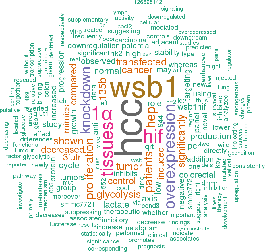
![]() 14 publications mentioning hsa-mir-592Open access articles that are associated with the species Homo sapiens and mention the gene name mir-592. Click the [+] symbols to view sentences that include the gene name, or the word cloud on the right for a summary. |
|
1 |
![]()
Other miRNAs from this paper: hsa-mir-93, hsa-mir-133a-1, hsa-mir-133a-2, hsa-mir-138-2, hsa-mir-143, hsa-mir-138-1, hsa-mir-133b
Overexpression of WSB1 protein in HCC cells also significantly abolished the suppression of miR-592 on glucose consumption (Figure 4D), lactate production (Figure 4E), and ATP level (Figure 4F), suggesting that miR-592 may suppress glycolysis by directly decreasing WSB1 expression in HCC cells.
[score:10]
It was found that the overall survival of HCC patients with low miR-592 expression was shorter than that of patients with high miR-592 expression (Figure 1E) Together, our data indicate that downregulation of miR-592 expression closely associates with poor HCC prognosis, suggesting that miR-592 participates in HCC progression.
[score:10]
Therefore, although miR-592 overexpression or WSB knockdown increased pVHL stability, CoCl [2] inhibited PHD activity and disrupted the interaction between PHD and pVHL, thus miR-592 overexpression or WSB knockdown could not block the CoCls -induced lactate production.
[score:9]
Herein, we report that the expression of miR-592 in HCC cells and tissues is downregulated, and miR-592 directly targets WSB1 mRNA.
[score:9]
It was found that overexpression of miR-592 remarkably suppressed the luciferase activity of the reporter gene with the wild-type construct, but not with the mutant WSB1 3′-UTR construct in both SK-Hep-1 and SMMC7721 cells (Figure 3B), suggesting that miR-592 directly targeted the WSB1 3′-UTR.
[score:8]
Although it was observed thatthe decreasing of lactate production upon overexpression of miR-592 or knockdown of WSB1 statistically significant compared to control in DMSO treated group, neither miR-592 overexpression nor WSB1 knockdown blocked the CoCl [2] or HIF-1α -induced increase of HK2 and Glut1 expression and lactate production (Figure 5F, lower panel).
[score:8]
In addition, Given that CoCl [2] only partially rescued miR-592 overexpression- caused cell proliferation retardation, and so did WSB-1 overexpression, this suggests that there may be alternative targets of miR592 that contributed to the growth retardation effect of miR-592.
[score:7]
Knockdown of miR-592 also increased the expression of Glut1 and HK2 and lactate production; however, YC-1 rescued Glut1 and HK2 expression and lactate production enhanced by miR-592 knockdown (Figure 5E).
[score:7]
Our results indicate that miR-592 is a robust inhibitor of the Warburg effect by targeting WSB1/HIF-1a axis, and more importantly, the newly identified miR-592 and its downstream pathway might serve as potential therapeutic targets and novel prognosis indicators in HCC.
[score:7]
Indeed, we observed that the miR-592 knockdown -mediated upregulation of the WSB1 level increased the expression of HIF-1α, which plays an important role in critical aspects of HCC cancer biology, especially glucose metabolism [16, 17].
[score:7]
In the present study, we showed that miR-592, by suppressing WSB1, but not key glycolytic genes, Myc, HK1 HIF-1α, HK2, stat3, or PKM2, effectively inhibits both glucose metabolism in HCC cells and the growth of xenografted HCC tumors, indicating a strong rationale for developing miR-592 as a novel metabolism -targeting therapeutic agent against HCC.
[score:7]
These data further support mechanism postulating that miR-592 knockdown directly promotes the expression of WSB1, thereby enhancing HIF-1α protein expression to maintain increased glycolysis in HCC (Figure 6E).
[score:7]
Together, our findings indicate that HIF-1α is responsible for the changes in glycolysis induced by miR-592 downregulation or WSB1 upregulation.
[score:7]
According to the median level of miR-592 expression, the patients (90 cases) were divided into two groups as either high expression (45 patients) or low expression (45 patients).
[score:7]
In addition, miR-592 knockdown increased, whereas miR-592 overexpression decreased HIF-1α protein expression, but not mRNA level (Figure 5D).
[score:6]
F. The effect of CoCl2 on the expression of HK2 and Glut1 (left panel), and the effects of CoCl2 on lactate production (right panel) in SK-Hep-1 cells with WSB1 knockdown or miR-592 overexpression.
[score:6]
Moreover, the inhibitory role of miR-592 in proliferation and cell cycle progression was reverted under the condition of mutant WSB1 overexpression (Figure 3E and Figure 3F).
[score:5]
Conversely, suppression of miR-592 enhanced the expression of WSB1 in both SK-Hep-1 and SMMC7721 cells (Figure 3C).
[score:5]
To uncover the underlying mechanism accounting for miR-592 -induced inhibition of HCC cells proliferation, bioinformatics research was performed to predict miR-592 targets using PITA and miRanda [11].
[score:5]
In order to validate the therapeutic value of miR-592, tumor cells with miR-592 overexpression were injected into mice to examine the inhibition of tumor growth.
[score:5]
In addition, two recent studies indicating that miR-592 inhibits prostate cancer and colorectal cancer growth [27, 28], further support miR-592 as a strongly general target candidate for anticancer therapies.
[score:5]
Our data suggest that decreased WSB1/HIF-1α expression via overexpression of miR-592 may decrease proliferation, glycolysis, and lactate production in HCC cells, which provides a new approach for HCC therapeutics.
[score:5]
As shown in supplementary Figure 1A, 1B, 1C, 1D and 1E, miR-592 overexpression significantly inhibited HCC growth in mice mo dels.
[score:5]
B. Correlationship between miR-592 expression and WSB1 and HIF-1a expression in HCC patients tissues.
[score:5]
To investigate whether the tumor suppressor role of miR-592 on HCC cell proliferation is mediated by inhibiting the expression of WSB1, SK-Hep-1 cells were co -transfected with miR-592 mimics and WSB1 plasmid without 3′-UTR.
[score:5]
As shown in Figure 6A, levels of both WSB1 and HIF-1α in human HCC tissues with high miR-592 expression were lower than those in HCC with low miR-592 expression.
[score:5]
In conclusion, the present study demonstrated a novel inhibitory role of miR-592 in HCC glycolysis by targeting WSB1/HIF-1α axis.
[score:5]
Consistently, we observed an inverse expression pattern between miR-592 and WSB1 or HIF-1α, whereas a positive expression pattern between WSB1 and HIF-1a was observed (Figure 6B).
[score:5]
To uncover the molecular mechanism of miR-592 HCC inhibitory potential, we hypothesized that decreased WSB1 expression by miR-592 would reduce the HIF-1α level in HCC because HIF-1α signaling has been demonstrated to be enhanced by WSB1 in various cancers [10].
[score:5]
E. The expression of HK2 and Glut1 in SK-Hep-1 cells treated with YC-1 and/or miR-592 knockdown (left panel); lactate production in HCC (right panel).
[score:4]
As shown in Figure 3D, miR-592 -induced WSB1 downregulation was rescued following co-transfection.
[score:4]
D. The protein level (left panel) or mRNA level (right panel) of HIF-1α with overexpression or knockdown of miR-592.
[score:4]
miR-592 is frequently downregulated in human HCC tissues and cell lines, which associates with low postoperative survival rate.
[score:4]
B. Measurement of the cellular G6P level in HCC cells with miR-592 overexpression or downregulation.
[score:4]
The new finding of this study is that downregulation of miR-592 may facilitate HCC pathogenesis through the WSB1/HIF-1α axis.
[score:4]
Consistently, overexpression of miR-592 greatly reduced the cellular G6P level in HCC cells, whereas knockdown of the endogenous miR-592 increased the G6P level (Figure 4B).
[score:4]
miR-592 directly targets WSB1 in HCC cells.
[score:4]
Another important aspect of the current study is the novel role of miR-592 in inhibition of glycolytic phenotypes.
[score:3]
Given that WSB1 increases one of the most important glycolytic proteins-HIF-1α and contributes to human malignancy [10], thus WSB1 was predicted as a potentially functional target gene of miR-592.
[score:3]
As shown in Figure 6C and Figure 6D, the inhibitory role of miR-592 in proliferation and cell cycle progression was reverted under the condition of treatment with CoCl [2].
[score:3]
WSB1 is a bona fide target gene of miR-592 and functional downstream of miR-592 in HCC cells.
[score:3]
Compared with the negative controls, overexpression of miR-592 in SK-Hep-1 and SMMC7721 cells significantly decreased cell number (Figure 2C and Figure 2D) whereas knockdown of the endogenous miR-592 markedly increased the viability of these cells (Figure 2E and Figure 2F).
[score:3]
A. Representative images of IHC staining for WSB1 and HIF-1a in HCC patients with high and low miR-592 expression.
[score:3]
In addition, as shown in supplementary Figure 1F, miR-592 inhibits the migration of HCC cells in vitro.
[score:3]
Therefore, the newly identified miR-592/WSB1/HIF-1α axis provides new insight into the pathogenesis of HCC, suggesting a novel, potential therapeutic target for the treatment of HCC.
[score:3]
E. miR-592 expression was inversely associated with the overall survival of 90 HCC patients.
[score:3]
K. The expression of miR-592 in tumor tissues was analyzed by qRT-PCR.
[score:3]
The miR-592/WSB1 signal pathway inhibits glycolysis in HCC cells.
[score:3]
Figure 6 A. Representative images of IHC staining for WSB1 and HIF-1a in HCC patients with high and low miR-592 expression.
[score:3]
Figure 1 A. qRT-PCR analysis of miR-592 expression in 40 pairs HCC tissues and their adjacent normal tissues.
[score:3]
Clinical significance of miR-592, WSB1 and HIF-1a expression in patients with HCC.
[score:3]
To determine the expression of miR-592 in HCC, we analyzed 40 cases of HCC tissues and adjacent non-tumorous liver tissues using quantitative real-time PCR.
[score:3]
Given that WSB1 promotes VHL ubiquitination and proteasomal degradation, thereby stabilizing HIF under both normoxic and hypoxic conditions, and HIF-1α has been shown to be a key transcription factor promoting glycolytic energy metabolism in cancer cells [10], we wondered if this newly discovered miR-592/WSB1 axis might inhibit glycolysis in HCC cells.
[score:3]
Moreover, overexpression of miR-592 also repressed the protein levels of WSB1 in both SK-Hep-1 and SMMC7721 cells.
[score:3]
The miR-592/WSB1 axis inhibits glycolysis in HCC cells.
[score:3]
D. SK-Hep-1 cells were co -transfected with miR-592 mimics or NC and WSB1 plasmid without 3′-UTR, the expression levels of WSB1 were analyzed using western blot.
[score:3]
Because miR-529 presents as the sole upstream regulator of WSB1, it will be possible that mir-592 is a potential key regulator keeping the equilibrium of WSB1/HIF-1α signaling.
[score:3]
miR-592 was recently proposed to be a new prognosis predictor for HCC patients and a new prospective target for HCC [14, 15].
[score:3]
Clinical relevance of miR-592, WSB1 and HIF-1a expression in patients with HCC.
[score:3]
MiR-592 is frequently down-regulated in human HCC and associates with poor clinicopathologic features.
[score:3]
To demonstrate whether the 3′-UTR of WSB1 could be a genuine target of miR-592, a dual-luciferase reporter system was constructed.
[score:3]
The miR-592 inhibitors (Cat.
[score:3]
The expression of miR-592 was confirmed by qRT-PCR.
[score:3]
A. qRT-PCR analysis of miR-592 expression in 40 pairs HCC tissues and their adjacent normal tissues.
[score:3]
The significance of this study is also underscored by the fact that miR-592 is frequently decreased in human HCCs, and that miR-592 knockout significantly promoted mouse HCC development.
[score:3]
qRT-PCR analysis of tumor tissues confirmed that the expression of miR-592 was significantly decreased (Figure 2K).
[score:3]
As shown in supplementary Figure 2A and 2B, miR-592 significantly decreases HIF-1α stability which was reverted by WSB1 overexpression.
[score:3]
C. The expression of miR-592 in HCC tissues, tissues with lymph node metastases, and their adjacent normal tissues, n=10/group.
[score:3]
In addition, we analyzed the correlationship between miR-592 expression and the patients' prognoses.
[score:3]
B. The expression of miR-592 in HCC tissues was significantly lower than in corresponding normal tissues.
[score:3]
Does HIF-1α as a transcription factor increase or decrease the expression of miR-592 in HCC?
[score:3]
Knockdown of miR-592 enhances glycolysis by increasing WSB1 -induced HIF-1α stability.
[score:2]
Because WSB1 can increase the stability of HIF-1α protein [10] and HIF-1α is one of key transcription factors in enhancing glycolysis [12], we hypothesized that HIF-1α may dictate the miR-592 knockdown -induced glycolysis in HCC cells.
[score:2]
To confirm this role of miR-592 applied in vivo, we tested whether miR-592 knockdown would promote tumour growth in SK-Hep-1 orthotopic tumour mo dels and mouse subcutaneous xenograft mo dels.
[score:2]
E. Schematic diagram of the regulatory pathway from miR-592/WSB1 / HIF-1a axis to glycolysis in HCC.
[score:2]
MiR-592 inhibits HCC cell proliferation and tumor growth.
[score:2]
Moreover, compared with primary HCC tissues and the adjacent normal tissues, tissues from lymph node metastases expressed lower levels of miR-592 (Figure 1C).
[score:2]
Consistent with these clinical findings, the expression of miR-592 was significantly decreased in five HCC cell lines compared with two normal liver cell lines (LO2 and 7701) (Figure 1D).
[score:2]
Compared with the control tissues, the expression of miR-592 was significantly decreased in 33 of 40 HCC tissues (Figure 1A).
[score:2]
To further confirm that miR-592 decreases HIF-1α stability via WSB1, the protein degradation of endogenous HIF-1α was measured in HCC cells with overexpression and knockdown of miR-592 and WSB1.
[score:2]
The effect of miR-592 and CoCl2 on C. SK-Hep-1 cell proliferation and D. cell cycle progression.
[score:1]
Fold changes for the expression levels of miR-592 were calculated using the comparative cycle threshold method (2 [−ΔCT]).
[score:1]
HSTUD0817, Sigma), miR-592 mimics (Cat.
[score:1]
C. and D. Overexpression of miR-592 significantly decreased cell proliferation of SK-Hep-1 and SMMC772 cells compared to the negative controls using CCK-8 assay.
[score:1]
Importantly, reintroduction of WSB1 reverted the miR-592 -induced decrease in the cellular G6P level in HCC cells (Figure 4C).
[score:1]
To further validate the inverse relationship between WSB1, HIF-1α and miR-592 in HCC cells, we examined miR-592, WSB1 and HIF-1α levels in the same human HCC tissues.
[score:1]
SK-Hep-1 cells transfected with anti-miR-592 or negative control (anti-NC) were subcutaneously injected into the flank region of nude mice.
[score:1]
Figure 3 A. Sequence alignment of wild-type (WT) and mutated (Mut) putative miR-592 -binding sites in the 3′-UTR of WSB1.
[score:1]
Given that many of the HCC patients are often being excluded from chemotherapy or surgical resection due to their poor liver functions, thus, therapeutic against miR-592 may selectively provide a novel approach.
[score:1]
A. Sequence alignment of wild-type (WT) and mutated (Mut) putative miR-592 -binding sites in the 3′-UTR of WSB1.
[score:1]
WSB1 3′-UTR sequences containing wild type or mutated binding site of miR-592 were cloned into the psiCHECK2 vector, respectively, and co -transfected with the miR-592 mimics or NC into HCC cells.
[score:1]
Collectively, our findings suggest that miR-592 decreases HCC cell proliferation in vitro and tumorigenicity in vivo.
[score:1]
L. and M. Cell cycle distribution of SK-Hep-1 and SMMC772 cells transfected with miR-592 mimics or NC was examined using flow cytometry.
[score:1]
As shown in Figure 2L and Figure 2M, compared to cells transfected with negative controls, there was a significant increase in G0/G1 phase cells and decrease in S phase cells in miR-592 mimics -transfected SK-Hep-1 and SMMC772 cells, whereas miR-592 knockdown cells displayed the opposite data (data not shown).
[score:1]
And the median level of miR-592 in HCC tissues was obviously lower than in corresponding normal tissues (Figure 1B).
[score:1]
A. and B. SK-Hep-1 and SMMC772 cells were transfected with hsa-miR-592 mimics, anti-miR-592 or negative control, respectively.
[score:1]
To further investigate whether the tumor suppressor role of miR-592 on HCC cells proliferation is mediated through disrupting the enhanced stability of HIF-1α, SK-Hep-1 cells were co -transfected with miR-592 mimics and treated with or without CoCl [2].
[score:1]
However, the molecular mechanisms by which miR-592 exerts its role in tumor are not frequently studied.
[score:1]
Together, these results suggest that WSB1/HIF-1α axis is functional downstream of miR-592 in HCC.
[score:1]
B. Relative luciferase activities of plasmids carrying WT or mut WSB1 3′-UTR in SK-Hep-1 and SMMC7721 cells cotransfected with miR-592 mimics or NC.
[score:1]
In this study, for the first time, we investigated the aberrant status of miR-592 and its potential target gene to explore this miRNA's cancer -associated functions in HCC.
[score:1]
Secondly, our in vivo orthotopic tumour study also further reinforced our in vitro results, and established the potential significance of miR-592 in HCC progression (Figure 2J).
[score:1]
To measure the expression levels of mature miR-592, real-time quantitative RT-PCR was performed.
[score:1]
Stably transfected cells (1.5 × 10 [6] in 0.2 mL) transfected with anti-miR-592 or negative control were injected subcutaneously into the flank region of 6 week-old male severe combined immunodeficiency mice (SCID; Institute of Laboratory Animal Sciences, Chinese Academy of Medical Sciences).
[score:1]
Figure 2 A. and B. SK-Hep-1 and SMMC772 cells were transfected with hsa-miR-592 mimics, anti-miR-592 or negative control, respectively.
[score:1]
In future studies, it will be interesting to determine whether this newly discovered miR-592/WSB1/HIF-1α axis is distinct in HCC etiology.
[score:1]
As shown in Figure 3A, one predicted binding site in the WSB1 3′-UTR with a perfect complementarity to the seed region of the miR-592 was identified.
[score:1]
The reaction mixture was used for real-time RT-PCR of miR-592.
[score:1]
The effect of miR-592 and WSB1 on E. HCC cell proliferation and F. cell cycle progression.
[score:1]
[1 to 20 of 109 sentences]
|
2 |
![]()
Other miRNAs from this paper: hsa-mir-17, hsa-mir-31, hsa-mir-106a, hsa-mir-211, hsa-mir-222, hsa-mir-142, hsa-mir-9-1, hsa-mir-9-2, hsa-mir-9-3, hsa-mir-194-1, hsa-mir-194-2, hsa-mir-369, hsa-mir-135b, hsa-mir-489, hsa-mir-455, hsa-mir-552, hsa-mir-876, hsa-mir-944
When analyzing the expression levels of the microRNAs selected for validation (Table 2), we observed that 5 of them (hsa-miR-135b *, hsa-miR-592, hsa-miR-31, hsa-mir-135b, hsa-miR-944) have similar patterns of expression, i. e., there are statistically significant differences between the UAs and the control group.
[score:5]
On the SA group, we observed differences in the expression between SAs and the control group in seven out of the 13 microRNAs selected for biological validation (p <0.05), (hsa-miR-135b *, hsa-miR- 489, hsa-miR-592, hsa-miR-369-5p, hsa-miR-31, hsa-mir-135b, hsa-miR-944).
[score:3]
Liu et al. [39] described this overexpression of miR-592 in colorectal cancer associating it to the size of the tumor, the distance of the metastases and the patient survival.
[score:3]
With these data, we consider that alterations in miR-592 expression in ameloblastomas would be mainly linked with tumor growth and progression.
[score:3]
When analyzing the expression of each miRNA, we observed statistically significant differences in 6 microRNAs among the UAs and the control group, (p <0.05) (hsa-miR-135b *, hsa-miR-592, hsa-miR-31, hsa-mir-135b, hsa-miR-944, hsa-miR-142-5p)).
[score:3]
MicroRNAs with a differential expression in both SA and UA when compared to the control group are the microRNA miR-135b, miR-135b *, miR-31, miR-592 and miR-944.
[score:2]
Different studies [36, 37] linked the alterations of miR-592 to neoplastic development in humans.
[score:2]
After applying the inclusion criteria (|FC| <0.2 or> 5 and p adjusted <0.05), as previously mentioned, biological validation was performed by RT-qPCR of the 13 differently regulated miRNAs (hsa-miR-9, hsa-miR-135b*, hsa-miR-194*, hsa-miR-489, hsa-miR-592, hsa-miR-369-5p, hsa-miR-876-5p, hsa-miR-31, hsa-miR-135b, hsa-miR-211, hsa-miR-944, hsa-miR-142-5p, hsa-miR-455-3p), in an independent set of 46 samples corresponding to 19 SA, 8 UA and 19 controls.
[score:2]
miR-592 and miR-552 can distinguish between primary lung adenocarcinoma and colorectal cancer metastases in the lung.
[score:1]
[1 to 20 of 9 sentences]
|
3 |
![]()
Other miRNAs from this paper: hsa-mir-15a, hsa-mir-21, hsa-mir-28, hsa-mir-29a, hsa-mir-32, hsa-mir-29b-1, hsa-mir-29b-2, hsa-mir-34a, hsa-mir-181a-2, hsa-mir-181b-1, hsa-mir-181c, hsa-mir-181a-1, hsa-mir-1-2, hsa-mir-125b-1, hsa-mir-125b-2, hsa-mir-206, hsa-mir-200c, hsa-mir-1-1, hsa-mir-181b-2, hsa-mir-29c, hsa-mir-200a, hsa-mir-365a, hsa-mir-365b, hsa-mir-193b, hsa-mir-181d, hsa-mir-550a-1, hsa-mir-550a-2, hsa-mir-617, hsa-mir-550a-3, hsa-mir-1207, hsa-mir-550b-1, hsa-mir-550b-2
NRF2 mediated drug resistance has been shown in colon cancer and is up-regulated in hepatocyte carcinoma [38, 52], indicating a possible link between the downregulation of miR-592 in these disorders and NRF2 activity.
[score:7]
Interestingly, miR-592 down-regulation has also been observed in colon cancers with deficient DNA mismatch repair (dMMR), a common mutation in colorectal tumors.
[score:5]
miR-592 was also down-regulated in hepatitis B hepatocellular carcinoma [89].
[score:4]
For example, in colorectal cancer, low levels of miR-592 expression have shown promise as a biomarker, identifying patients who will react poorly to anti-EGFR chemotherapies [88].
[score:3]
miRNA Location Targets References miR 193b/365Chr16,14397824-14397906 (miR193b)14403142-14403228 (miR-365)TTf1 - oncogenicBCL2 – TSGCyclin D- TSG, uPa(Qi et al., 2012)(Nie et al., 2012)(Li et al., 2009) Chr7, 130562218-130562298Sp-1MCL-1 - oncogenicTCL1 – oncogenic(Amodio et al., 2012)(Mott et al., 2007)(Pekarsky et al., 2006) Chr19, 13985513-13985622SIRT1- oncogenic and TSGKRAS - oncogenicTGFβ - TSGTNF - TSGNOTCH - oncogenic and TSG(Zhang et al., 2012)(Schonrock et al., 2012)(Hashimoto et al., 2010) miR-617 Chr12, 81226312-81226408 N/A miR-592 Chr7, 126698142-126698238 N/A miR-1207 Chr8 129061398-129061484.
[score:3]
miR-592 is located on chromosome 7 at position 126698142-126698238 and has been associated with a number of malignancies including, colorectal and liver carcinoma.
[score:1]
[1 to 20 of 6 sentences]
|
4 |
![]()
Other miRNAs from this paper: hsa-mir-34a, hsa-mir-137, hsa-mir-138-2, hsa-mir-134, hsa-mir-138-1, hsa-mir-195, hsa-mir-34b, hsa-mir-34c, hsa-mir-503, hsa-mir-4779
Moreover, miR-138 reportedly induced cell cycle arrest by targeting CCND3 in hepatocellular carcinoma cells [37] and miR-592 inhibited cell proliferation by suppressing CCND3 expression in CRC [38] Similarly, miR-4779 efficiently induced cell cycle arrest and apoptosis by targeting CCND3, and further contributed to cell cycle arrest by suppressing PAK2 expression.
[score:15]
Liu Z MiR-592 inhibited cell proliferation of human colorectal cancer cells by suppressing of CCND3 expressionInt.
[score:6]
[1 to 20 of 2 sentences]
|
5 |
![]()
Other miRNAs from this paper: hsa-let-7a-1, hsa-let-7a-2, hsa-let-7a-3, hsa-mir-21, hsa-mir-30a, hsa-mir-31, hsa-mir-32, hsa-mir-33a, hsa-mir-96, hsa-mir-147a, hsa-mir-10b, hsa-mir-181c, hsa-mir-182, hsa-mir-183, hsa-mir-224, hsa-mir-1-2, hsa-mir-133a-1, hsa-mir-133a-2, hsa-mir-135a-1, hsa-mir-135a-2, hsa-mir-137, hsa-mir-143, hsa-mir-9-1, hsa-mir-9-2, hsa-mir-9-3, hsa-mir-1-1, hsa-mir-363, hsa-mir-378a, hsa-mir-328, hsa-mir-135b, hsa-mir-196b, hsa-mir-20b, hsa-mir-486-1, hsa-mir-503, hsa-mir-552, hsa-mir-551b, hsa-mir-622, hsa-mir-625, hsa-mir-33b, hsa-mir-542, hsa-mir-378d-2, hsa-mir-147b, hsa-mir-378b, hsa-mir-378c, hsa-mir-378d-1, hsa-mir-378e, hsa-mir-378f, hsa-mir-378g, hsa-mir-378h, hsa-mir-378i, hsa-mir-378j, hsa-mir-486-2
In addition, miR-552 and miR-592 expression was up-regulated in pMMR tumors and down-regulated in dMMR tumors.
[score:9]
Significant differences were also seen in 6 miRNAs including miR-31 and miR-592, in the direct comparison of tumors that were deficient or proficient for mismatch repair.
[score:2]
miRNAs that showed decreased levels in pMMR relative to dMMR tumors included miR-552, miR-592, miR-181c and miR-196b.
[score:1]
Figures Array data was validated by by qRT-PCR for 10 miRNAs (mir-1, miR-10b, miR-135b, miR-147, miR-31, miR-33, miR-503, miR-552, miR-592, miR-622).
[score:1]
[1 to 20 of 4 sentences]
|
6 |
![]()
One might hypothesize that the SNP may potentially affect posttranslational modifications of GRM8 via regulating the expression of microRNA592.
[score:5]
[1 to 20 of 1 sentences]
|
7 |
![]()
Other miRNAs from this paper: hsa-mir-26a-1, hsa-mir-92a-1, hsa-mir-92a-2, hsa-mir-203a, hsa-mir-122, hsa-mir-200c, hsa-mir-26a-2, hsa-mir-519d, hsa-mir-1236, hsa-mir-203b
miR-592 functioned as a tumor suppressor in lung cancer by targeting SOX9 [29].
[score:5]
[1 to 20 of 1 sentences]
|
8 |
![]()
Other miRNAs from this paper: hsa-let-7b, hsa-mir-18a, hsa-mir-20a, hsa-mir-21, hsa-mir-29a, hsa-mir-92a-1, hsa-mir-92a-2, hsa-mir-93, hsa-mir-106a, hsa-mir-7-1, hsa-mir-7-2, hsa-mir-7-3, hsa-mir-10b, hsa-mir-181b-1, hsa-mir-203a, hsa-mir-215, hsa-mir-125b-1, hsa-mir-133a-1, hsa-mir-133a-2, hsa-mir-141, hsa-mir-143, hsa-mir-125b-2, hsa-mir-126, hsa-mir-150, hsa-mir-195, hsa-mir-206, hsa-mir-181b-2, hsa-mir-301a, hsa-mir-375, hsa-mir-378a, hsa-mir-494, hsa-mir-615, hsa-mir-378d-2, hsa-mir-760, hsa-mir-378b, hsa-mir-378c, hsa-mir-378d-1, hsa-mir-378e, hsa-mir-378f, hsa-mir-378g, hsa-mir-378h, hsa-mir-378i, hsa-mir-203b, hsa-mir-378j
They characterized low expression of hsa-miR-592 and high expression of hsa-miR-10b-5p and hsa-miR-615-3p were associated with tumors located in the right colon relative to the left colon and rectum and high expression of hsa-miR-615-3p [14].
[score:5]
[1 to 20 of 1 sentences]
|
9 |
![]()
Other miRNAs from this paper: hsa-let-7a-1, hsa-let-7a-2, hsa-let-7a-3, hsa-let-7b, hsa-let-7c, hsa-let-7d, hsa-let-7e, hsa-let-7f-1, hsa-let-7f-2, hsa-mir-30a, hsa-mir-31, hsa-mir-96, hsa-mir-100, hsa-mir-106a, hsa-mir-139, hsa-mir-182, hsa-mir-183, hsa-mir-203a, hsa-let-7g, hsa-let-7i, hsa-mir-1-2, hsa-mir-133a-1, hsa-mir-133a-2, hsa-mir-137, hsa-mir-141, hsa-mir-143, hsa-mir-145, hsa-mir-9-1, hsa-mir-9-2, hsa-mir-9-3, hsa-mir-125a, hsa-mir-1-1, hsa-mir-200a, hsa-mir-130b, hsa-mir-363, hsa-mir-378a, hsa-mir-135b, hsa-mir-133b, hsa-mir-422a, hsa-mir-429, hsa-mir-490, hsa-mir-574, hsa-mir-378d-2, hsa-mir-874, hsa-mir-374b, hsa-mir-1290, hsa-mir-378b, hsa-mir-378c, hsa-mir-378d-1, hsa-mir-378e, hsa-mir-378f, hsa-mir-378g, hsa-mir-378h, hsa-mir-378i, hsa-mir-203b, hsa-mir-4770, hsa-mir-378j
Twelve miRNAs (hsa-miR-130b, hsa-miR-203, hsa-miR-1974, hsa-miR-592, hsa-miR-200a, hsa-miR-429, hsa-miR-183, hsa-miR-182, hsa-miR-1290, hsa-miR-141, hsa-miR-135b, and hsa-miR-96) were overexpressed, whereas 84 miRNAs (hsa-miR-1, hsa-miR-145, hsa-miR-145*, and so on) were downexpressed in tumor tissues compared with those in normal tissues.
[score:4]
[1 to 20 of 1 sentences]
|
10 |
![]()
Other miRNAs from this paper: hsa-let-7a-1, hsa-let-7a-2, hsa-let-7a-3, hsa-let-7b, hsa-let-7c, hsa-let-7d, hsa-let-7e, hsa-let-7f-1, hsa-let-7f-2, hsa-mir-15a, hsa-mir-16-1, hsa-mir-21, hsa-mir-26a-1, hsa-mir-26b, hsa-mir-31, hsa-mir-32, hsa-mir-96, hsa-mir-98, hsa-mir-29b-1, hsa-mir-29b-2, hsa-mir-103a-2, hsa-mir-103a-1, hsa-mir-107, hsa-mir-16-2, hsa-mir-192, hsa-mir-148a, hsa-mir-10b, hsa-mir-183, hsa-mir-203a, hsa-mir-212, hsa-mir-214, hsa-mir-222, hsa-let-7g, hsa-let-7i, hsa-mir-15b, hsa-mir-23b, hsa-mir-122, hsa-mir-124-1, hsa-mir-124-2, hsa-mir-124-3, hsa-mir-125b-1, hsa-mir-128-1, hsa-mir-132, hsa-mir-135a-1, hsa-mir-135a-2, hsa-mir-138-2, hsa-mir-140, hsa-mir-141, hsa-mir-143, hsa-mir-144, hsa-mir-145, hsa-mir-152, hsa-mir-191, hsa-mir-125b-2, hsa-mir-126, hsa-mir-134, hsa-mir-150, hsa-mir-184, hsa-mir-186, hsa-mir-193a, hsa-mir-320a, hsa-mir-200c, hsa-mir-155, hsa-mir-128-2, hsa-mir-29c, hsa-mir-200a, hsa-mir-34b, hsa-mir-130b, hsa-mir-26a-2, hsa-mir-369, hsa-mir-381, hsa-mir-326, hsa-mir-148b, hsa-mir-424, hsa-mir-493, hsa-mir-432, hsa-mir-524, hsa-mir-516b-2, hsa-mir-516b-1, hsa-mir-516a-1, hsa-mir-516a-2, hsa-mir-503, hsa-mir-570, hsa-mir-574, hsa-mir-582, hsa-mir-601, hsa-mir-603, hsa-mir-548c, hsa-mir-629, hsa-mir-542, hsa-mir-320b-1, hsa-mir-320c-1, hsa-mir-766, hsa-mir-320b-2, hsa-mir-765, hsa-mir-300, hsa-mir-450b, hsa-mir-744, hsa-mir-938, hsa-mir-103b-1, hsa-mir-103b-2, hsa-mir-320d-1, hsa-mir-320c-2, hsa-mir-320d-2, hsa-mir-320e, hsa-mir-203b
Six miRNAs of them (miR-450b-5p, miR-424, miR-503, miR-542-3p, miR-629, and miR-214) were significantly underexpressed, while one miRNA (miR-592) was significantly overexpressed in NFA compared to normal pituitary tissues.
[score:4]
[1 to 20 of 1 sentences]
|
11 |
![]()
Other miRNAs from this paper: hsa-let-7a-1, hsa-let-7a-2, hsa-let-7a-3, hsa-mir-20a, hsa-mir-21, hsa-mir-22, hsa-mir-24-1, hsa-mir-29a, hsa-mir-33a, hsa-mir-96, hsa-mir-29b-1, hsa-mir-29b-2, hsa-mir-196a-1, hsa-mir-199a-1, hsa-mir-129-1, hsa-mir-30d, hsa-mir-7-1, hsa-mir-7-2, hsa-mir-7-3, hsa-mir-34a, hsa-mir-182, hsa-mir-183, hsa-mir-196a-2, hsa-mir-199a-2, hsa-mir-204, hsa-mir-205, hsa-mir-212, hsa-mir-214, hsa-mir-217, hsa-mir-223, hsa-mir-23b, hsa-mir-27b, hsa-mir-124-1, hsa-mir-124-2, hsa-mir-124-3, hsa-mir-132, hsa-mir-133a-1, hsa-mir-133a-2, hsa-mir-135a-1, hsa-mir-135a-2, hsa-mir-137, hsa-mir-138-2, hsa-mir-140, hsa-mir-142, hsa-mir-145, hsa-mir-153-1, hsa-mir-153-2, hsa-mir-9-1, hsa-mir-9-2, hsa-mir-9-3, hsa-mir-126, hsa-mir-129-2, hsa-mir-134, hsa-mir-138-1, hsa-mir-149, hsa-mir-150, hsa-mir-190a, hsa-mir-194-1, hsa-mir-206, hsa-mir-155, hsa-mir-194-2, hsa-mir-29c, hsa-mir-34b, hsa-mir-370, hsa-mir-342, hsa-mir-135b, hsa-mir-338, hsa-mir-339, hsa-mir-133b, hsa-mir-18b, hsa-mir-486-1, hsa-mir-146b, hsa-mir-494, hsa-mir-518b, hsa-mir-517a, hsa-mir-520h, hsa-mir-519a-1, hsa-mir-519a-2, hsa-mir-504, hsa-mir-506, hsa-mir-33b, hsa-mir-421, hsa-mir-671, hsa-mir-802, hsa-mir-190b, hsa-mir-1291, hsa-mir-486-2
In another report, miR-592 promotes metastasis, in part, by targeting FOXO3A in human colorectal cancer [79].
[score:3]
[1 to 20 of 1 sentences]
|
12 |
![]()
Other miRNAs from this paper: hsa-let-7b, hsa-mir-17, hsa-mir-25, hsa-mir-15b, hsa-mir-339, hsa-mir-488, hsa-mir-877, hsa-mir-4745, cja-let-7b, cja-mir-17, cja-mir-15b, cja-mir-339, cja-mir-488, cja-mir-4745, cja-mir-25
10 miRNAs were consistently validated (Fig. 6) to be differentially expressed by HP diet, namely miR-592, miR-488-3p, miR-339-3p, miR-17-5p-1, miR-877-5p, miR-15b-5p, miR-484-1, miR-4745-5p, let-7b-5p and miR-25-3p.
[score:3]
[1 to 20 of 1 sentences]
|
13 |
![]()
Other miRNAs from this paper: hsa-let-7a-1, hsa-let-7a-2, hsa-let-7a-3, hsa-let-7e, hsa-let-7f-1, hsa-let-7f-2, hsa-mir-17, hsa-mir-22, hsa-mir-26a-1, hsa-mir-26b, hsa-mir-93, hsa-mir-98, hsa-mir-34a, hsa-mir-183, hsa-mir-218-1, hsa-mir-218-2, hsa-let-7i, hsa-mir-1-2, hsa-mir-15b, hsa-mir-23b, hsa-mir-135a-1, hsa-mir-135a-2, hsa-mir-125a, hsa-mir-136, hsa-mir-146a, hsa-mir-188, hsa-mir-195, hsa-mir-1-1, hsa-mir-296, hsa-mir-26a-2, hsa-mir-326, hsa-mir-135b, hsa-mir-335, hsa-mir-424, hsa-mir-491, hsa-mir-503, hsa-mir-455, hsa-mir-1301
In the cerebellum, novel_circRNA_007362 was predicted to combine with 24 miRNAs (tch-let-7e-5p, tch-let-7i-5p, tch-let-7f-5p, tch-miR-125a-5p, tch-miR-1301, tch-miR-135a-5p, tch-miR-135b-5p, tch-miR-15b-5p, tch-miR-195-5p, tch-miR-1-5p, tch-miR-218-5p, tch-miR-22-3p, tch-miR-26a-5p, tch-miR-26b-5p, tch-miR-296-3p, tch-miR-335-5p, tch-miR-34a-5p, tch-miR424-5p,tch-miR-491-5p, tch-miR-455-3p,tch-miR-503, tch-miR-592, tch-miR-9771e, and tch-miR-98-5p).
[score:1]
[1 to 20 of 1 sentences]
|
14 |
![]()
Other miRNAs from this paper: hsa-mir-19a, hsa-mir-19b-1, hsa-mir-19b-2, hsa-mir-20a, hsa-mir-124-1, hsa-mir-124-2, hsa-mir-124-3, hsa-mir-128-1, hsa-mir-137, hsa-mir-9-1, hsa-mir-9-2, hsa-mir-9-3, hsa-mir-128-2, hsa-mir-20b, hsa-mir-451a, hsa-mir-451b
Other miRNAs controlling both proliferation and differentiation of adult NSPCs are miR-137 [122] and rno-miR-592 [123].
[score:1]
[1 to 20 of 1 sentences]
|