![]() |
miRBase |
![]() |
![]() |
![]() 46 publications mentioning hsa-mir-513a-1Open access articles that are associated with the species Homo sapiens and mention the gene name mir-513a-1. Click the [+] symbols to view sentences that include the gene name, or the word cloud on the right for a summary. |
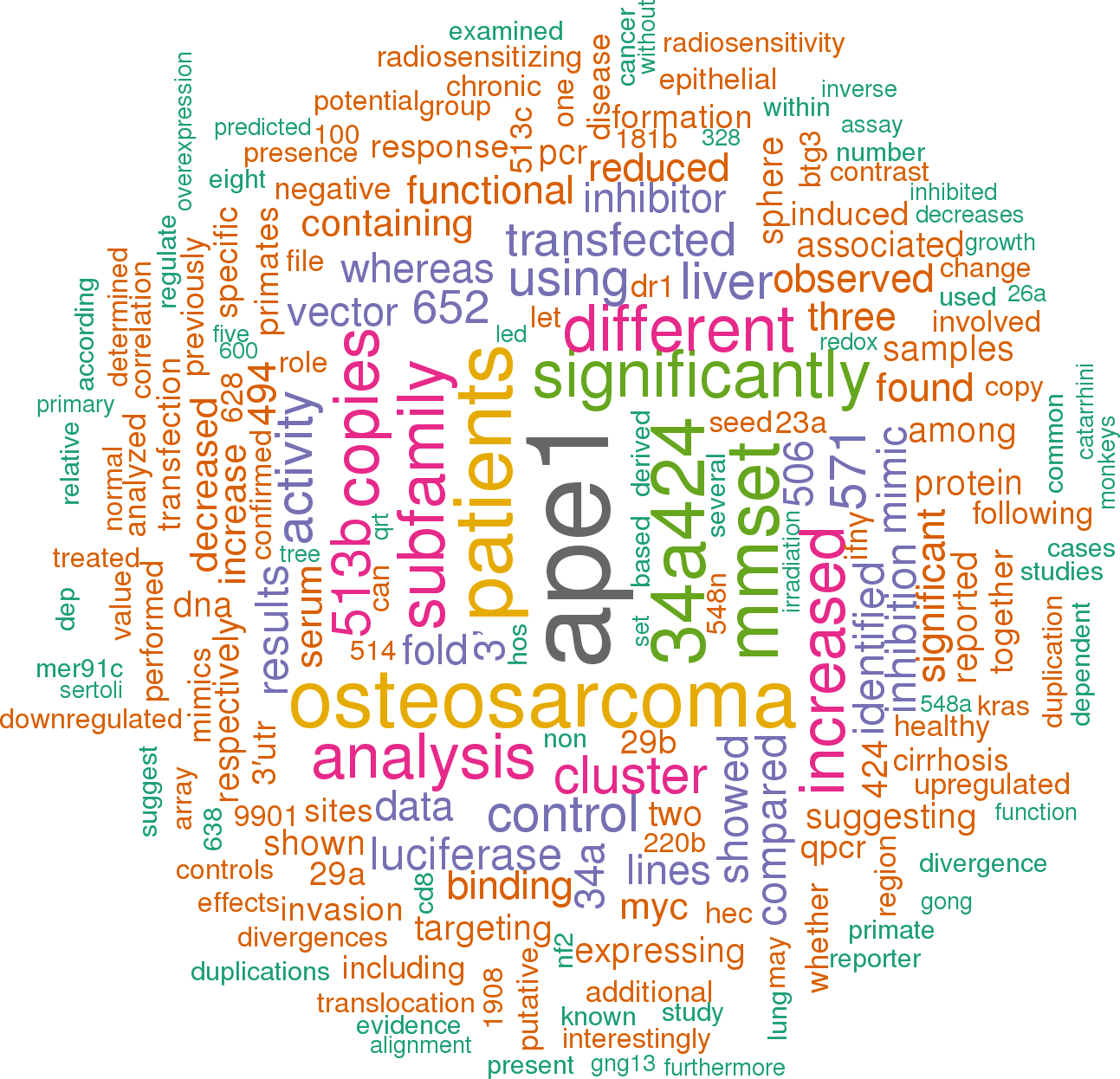
|
1 |
![]()
Other miRNAs from this paper: hsa-let-7a-1, hsa-let-7a-2, hsa-let-7a-3, hsa-mir-451a, hsa-mir-513a-2, hsa-mir-451b
Four Gy IR causes a significant number of DNA base lesions and SSBs which can be effectively repaired by all three vector -expressing osteosarcoma cell lines, while these lesions were left unrepaired or repaired more slowly in miR-513a-5p -expressing cells (Figure 4B and 4C), suggesting that the AP endonuclease activity of APE1, together with the BER and SSBR capacity, were impaired in stably -expressing miR-513a-5p osteosarcoma cells.
[score:7]
In addition, our recent clinicopathological correlation study of PD-L1 and APE1 expression in gastric cancer demonstrated a positive correlation of PD-L1 and APE1 protein expression in cancerous tissue (Qing et al. in press), suggesting that this positive correlation could be due to regulation by a common miRNA regulator, miR-513a-5p.
[score:7]
org database) containing the target sites for miR-513a-5p was synthesized and inserted into the pmirGLO Dual-Luciferase miRNA Target Expression Vector (Promega, USA) at XhoI and Xbal restriction enzyme sites.
[score:7]
Based on the studies of miR-513a-5p in osteosarcoma, we hypothesized that the downregulation of miR-513a-5p in osteosarcoma cells could be associated with the intrinsic radioresistance of osteosarcoma which has been reported to be due to the high expression of certain DNA repair proteins, including APE1.
[score:6]
Figure 6Osteosarcoma cells with miR-513a-5p expression and control cells were first transfected with wildtype APE1 expression vector and irradiated with 4 Gy X-ray.
[score:5]
APE1 expression is inhibited by miR-513a-5p.
[score:5]
To our knowledge, this is the first report on the miRNA suppression of APE1 gene expression and the establishment of the activity of miR-513a-5p in increasing radiosensitivity in osteosarcoma.
[score:5]
Transfecting an APE1 -overexpressing lentivirus in which the 3′UTR is absent makes the exogenous APE1 expression independent of control by miR-513a-5p, and in this case we observed that the apoptotic population was significantly reduced after IR (Figure 6C).
[score:5]
We increased or decreased APE1 expression utilizing miR-513a-5p in osteosarcoma cells, and thereby provided evidence that miR-513a-5p can increase radiosensitization through decreasing APE1 expression.
[score:5]
The DNA damage response marker Υ-H2AX was significantly increased in miR-513a-5p expressing 9901 cells while miR-513a-5p inhibitor transfection restored the genome integrity (Figure 4D and 4E).
[score:5]
Considering APE1’s suppression of radiosensitivity and the inverse relationship between APE1 and miR-513a-5p, we assumed APE1 could be an important intracellular target of miR-513a-5p in radiosensitization.
[score:5]
Osteosarcoma cells with miR-513a-5p expression and control cells were first transfected with wildtype APE1 expression vector and irradiated with 4 Gy X-ray.
[score:5]
MiR-513a-5p, minimally expressed in osteosarcoma, is a radiosensitizing miRNA that decreases the expression of APE1, previously reported as a radioresistance gene.
[score:5]
The APE1-3′UTR sequence (258 bp) containing the putative miR-513a-5p binding site and mutant sites was synthesized and inserted respectively into the pmirGLO Dual-Luciferase miRNA target expression vector at XhoI and Xbal restriction enzyme sites.
[score:5]
We increased miR-513a-5p expression in osteosarcoma cells using a lentivirus expressing miR-513a-5p with a puromycin selection marker.
[score:5]
We observed a significantly decreased APE1 protein level, but not mRNA level in the miR-513a-5p mimic transfected HOS cells, whereas increased APE1 expression was observed after transfection with the miR-513a-5p inhibitor (Figure 3C).
[score:5]
APE1 expression is suppressed by miR-513a-5p.
[score:5]
As determined by EMSA at 24 h post irradiation, we found the amount of DNA binding complexes of NF-KB, p53 or AP-1 were significantly inhibited in osteosarcoma cells with increased miR-513a-5p, whereas the bound complexes were restored in miR-513a-5p inhibitor transfected cells (Figure 4F).
[score:5]
On the other hand, despite the low expression profile of miR-513a-5p in all three osteosarcoma cell lines, radioresistance following IR was observed in the miR-513a-5p inhibitor -transfected osteosarcoma cell lines.
[score:5]
Figure 4(A) AP endonuclease activities of APE1 protein in the control cell line, stably -expressing miR-513a-5p cells, and inhibitor treated HOS, U2OS and 9901 cells were detected by abasic site incision assay.
[score:4]
To confirm that the radiosensitizing effect of miR-513a-5p results from decreased APE1 levels, we introduced either an APE1 overexpression or knockdown lentivirus to increase and decrease APE1 levels.
[score:4]
In contrast, considering its pivotal activity in DNA repair and transcription factor regulation the newly reported suppression of APE1 by miR-513a-5p provides a more plausible explanation to its radiosensitizing, or chemosensitizing, effects.
[score:4]
As shown in Figure 2A, osteosarcoma cell lines with stable miR513a-5p expression were significantly more sensitive to IR when compared to the vector -expressing controls (all p < 0.01).
[score:4]
Intriguingly, it has been previously reported that B7-H1, also known as Programmed Death-Ligand 1 (PD-L1), is the regulatory target for miR-513a-5p as well.
[score:4]
Based on these data, we hypothesized that miR-513a-5p decreases APE1 expression and thus increases the radiosensitivity of osteosarcoma.
[score:3]
We further validated miR-513a-5p expression in established osteosarcoma cell lines and a normal osteoblast line.
[score:3]
Future studies are required to correlate miR-513a-5p and APE1 expression in different tissues.
[score:3]
As shown in Figure 4A, AP endonuclease activity was significantly decreased in HOS-513, U2-OS-513 and 9901-513 cells after irradiation, whereas it was increased significantly after miR-513a-5p inhibitor transfection.
[score:3]
The exogenous miR-513a-5p was over-expressed 146-fold, 406-fold and 95-fold in HOS-513, U2-OS-513 and 9901-513 cells, respectively.
[score:3]
Figure 2(A) The stable expression of miR-513a-5p in osteosarcoma cells and control cells was determined by quantitative RT-PCR and shown in the bar graph.
[score:3]
We performed in vitro experiments to correlate miR-513a-5p expression in osteosarcoma cells to radioresistance.
[score:3]
As shown in Figure 1A, miR-513a-5p expression is significantly lower in samples from osteosarcoma patients than those from healthy donors (p < 0.0001).
[score:3]
miR-513a-5p is decreased in osteosarcoma and inversely correlated to APE1 expression.
[score:3]
The radiosensitizing effect of miR-513a-5p is increased by suppressing APE1.
[score:3]
An enzyme-resistant miR-513a-5p inhibitor was introduced to increase miR-513a-5p.
[score:3]
Radiosensitizing effect of miR-513a-5p is increased after suppressing APE1.
[score:3]
Based on the results displayed in Figure 2A, total cell viability was significantly reduced in stably -expressing miR-513a-5p cells following IR.
[score:3]
We observed an inverse correlation between APE1 protein level and miR-513a-5p expression throughout the clinical samples we studied (r = −0.493, p = 0.001).
[score:3]
D [0] and D [q] values of miR-513a-5p expressed osteosarcoma cell lines.
[score:3]
We initially detected the expression of miR-513a-5p using quantitative PCR in formalin fixed paraffin embedded (FFPE) tissue from both osteosarcoma patients and healthy donors.
[score:3]
To explore this mechanism, we initially attempted to confirm whether APE1 expression is decreased by miR-513a-5p.
[score:3]
Although the biological functions and intracellular targets of miR-513a-5p remain to be determined, previous studies suggest that miR-513a-5p is associated with cellular sensitivity to etoposide- and cisplatin-containing chemotherapy in retinoblastoma and non-small cell lung cancer (NSCLC) [18, 19].
[score:3]
Luciferase activity was detected at 48∼72 h after transfection with or without scrambled mimics, miR-513a-5p mimics and miR-513a-5p inhibitor, respectively.
[score:3]
The D [0] and D [q] values of each group are listed in Table 2. In addition to the radiosensitizing effect, stable expression of miR-513a-5p resulted in slightly impaired cell viabilities in all three cell lines as evidenced by reduced plating efficiency (PE) from 42% (HOS), 64% (U2OS) and 53% (9901) to 22%, 45% and 44%, respectively.
[score:3]
The results demonstrated that the protein level of APE1 is elevated in osteosarcoma cells which further confirmed an inverse relationship with miR-513a-5p expression.
[score:3]
Production of lentiviral vector expressing hsa-miR-513a: A 506 bp genomic fragment containing hsa-miR-513a was cloned into lentiviral vector pLentis-CMV-GFP-MCS-PGK-PURO.
[score:3]
Taken together, these results further confirmed that APE1 expression is decreased by miR-513a-5p.
[score:3]
In the 9901-vec+IR group, intensive nuclear and cytoplasmic immunostaining of APE1 was observed in tumor cells, and the tumors in the 9901-513+IR group showed significant decreases in both nuclear and cytoplasmic immunostaining of APE1 and significant increases in miR-513a-5p expression (Figure 5B–5D).
[score:3]
Although previous studies of miR-513a-5p demonstrate suppression of PD-L1 in the context of an etoposide -induced cellular response [18], the overall evidence supporting the activity of PD-L1 in tumor chemo- or radiosensitivity is scarce.
[score:3]
We observed the same trend of radiosensitization by the miR-513a-5p mimic as determined by cells stably expressing miR-513a-5p using a colony formation assay (Figure 2B).
[score:2]
We also performed an MTT assay to determine the overall cell viabilities of different osteosarcoma cell lines transiently transfected with either an miR-513a-5p mimic or inhibitor following 4 Gy irradiation (Figure 2C).
[score:2]
We screened and further confirmed that seven miRNAs were increased in APE1 knockdown HOS cells, and among them, miR-513a-5p exhibited an approximately 3-fold increase.
[score:2]
The impact of miR-513a-5p level on the redox activity of APE1 was analyzed by EMSA using the probes containing the binding sequences of NF-κB, p53 and AP-1, which are transcription factors regulated by APE1 redox activity (F).
[score:2]
The clinical characteristics of the cohort studied are displayed in Table 1. Reduced expression of miR-513a-5p is observed in bone tissue from both osteosarcoma patients and healthy donors compared to other miRNAs which have been reported to be highly expressed in osteosarcoma, such as let-7a or miR-451(20) (data not shown).
[score:2]
As shown previously, miR513a-5p is increased in APE1 knockdown osteosarcoma cell lines, so we speculated that there could be an inverse correlation between miR-513a-5p and APE1.
[score:2]
In agreement with the in vivo data, the in vitro apoptosis assay (a representative flow cytometry graph is displayed in Figure 6A and the analysis in 6B) showed that miR-513a-5p stably -expressing osteosarcoma cells harbored more apoptotic cells at 24 hours following 4 Gy IR.
[score:2]
We cloned a segment from the 3’ UTR of APE1 (258bp) which contains the putative binding region for miR-513a-5p, with either wildtype sequence or a sequence with the key bases mutated, to a commercially available luciferase reporter vector, pmirGLO-dual, downstream of the luciferase reporter gene (named GLO-APE1-513 and GLO-APE1-513M, respectively) (Figure 3A).
[score:1]
Moreover, γ-H2AX levels, which represent the DNA damage response, were elevated in xenografts with increased levels of miR-513a-5p (Figure 5D).
[score:1]
The increase of miR-513a-5p in all three cell lines was statistically significant (p < 0.001).
[score:1]
We also determined if an increase in miR-513a-5p could alter the redox activity of APE1 and the DNA -binding activities of NF-κB, p53 and AP-1, which are transcription factors reported to be increased by APE1 redox activity.
[score:1]
The fold increase is shown at the top of the bar of the miR-513a-5p group.
[score:1]
Effects of increased miR-513a-5p on xenograft radiosensitivity.
[score:1]
Varying levels of miR-513a-5p alter radiosensitivity of osteosarcoma cells.
[score:1]
These findings established an inverse correlation between radiosensitization by miR-513a-5p and the intracellular level of APE1.
[score:1]
Additionally, the level of miR-513a-5p in osteosarcoma is reduced based on the data from cell lines and clinical samples.
[score:1]
miR-513a-5p is correlated with intracellular APE1 levels post irradiation.
[score:1]
Notably, in contrast with the in vitro cell viability results, the growth of xenografts derived from 9901-513 cells was similar to 9901-vec cells without irradiation, suggesting that miR-513a-5p exerts more complex effects on tumor growth in vivo.
[score:1]
These data further confirmed that miR-513a-5p impairs DNA repair capacity in osteosarcoma cells and increases the DNA damage response.
[score:1]
These results provide evidence for the radiosensitizing activity of miR-513a-5p in osteosarcoma both in vitro and in vivo.
[score:1]
This suggests a possible link between decreased miR-513a-5p and the radioresistance of osteosarcoma [17, 20].
[score:1]
org/), we found that the putative binding region and key binding base pairs for miR-513a-5p are at the 3’UTR of APE1 (Figure 3A).
[score:1]
miR-513a-5p decreases DNA repair and redox activity of APE1 in osteosarcoma cells.
[score:1]
We now report miR-513a-5p as the first validated miRNA for APE1.
[score:1]
When transfected with the GLO-APE1-513M reporter vector, the miR-513a-5p mimic did not decrease the luciferase activity suggesting that the mutated sites are critical to miR-513a-5p binding (Figure 3B).
[score:1]
Increasing miR-513a-5p levels induced radiosensitivity of osteosarcoma cells.
[score:1]
To link the radiosensitizing effects of miR-513a-5p with the biological function of APE1, we examined whether APE1 activities correlate with the intracellular miR-513a-5p level.
[score:1]
[1 to 20 of 76 sentences]
|
2 |
![]()
Other miRNAs from this paper: hsa-mir-26a-1, hsa-mir-31, hsa-mir-34a, hsa-mir-203a, hsa-mir-126, hsa-mir-26a-2, hsa-mir-424, hsa-mir-513a-2, hsa-mir-513b, hsa-mir-513c, hsa-mir-203b
Consistently, inhibiting MMSET expression using siRNA in Ishikawa cells transfected with miR-34a/miR-424/miR-513 inhibitor reduced the expression of Twist1 and Vimentin but elevated the expression of E-cadherin (Figure 5D).
[score:11]
miR-34a, miR-424 and miR-513 directly target MMSET 3′-UTR to downregulate MMSET expression in EC cells.
[score:9]
Consistently, our western blot data showed that transfection of miR-34a/miR-424/miR-513 reduced MMSET expression in HEC-1 cells, whereas transfection of miR-34a/miR-424/miR-513 inhibitor enhanced MMSET expression in Ishikawa cells (Figure 4D), suggesting that miR-34a/miR-424/miR-513 directly repress MMSET.
[score:8]
Taken together, our data suggest that miR-34a/miR-424/miR-513 inhibits the invasive and stem cell-like properties of EC cells by suppressing MMSET expression via interacting with its 3′-UTR.
[score:7]
We measured Twist1, Vimentin or E-cadherin mRNA by qPCR assay, and found that rescuing MMSET expression with MMSET ORF in the presence of miR-34a/miR-424/miR-513 mimic resulted in up-regulation of Twist1 and Vimentin and downregulation of E-cadherin (Figure 5C).
[score:6]
The overexpression of MMSET ORF in HEC-1 cells partially rescued miR-34a/miR-424/miR-513 mimic -suppressed invasion and sphere formation (Figure 5A).
[score:5]
Ectopic expression of miR-34a/miR-424/miR-513 in HEC-1 cells resulted in a significant decrease in the relative luciferase activity of MMSET 3′-UTR, but there was no inhibition of luciferase activity when the cells were transfected with miR-34a/miR-424/miR-513 mimic and mutant MMSET 3′-UTR (Figure 4C).
[score:5]
To further validate the above observations, we investigated whether transient over -expression of a MMSET open reading frame (ORF) could reverse the inhibitory effects of miR-34a/miR-424/miR-513 mimic on EC cell invasion and sphere formation, or whether silencing of MMST with specific siRNA could repress the miR-34a/miR-424/miR-513 inhibitor -induced EC cell invasion and sphere formation.
[score:5]
Our results demonstrated that the expression of miR-34a/miR-424/miR-513 is frequently lost in human EC tissues, and ectopic miR-34a/miR-424/miR-513 expression reduced EC cell sphere formation and invasion.
[score:5]
In this study, we identified miR-34a/miR-424/miR-513 that could directly regulate MMSET expression in EC cells.
[score:5]
For a set of 50 patients with EC, reduced expression of miR-34a/miR-424/miR-513 was significantly associated with a poorer prognosis of EC patients (Figure 6B), To explore whether the miR-34a/miR-424/miR-513-MMSET axis is clinically relevant, we assessed the correlation between the expression of miR-34a/miR-424/miR-513 and MMSET in EC tissues using qPCRs.
[score:5]
EC cells (50% confluence) were transfected with miRNA mimics and miRNA inhibitors for miR-34a, miR-424 and miR-513 (40 nM, Ambion, Austin, TX), siRNAs against MMSET or Twist1 (5 nM, Ambion, Austin, TX) and the expression vector for MMSET (OriGene, Rockville, MD) using Lipofectamine 3000 (Invitrogen, Carlsbad, CA) according to the manufacturer’s instructions.
[score:5]
Importantly, overexpression of miR-34a/miR-424/miR-513 inhibited invasion and sphere formation of HEC-1 cells (Figure 4E and 4F).
[score:5]
Furthermore, in the present work, we provide new evidence that miR-513 can suppress EC cell sphere formation and invasiveness by targeting MMSET.
[score:5]
miR-34a, miR-424 and miR-513 inhibit EMT, invasion and the sphere-forming ability of EC cells through targeting MMSET.
[score:5]
We also found that repression of miR-34a/miR-424/miR-513 upregulates MMSET levels, thus promoting the invasion and sphere-forming ability of EC cells.
[score:4]
Using qPCR analysis, we found that invasive HEC-1 cells had very low miR-34a/miR-424/miR-513 expression compared with less invasive Ishikawa cells (Figure 4B), indicating that the levels of miR-34a/miR-424/miR-513 were inversely correlated with MMSET expression in EC cells (Figure 1A).
[score:4]
To determine if reduced miR-34a/miR-424/miR-513 expression is associated with any change in survival probability, we compared Kaplan-Meier plots for high and low expression of miR-34a/miR-424/miR-513.
[score:4]
Moreover, the miR-34a/miR-424/miR-513 inhibitor -induced Ishikawa cell invasion and sphere formation were significantly reduced by MMSET siRNA (Figure 5B).
[score:3]
However, Ishikawa cells transfected with miR-34a/miR-424/miR-513 inhibitor exhibited significantly increased cell invasion and sphere formation (Figure 4E and 4F).
[score:3]
We detected a significant negative association between miR-34a/miR-424/miR-513 and MMSET mRNA expression (Figure 6C).
[score:3]
We show that MMSET is a tumor promoter in EC, and the loss of miR-34a, miR-424 and miR-513 contributes to the overexpression of MMSET and aggressive phenotypes of EC cells.
[score:3]
These observations suggest that miR-34a/miR-424/miR-513 are suppressors of EC cell invasion and sphere formation.
[score:3]
To address the relevance of miR-34a/miR-424/miR-513 expression to human EC, we examined the levels of these miRNAs in primary ECs using qPCR analysis.
[score:3]
In conclusion, our results demonstrate that MMSET exerts tumor-promoting effects in EC cells, and the loss of miR-34a, miR-424 and miR-513 enhances MMSET expression in EC.
[score:3]
Figure 6(A) Relative miR-34a/miR-424/miR-513 expression in EC tissues and matched adjacent normal endometrial tissues.
[score:3]
Mutation in the miR-34a, miR-424 or miR-513 -binding sequence was generated by using the QuickChange Mutagenesis Kit (Stratagene, La Jolla, CA).
[score:2]
To test whether MMSET is directly regulated by miR-34a/miR-424/miR-513, we performed luciferase reporter assays by transfecting the reporter vector containing the full-length 3′-UTR of human MMSET into HEC-1 cells, together with miR-34a, miR-424, miR-513 or control mimic, respectively.
[score:2]
Then, these predicted miRNAs were overlapped with a set of miRNAs that are significantly repressed in highly invasive EC cells [16], leading to the identification of 3 miRNAs (miR-34a, miR-424 and miR-513) (Figure 4A).
[score:1]
Firefly luciferase reporter plasmid (100 ng) plus Renilla luciferase vector (10 ng), together with miR-34a/miR-424/miR-513 mimic or the negative control mimic, were transfected into HEC-1 cells using Lipofectamine 3000 (Invitrogen, Carlsbad, CA).
[score:1]
miR-34a, miR-424 and miR-513 expression was measured using the NCode miRNA qRT-PCR analysis (Invitrogen, Carlsbad, CA) following manufacturer-recommended protocols.
[score:1]
Figure 4(A) Alignment of miR-34a, miR-424 and miR-513 and their corresponding complementary binding sequences in MMSET 3′-UTR by bioinformatics algorithms.
[score:1]
Figure 5(A) miR-34a/miR-424/miR-513 mimic or control mimic was co -transfected into HEC-1 cells, together with (or without) MMSET cDNA vector lacking the 3′-UTR region.
[score:1]
Taken together, these results supported an existence of the miR-34a/miR-424/miR-513-MMSET axis in human EC.
[score:1]
miR-34a/miR-424/miR-513 repression was associated with poorer prognosis of EC patients.
[score:1]
[1 to 20 of 35 sentences]
|
3 |
![]()
Other miRNAs from this paper: hsa-mir-513a-2, hsa-mir-514a-1, hsa-mir-514a-2, hsa-mir-514a-3, mml-mir-513b-1, mml-mir-513b-2, mml-mir-513a-1, mml-mir-513a-2, mml-mir-513a-3, mml-mir-514a, hsa-mir-513b, hsa-mir-513c, hsa-mir-514b, mml-mir-514b
In contrast to one or several miRNA binding sites in the 3′ UTRs of target genes, there are 13 target sites of miR-513a in the 3′ UTR of GNG13, because the 3′ UTR of GNG13 contains 13 repeat elements matching the recognition site of miR-513a, resulting in a strong miR-513a -dependent inhibition of luciferase expression.
[score:9]
However, over the course of our study we observed significant up-regulation of both DR1 and BTG3 in the testis after sexual maturation of male rhesus macaques, and a negative correlation between the expression of DR1/BTG3 and miR-513, suggesting miR-513′s role on testicular development via its regulation of DR1 and BTG3.
[score:8]
Notably, the miRNA -dependent inhibition of luciferase expression is specific for each member of the miR-513 subfamily (Figure 4), suggesting that different copies (miR-513a/b/c) with diverged seed sequences developed functional divergence by targeting different genes.
[score:7]
To identify potential targets of miR-513 sequences, we used TargetScanS [15], and eight genes with top rank values were selected for functional testing.
[score:5]
Previously, Zhang et al. reported that the miR-513 subfamily members are preferentially expressed in testis, and that expression decreases sharply during the course of sexual maturation in male rhesus macaques, suggesting that miR-513 may play some unknown functional roles in testis [6].
[score:5]
To further demonstrate whether miR-513b and miR-513c can inhibit the expression of DR1 and BTG3, we measured both mRNA and protein expression levels of endogenous DR1 and BTG3 in HeLa cells, after introducing the synthetic miR-513a/b/c and the negative control miRNAs.
[score:5]
The results showed that miR-513a, miR-513b and miR-513c are all capable of targeting their predicted sites in the 3′UTRs of GNG13, DR1 and BTG3 respectively (Figure 4 and Additional file 1: Figure S5), while the remaining five genes showed no evidence of significant regulation (Additional file 1: Figure S6).
[score:4]
Additionally, Gong et al. reported that miR-513a could regulate B7-H1’s translation and is involved in the IFN-gamma signal pathway in cholangiocyte [9].
[score:4]
Luciferase analysis indicated that GNG13, DR1 and BTG3 are the respectively potential target genes of miR-513a, miR-513b and miR-513c.
[score:3]
Our results showed that miR-513a, miR-513b and miR-513c are capable of specifically targeting their predicted sites in the 3′UTRs of GNG13, DR1 and BTG3 respectively.
[score:3]
Since the seed region of miRNA is critical for gene targeting, the between-copy nucleotide differences may potentially lead to functional divergence among gene copies of the miR-513 subfamily.
[score:3]
Alignments between miR-513a/b/c and their binding sites of the target genes.
[score:3]
The Y-axis represents the ratio of Renilla luciferase (with candidate target gene’s 3′ UTR) activity to firefly luciferase activity after treated with negative control or miR-513 mimics.
[score:3]
These observations collectively suggest that members of the miR-513 subfamily may play different roles in gene regulation.
[score:2]
Interestingly, a significantly increased expression of DR1 and BTG3 in testis of adult macaques was observed as compared with the infant macaques measured by real-time quantitative PCR (Figure 5), which is negatively correlated with the expression of the miR-513 members [6].
[score:2]
Across primate species, there have been several duplication events and different species each possess a variety of miR-513 copies, indicating it underwent rapid evolution.
[score:1]
Different copies of the miR-513 subfamily (miR-513a/b/c) have different seed sequences, due to after-duplication sequence divergences, which eventually led to functional divergences.
[score:1]
These findings suggest that the functional role of miR-513a is likely diverged from miR-513b/c and may not be related to male reproduction—an implication of neo-functionalization.
[score:1]
Alignment of the miR-513 precursor sequences.
[score:1]
This inference was confirmed by the seed sequence comparison showing the same seed region in miR-513b with its orthologous copy in rats and an ancestral copy in the mouse lemur while both miR-513a and miR-513c have a single-base difference from miR-513b in the seed region (Additional file 1: Figure S4).
[score:1]
Different copies of the miR-513 subfamily (miR-513a/b/c) with diverged seed sequences seem to have led to functional divergences.
[score:1]
Together, these results indicated that the miR-513 subfamily has a unique evolutionary history, quite different from other members in the same miRNA cluster.
[score:1]
By contrast, the initial duplication event of the miR-513 subfamily seems to have occurred earlier in the common ancestor of Catarrhini.
[score:1]
Functional divergence among gene copies of the miR-513 subfamily.
[score:1]
MiR-513 is a rapidly evolving miRNA subfamily in primates.
[score:1]
The ML tree of the miR-513 subfamily.
[score:1]
Sequence comparisons showed that the duplicated copies of miR-513 were derived from transposable element (MER91C).
[score:1]
Other genes of the miR-506-514 cluster, however, are not similar with MER91C, suggesting that the mechanism behind gene duplications in the miR-513 subfamily is different from the others.
[score:1]
As reflected in the phylogenetic tree and sequence alignment of mature miR-513 sequences (Figure 2 and Additional file 1: Figure S4), miR-513b is probably the ancestral copy in Catarrhini, while miR-513a and miR-513c are derived copies generated by duplications.
[score:1]
In the miR-506-514 cluster, the miR-513 subfamily has the greatest diversity in terms of both copy number and sequence variations [6].
[score:1]
All synthesized miR-513a/b/c mimics (miR10002877, miR1000578 and miR10005789) and the mimic negative control (miR01201) were purchased from RiboBio (China).
[score:1]
The miR-513 copies co-localized with MER91C and their sequence similarity.
[score:1]
In summary, we demonstrated that the miR-513 subfamily underwent multiple independent gene duplications among different lineages of primates.
[score:1]
The chimeric gene tree of the miR-513 subfamily constructed by AnGST.
[score:1]
All miR-513 copies are co-localized with MER91C and their sequences are similar (>50 identify; Additional file 1: Table S2).
[score:1]
Instead, copies with same or similar mature miRNA sequences cluster together (Figure 2), indicating ancient duplications of miR-513 in the common ancestor of Catarrhini after the divergence from Platyrrhini (New World monkeys), but before the Old World species split.
[score:1]
According to the miR-513 annotation in miRBase (http://www.
[score:1]
By contrast, in Catarrhini (Old World monkeys, lesser apes, great apes and humans) the clustering of miR-513 copies is not in agreement with the accepted species phylogeny.
[score:1]
Likewise, sequence divergences among the miR-513 copies have occurred, consistent with the previously proposed rapid evolution of the miR-506-514 cluster.
[score:1]
MiR-513 is an X-linked miRNA subfamily with multiple copies that has undergone rapid evolution in primates.
[score:1]
The after-duplication sequence divergences among the different copies of miR-513 led to functional divergence of these copies in primates.
[score:1]
Gene copies of the miR-513 subfamily are named from “a” to “e” based on their sequences in miRBase.
[score:1]
Sequence comparisons revealed that the duplicated copies of miR-513 were derived from transposable elements (MER91C), different from other members within the miR-506-514 cluster.
[score:1]
Figure 2 Phylogenetic tree of the miR-513 subfamily.
[score:1]
The miR-513 subfamily belongs to the miR-506-514 cluster located on the X chromosome.
[score:1]
Figure 1 Distribution of the miR-513 subfamily in primates.
[score:1]
Interestingly, most copies from the spider monkey cluster together, and the gene order of the eight miR-513 copies suggests a tandem duplication of a fragment containing three copies (e1-bL2-cL1/e2-bL3-cL2).
[score:1]
Moreover, duplication events of the miR-513 subfamily seem to have occurred independently in Platyrrhini (New World monkeys) and Catarrhini (Old World monkeys, apes and humans) after they diverged.
[score:1]
We analyzed the evolutionary pattern of gene duplications and their functional consequence for the miR-513 subfamily in primates.
[score:1]
org) and remapping the miR-513 precursor sequences to primate species’ genomes that are sequenced (Additional file 1: Table S1), aside from one orthologous copy in the rat genome (rno-miR-3585), miR-513 exists only in primates.
[score:1]
Interestingly, after alignment with known repeat sequences, the miR-513 copies seem likely to have been derived from MER91C (Additional file 1: Table S2), a DNA transposable element [7].
[score:1]
The miR-513 subfamily underwent multiple independent gene duplications among five different lineages of primates.
[score:1]
The single miR-513aL copy in orangutans was derived from miR-513a according to the phylogenetic tree and sequence alignment.
[score:1]
hsa-miR-513a and hsa-miR-513b were used as reference sequences.
[score:1]
More importantly, the after-duplication sequence divergences among the copies of miR-513 have led to functional divergence of these copies in primates.
[score:1]
The miR-513 subfamily belongs to an X-linked primate-specific miR506-514 cluster.
[score:1]
[1 to 20 of 56 sentences]
|
4 |
![]()
Other miRNAs from this paper: hsa-mir-20a, hsa-mir-93, mmu-mir-1a-1, mmu-mir-27b, mmu-mir-10b, hsa-mir-198, hsa-mir-10b, hsa-mir-1-2, hsa-mir-27b, mmu-mir-20a, mmu-mir-93, hsa-mir-1-1, mmu-mir-1a-2, hsa-mir-381, mmu-mir-381, hsa-mir-513a-2, hsa-mir-576, hsa-mir-580, hsa-mir-548a-1, hsa-mir-548a-2, hsa-mir-548a-3, hsa-mir-600, hsa-mir-628, hsa-mir-639, hsa-mir-640, hsa-mir-641, hsa-mir-647, hsa-mir-662, hsa-mir-1224, mmu-mir-1224, hsa-mir-888, hsa-mir-887, mmu-mir-1b, hsa-mir-1206, hsa-mir-1304, hsa-mir-548f-1, hsa-mir-548f-2, hsa-mir-548f-3, hsa-mir-548f-4, hsa-mir-548f-5, hsa-mir-1260a, hsa-mir-1261, hsa-mir-1268a, hsa-mir-548h-1, hsa-mir-548h-2, hsa-mir-548h-3, hsa-mir-548h-4, hsa-mir-513b, hsa-mir-1908, hsa-mir-1260b, hsa-mir-548aa-1, hsa-mir-548aa-2, hsa-mir-1268b, hsa-mir-548h-5, hsa-mir-548ab, hsa-mir-548ac, hsa-mir-548ad, hsa-mir-548ae-1, hsa-mir-548ae-2, hsa-mir-548ag-1, hsa-mir-548ag-2, hsa-mir-548ah, hsa-mir-548ai, hsa-mir-548aj-1, hsa-mir-548aj-2, hsa-mir-548ak, hsa-mir-548al, hsa-mir-548am, hsa-mir-548an, hsa-mir-548ao, hsa-mir-548ap, hsa-mir-548aq, hsa-mir-548ar, hsa-mir-548as, hsa-mir-548at, hsa-mir-548au, hsa-mir-548av, hsa-mir-548aw, hsa-mir-548ax, hsa-mir-548ay, hsa-mir-548az
B. Histograms of percent expression (mean and standard deviation) of target positivity (A/B*100, where A is the number of Alexa Fluor positive cells and B is the number of total (DAPI) cells per given field) obtained from Columbus analysis showing alterations in the expression of kRAS, FOXP2, FOSL2 and MEGF10 after hsa-miR-1206 inhibition; KI-67 after hsa-miR-600 inhibition; MYCN and DCLK1 (after hsa-miR-548a-5P inhibition; IFNγ, CD8, NF2, GRB10, kRAS after hsa-miR-513a-5P inhibition and, CCND1, NKX3.2, PhPT1, CD8, NF2 after hsa-miR-1908 inhibition.
[score:17]
We examined the cellular localization and expression levels of kRAS, FOXP2, FOSL2, MEGF10 (after hsa-miR-1206 inhibition), CCND1, NKX3.2, PhPT1, CD8, NF2 (after hsa-miR-1908 inhibition), IFNγ, CD8, NF2, GRB10, kRAS (after hsa-miR-513a-5P inhibition), KI-67 (after hsa-miR-600 inhibition), MYCN and DCLK1, (after hsa-miR-548a-5P inhibition) in SH-SY5Y cells using Operetta high content quantitative confocal imaging.
[score:13]
To verify the function of identified circulating miRNAs on the putative target proteins, we inhibited five human specific miRNAs, 2 upregulated (hsa-miR-1908; hsa-miR-513a-5P) and 3 downregulated (hsa-miR-1206; hsa-miR-548a-5P; hsa-miR-600) and examined for the miRNA -dependent modulations in protein targets.
[score:13]
For this, we selectively silenced five human (non-homologous) specific miRNAs, 2 upregulated (hsa-miR-1908; hsa-miR-513a-5P) and 3 downregulated (hsa-miR-1206; hsa-miR-548a-5P; hsa-miR-600) and examined for the alterations in 14 different critical protein targets including kRAS, CCND1, MYCN, IFNγ, CD8, NF2, KI-67, FOXP2, MYCN, FOSL2, GRB10, NKX3.2, DCLK1, PhPT1 and MEGF10 (Figure 7A).
[score:9]
Transient transfection of parental SH-SY5Y cells with hsa-miR-1908-, hsa-miR-513a-5P-, hsa-miR-1206-hsa-miR-548a-5P-, hsa-miR-600 -inhibitors (MISSION [®] Synthetic miRNA Inhibitors, Sigma-Aldrich) were carried out by using Neon electroporation transfection system (Life Technologies).
[score:5]
Of the 34 upregulated miRNAs examined, we identified 11 human-specific non-homologous miRNAs, including miR-1261, miR-1268, miR-1280, miR-1304, miR-1308, miR-1908, miR-198, miR-513a-5p, miR-513b, miR-548h, and miR-580 (Supplementary Table 1A).
[score:4]
Interestingly, inhibition of miR-513a-5P resulted in the significant activation of kRAS, IFNγ, CD8, GRB10 and NF2 (Figure 7B).
[score:3]
[1 to 20 of 7 sentences]
|
5 |
![]()
Other miRNAs from this paper: hsa-mir-16-1, hsa-mir-21, hsa-mir-29a, hsa-mir-99a, hsa-mir-29b-1, hsa-mir-29b-2, hsa-mir-16-2, hsa-mir-10a, hsa-mir-34a, hsa-mir-221, hsa-mir-122, hsa-mir-132, hsa-mir-140, hsa-mir-142, hsa-mir-29c, hsa-mir-99b, hsa-mir-30e, hsa-mir-340, hsa-mir-196b, hsa-mir-513a-2, hsa-mir-571, hsa-mir-652, hsa-mir-513b, hsa-mir-513c
0032999.g004 Figure 4(A) Expression of miR-513-3p, miR-571 and miR-652 was analyzed by qPCR in liver samples of patients with liver cirrhosis (n = 13) and livers from patients without chronic liver disease (n = 10) as control.
[score:5]
Serum level of miR-513-3p, miR-571 and miR-652 were analyzed in a second collective of patients with chronic liver disease and patients without chronic liver disease (n = 13 each) in order to confirm that the alterations remain stable when patients instead of healthy individuals were used as control.
[score:5]
0032999.g002 Figure 2(A) Serum levels of the three significantly regulated miRNAs (miR-513-3p; miR-571; miR-652) were analyzed by qPCR in a collective of 17 healthy controls and 67 patients with chronic liver disease and liver cirrhosis.
[score:4]
Finally, ROC curve analysis showed that miR-513-3p, miR-571 and miR-652 were highly predictive for the presence of liver disease/cirrhosis (Figure 2 B).
[score:3]
We next analyzed our array data with respect to miRNA regulations in serum between either different stages of liver cirrhosis or different etiologies in order to identify more specific stage- or etiology -dependent miRNAs, with a focus on the regulation of miR-652, miR-571 and miR-513-3p.
[score:3]
Serum levels of miR-513-3p, miR-571 and miR-652 are significantly altered in the serum of patients with chronic liver disease and liver cirrhosis.
[score:3]
Importantly, miR-652, miR-571 and miR-513-3p did not differ dependent on disease etiology (Figure 3 F).
[score:3]
On one hand, increased levels of miRNAs such as miR-513-3p or miR-571 might reflect an epiphenomenon resulting from increased hepatic cell death in chronic liver disease, a hypothesis that cannot be ruled out on basis of our data.
[score:3]
Next, we analyzed serum levels of miR-513-3p, miR-571 and miR-652 in a cohort of 17 healthy controls and 67 patients with chronic liver diseases by qPCR analysis (Table 1).
[score:3]
Thus, the confirmation of our array results in a large cohort of liver disease patients by qPCR indicated an involvement of miR-513-3p, miR-571 and miR-652 in the pathogenesis of liver cirrhosis.
[score:3]
Furthermore, to exclude a bias in regulation of these miRNA by co-morbidities or demographic differences, we correlated miR-652, miR-571 and miR-513-3p levels with age, gender, body mass index and serum creatinine concentration.
[score:2]
In this larger collective, we observed a significant alteration for miR-513-3p, miR-571 and miR-652 in patients with liver cirrhosis, which was concordant to the previous findings in the array analysis (Figure 2 A).
[score:1]
Interestingly, only levels of miR-571, but not of miR-652 or miR-513-3p, were concordantly regulated between serum and liver tissue from cirrhosis patients compared to healthy controls (Figure 4 A), suggesting that the liver is the primary source of serum miR-571 in cirrhosis patients.
[score:1]
Figure S3 Confirmation of alterations of serum levels of miR-513-3p, miR-571 and miR-652 in a second cohort of patients.
[score:1]
Furthermore, a hierarchical cluster analysis for miR-513-3p, miR-571 and miR-652 revealed two distinct subsets, one containing the control samples and the other containing all cirrhosis patients included into the array analysis (Figure 1 B), thus confirming the different abundance between patients and controls.
[score:1]
In conclusion, using a systematic array approach on serum samples from patients with chronic liver disease, we identified etiology-independent alterations of serum levels of miR-513-3p, miR-571 and miR-652, three previously uncharacterized miRNAs.
[score:1]
Of these three miRNAs, levels of miR-513-3p and miR-571 were significantly increased while levels of miR-652 were significantly decreased (Figure 1 A).
[score:1]
However, only miR-513-3p, miR-571 and miR-652 reached statistical significance (Figure 1 B).
[score:1]
[1 to 20 of 18 sentences]
|
6 |
![]()
Other miRNAs from this paper: hsa-let-7c, hsa-mir-29a, hsa-mir-29b-1, hsa-mir-29b-2, hsa-mir-29c, hsa-mir-513a-2, hsa-mir-628
c Validation of miRNA profiling was assessed by RT-qPCR, which confirmed differential expression of these miRNAs in the two groups, being hsa-miR-29a and hsa-miR-29b down-regulated and hsa-miR-513a-5p, and hsa-miR-628-3p up-regulated in MYC translocation -negative BL cases Table 2 miRNA profiling (p-value and fold change) TargetID p value Fold change (Absolute value) Regulation in MYC-neg hsa-miR-513a-5p 0,031124841 1,02109958 Down hsa-miR-628-3p 0,004815838 1,01011474 Down hsa-miR-29a 0,0142882 1,086645638 Up hsa-miR-29b 0,001516702 1,5403288 UpBy contrast, when we applied the previously described miRNA signature able to discriminate BL from diffuse large B-cell lymphomas (DLBCL) constituted by 30 miRNAs containing MYC-regulated and nuclear factor-kB pathways -associated miRNAs [36], we failed to discriminate BL cases according to the presence of MYC translocation, this ruling out bona fide the possible presence of DLBCLs morphologically mimicking classical BL in the present series (i. e. BL/DLBCL) [1].
[score:13]
c Validation of miRNA profiling was assessed by RT-qPCR, which confirmed differential expression of these miRNAs in the two groups, being hsa-miR-29a and hsa-miR-29b down-regulated and hsa-miR-513a-5p, and hsa-miR-628-3p up-regulated in MYC translocation -negative BL cases Table 2 miRNA profiling (p-value and fold change) TargetID p value Fold change (Absolute value) Regulation in MYC-neg hsa-miR-513a-5p 0,031124841 1,02109958 Down hsa-miR-628-3p 0,004815838 1,01011474 Down hsa-miR-29a 0,0142882 1,086645638 Up hsa-miR-29b 0,001516702 1,5403288 Up By contrast, when we applied the previously described miRNA signature able to discriminate BL from diffuse large B-cell lymphomas (DLBCL) constituted by 30 miRNAs containing MYC-regulated and nuclear factor-kB pathways -associated miRNAs [36], we failed to discriminate BL cases according to the presence of MYC translocation, this ruling out bona fide the possible presence of DLBCLs morphologically mimicking classical BL in the present series (i. e. BL/DLBCL) [1].
[score:13]
Interestingly, when we compared the miRNA profiling of MYC translocation -positive versus MYC translocation -negative BL cases, we identified four miRNAs differentially expressed, of which hsa-miR-513a-5p and hsa-miR-628-3p were up-regulated and two miR-29 family members (hsa-miR-29a and hsa-miR-29b) were down-regulated in BL cases lacking the MYC translocation.
[score:8]
Interestingly, two of the differentially expressed miRNAs (miR-513a-5p and miR-628-3p) have been recently reported dysregulated in human neuroblastomas, in which aberrant expression of MYCN is quite common [53, 54].
[score:6]
Validation of the results was performed on all the dysregulated miRNAs so identified (hsa-miR-29a, hsa-miR-29b, hsa-miR-513a-5p, and hsa-miR-628-3p).
[score:2]
Collectively, fold changes of hsa-miR-29a, hsa-miR-29b, hsa-miR-513a-5p, and hsa-miR-628-3p obtained by microarray results were confirmed by RT-qPCR (Fig. 4c).
[score:1]
Hsa-miR-628-3p and hsa-miR-513a-5p are less referred in the literature, whereas, more is known about the miR-29 family [41].
[score:1]
[1 to 20 of 7 sentences]
|
7 |
![]()
miRNA-513 was found to target the 3′ UTR of B7-H1, resulting in translational repression and ultimately leading to an inhibition of apoptosis when miR-513 was overexpressed (Gong et al. 2009).
[score:9]
We assessed the potential biological functions of select miRNAs by identifying putative miRNA targets using TargetScan and miRDB for miR-513a-5p, miR-494, miR-96, and miR-923.
[score:5]
To assess potential biological mechanisms of acute DEP exposure, we identified putative targets for miR-513a-5p, miR-494, and miR-96, three miRNAs whose expression was significantly altered after DEP exposure.
[score:5]
Overall, gene network analysis on putative targets for miR-494 and miR-513 lead us to speculate that these two miRNAs may play a role in the regulation of a cell’s inflammatory response.
[score:4]
In response to the proinflammatory cytokine IFN-γ, miR-513 expression levels decrease and B7-H1 protein levels increase, subsequently leading to cell death (Gong et al. 2009).
[score:3]
As observed in the array data, miR-513a-5p was the greatest induced miRNA, with a relative 3-fold increase over control cells, whereas miR-96 expression was reduced by half.
[score:3]
In agreement with our microarray data, each miRNA showed the same trend in regulation, specifically, miR-513a-5p, miR-494, and miR-923 each increased in response to DEP, whereas miR-96 decreased in DEP -treated samples (Figure 2).
[score:2]
It has been shown recently that miR-513 plays a role in regulating interferon-γ (IFN-γ) -induced apoptosis.
[score:2]
Although these studies were conducted in a different cell mo del from our study, these experimentally based findings support our computer-generated network analysis in that both types of analysis suggest that miR-513 family members may be participating in inflammatory signaling pathways.
[score:1]
[1 to 20 of 9 sentences]
|
8 |
![]()
Other miRNAs from this paper: hsa-mir-181a-2, hsa-mir-181b-1, hsa-mir-181c, hsa-mir-181a-1, hsa-mir-222, hsa-mir-127, hsa-mir-181b-2, hsa-mir-299, hsa-mir-193b, hsa-mir-181d, hsa-mir-513a-2, hsa-mir-548n
miR-220b inhibits the autoimmune regulator (AIRE) gene translation through the 3′UTR region of AIRE gene, which is responsible for autoimmune polyendocrinopathy-candidiasis-ectodermal dystrophy 37. miR-513a-3p has been reported to regulate expression of the luteinizing hormone/chorionic gonadotropin receptor (LHCGR), which is essential for normal male and female reproductive processes 38. miRNA-548n regulates host antiviral response by direct targeting of Interferon (IFN)-λ1 39.
[score:13]
Besides miRNA 181a, miR-220b, miR-513a-3p, miR-181b, miR-181c, miR-181d, miR-548n and miR-127-5p, are also predicted to target and regulate the expression of OPN.
[score:6]
OA patients have higher expression of miR-220b, miR-513a-3p and miR-548n, but lower expression of miR-181a, miR-181b, miR-181c, miR-181d and miR-127-5p, compared to non-OA patients (Fig. 1A).
[score:4]
Although the exact role of miR-220b, miR-513a-3p and miR-548n in the pathogenesis of OA is unknown, it is interesting to investigate the function of increased expression of miR-220b, miR-513a-3p and miR-548n in the establishment and development of OA.
[score:2]
In total, eight potential regulatory miRNAs, including miR-220b, miR-513a-3p, miR-181a, miR-181b, miR-181c, miR-181d, miR-548n and miR-127-5p, were identified by the five algorithms.
[score:2]
Further analysis found that expression of miR-220b, miR-513a-3p and miR-548n increase in OA patients compared to non-OA patients.
[score:2]
[1 to 20 of 6 sentences]
|
9 |
![]()
Other miRNAs from this paper: hsa-let-7a-1, hsa-let-7a-2, hsa-let-7a-3, hsa-let-7b, hsa-let-7c, hsa-let-7d, hsa-let-7e, hsa-let-7f-1, hsa-let-7f-2, hsa-mir-200b, hsa-let-7g, hsa-let-7i, hsa-mir-200c, hsa-mir-200a, hsa-mir-335, hsa-mir-513a-2, hsa-mir-506, hsa-mir-507, hsa-mir-508, hsa-mir-509-1, hsa-mir-514a-1, hsa-mir-514a-2, hsa-mir-514a-3, hsa-mir-509-2, hsa-mir-509-3, hsa-mir-513b, hsa-mir-514b
Three miRNAs belonging to the chrXq27.3 cluster, miR-513b, miR-506 and miR-513a-5p, were selected according to their different fold change expression observed by microarray and class comparison analysis on EOC samples (see Table 2) and their expression was forced by transient transfection in two EOC cell lines, S KOV3 and OAW42.
[score:5]
Forced expression of miR-513b caused a higher accumulation of cells in G2-M phase as compared to miR-506 and miR-513a-5p whereas forced expression of miR513a induced a more consistent increase of cell debris as compared to the other two miRNAs.
[score:3]
Forced expression of miR-506 induced a significant reduction of cell proliferation in both cell lines (39±3% and 57±7%, relative to control cells transfected with the scrambled miRNA, in S KOV3 and OAW42 respectively), whereas miR-513a-5p and miR-513b showed a less intense, albeit significant anti proliferative effect (37±5% and 17±6% of reduction respectively) on OAW42 cells only (Figure 5A).
[score:3]
The other two miRNAs (miR-513a-5p and miR-513b) were selected among the other miRNAs belonging to the cluster on the basis of their differential fold change expression.
[score:3]
At a DDP concentration corresponding to the IC [50] in scramble -transfected cells, cell growth was further inhibited following 54±5% following miR-513a-5p and 66±14% by miR-513b transfection (Figure 6C).
[score:3]
Ectopic expression of miR-506, miR-513a-5p and miR-513b was pursued by exposing EOC cell lines to 20 nM miRNA precursors, purchased as a pre-miR molecule (Ambion, Austin,TX).
[score:3]
Of note, two different analysis both performed on available unselected EOC samples (validation set and TCGA data) indicate that miR-513a-5p and miR-513b form a separate subcluster within the Xq.
[score:1]
In miR-513a-5p -transfected OAW42 cells, accumulation of cells in the G1-phase was evident and was accompanied with a slight increase (8%) of cells blocked in the G2-M phase.
[score:1]
Also miR-513a-5p and miR-513b caused a significantly increased DDP sensitivity.
[score:1]
[1 to 20 of 9 sentences]
|
10 |
![]()
Other miRNAs from this paper: hsa-mir-328, hsa-mir-494, hsa-mir-513a-2, hsa-mir-638, hsa-mir-1246, hsa-mir-1275, hsa-mir-513b, hsa-mir-513c, hsa-mir-1908, hsa-mir-1972-1, hsa-mir-1973, hsa-mir-1972-2
Table S5 Compilation of target genes and/or genes co-regulated with hsa-miR-328, hsa-mir-494, hsa-mir-513 and hsa-mir-638.
[score:4]
While lack of available experimental data precluded systematic questioning, we were able to analyze the target and/or co-regulated mRNAs for hsa-miR-328, hsa-miR-494, hsa-miR-513 and hsa-miR-638 (Table S5).
[score:4]
Ten most significantly enriched processes for the genes targeted by hsa-miR-328, hsa-miR-494, hsa-miR-513 and hsa-miR-638 were scored and ranked in respect to the obtained p-values.
[score:3]
Ontology enrichment analysis for target genes of hsa-miR-328, hsa-miR-494, hsa-miR-513 and hsa-miR-638.
[score:3]
We found that genomic locations of the mitomiRs, besides hsa-miR-513a and hsa-miR-1275 were all relevant to mitochondria.
[score:1]
We found that all four mitomiRs were significantly involved in mitochondrial homeostasis, e. g hsa-mir-494 and hsa-mir-513 are both involved in ATP synthesis coupled electron transport (Figure 6).
[score:1]
hsa-miR-513a (1) Chr X: 146295056–146295073 (−) Xq27.3 Intergenic Localization (1) and (2): none.
[score:1]
While 44 miRNAs showed a greater enrichment in the cytosolic Hy3-labeled RNA fraction, 13 miRNAs were significantly and reproducibly enriched in the mitochondrial Hy5-labeled RNA sample (ranging from 1.5- to 56-fold), namely hsa-miR-1973, hsa-miR-1275, hsa-miR-494, hsa-miR-513a-5p, hsa-miR-1246, hsa-miR-328, hsa-miR-1908, hsa-miR-1972, hsa-miR-1974, hsa-miR-1977, hsa-miR-638, hsa-miR-1978 and hsa-miR-1201 (Figure 5A).
[score:1]
[1 to 20 of 8 sentences]
|
11 |
![]()
Other miRNAs from this paper: hsa-mir-100, hsa-mir-181b-1, hsa-mir-181b-2, hsa-mir-513a-2, hsa-mir-3919, hsa-mir-4521, hsa-mir-6739
Among of these miRNAs, mir-4521, mir-181b-5p, mir-100-5p and mir-3919 were significantly up-regulated, and mir-513a-5p was down-regulated.
[score:7]
The results showed that mir-6739-5p, mir-4521, mir-181b-5p, mir-100-5p and hmir-3919 were up-regulated, while hsa-mir-513a-5p was down-regulated in arsenic -treated HaCaT cells (Figure 2C).
[score:7]
The following forward primers were used: miRNA Sequence mir-181b-5p 5’-GCAACATTCATTGCTGTCGGTGGGT-3’ mir-100-5p 5’-GCGCAACCCGTAGATCCGAACTTGT-3’ mMir-6739-5p 5’-GCGCAGTGGGAAAGAGAAAGAACAAGT-3’ mir-4521 5’-GCGCTAAGGAAGTCCTGTGCTCAG-3’ mir-3919 5’-GGGCAGAGAACAAAGGACTCAGT-3’ mir-513a-5p 5’-GCGTTCACAGGGAGGTGTCAT-3’ U6 5’-ACACGCAAATTCGTGAAGCGTTCC-3’ To obtain a wide view of the potential miRNA targets, we performed an in silico prediction analysis using three distinct software: Miranda (www.
[score:2]
[1 to 20 of 4 sentences]
|
12 |
![]()
Other miRNAs from this paper: hsa-mir-21, hsa-mir-23a, hsa-mir-24-1, hsa-mir-24-2, hsa-mir-199a-1, hsa-mir-30d, hsa-mir-7-1, hsa-mir-7-2, hsa-mir-7-3, hsa-mir-199a-2, hsa-mir-221, hsa-mir-155, hsa-mir-376a-1, hsa-mir-513a-2, hsa-mir-376a-2
Overexpression of miR-23a enhanced autophagyTo explore the role of miRNAs in autophagy, we performed qRT-PCR analysis for the expression levels of miR-24, miR-7, miR-513a-5p and miR-23a in MCF-7 and T47D cells treated with EBSS.
[score:5]
To determine whether miRNAs potentially participated in regulating autophagy, we identified several miRNAs potentially targeting XIAP by bioinformatic analysis, including miR-24, miR-7, miR-23a and miR-513a-5p.
[score:4]
To explore the role of miRNAs in autophagy, we performed qRT-PCR analysis for the expression levels of miR-24, miR-7, miR-513a-5p and miR-23a in MCF-7 and T47D cells treated with EBSS.
[score:3]
Figure 2Forced expression of miR-23a induces autophagic activity(A) MCF-7 and T47D cells were transfected with miR-24 mimics, miR-7 mimics, miR-23a mimics and miR-513a-5p mimics.
[score:3]
Shown is the qRT-PCR analysis for miR-24, miR-7, miR-513a-5p and miR-23a.
[score:1]
[1 to 20 of 5 sentences]
|
13 |
![]()
Other miRNAs from this paper: hsa-mir-29a, hsa-mir-101-1, hsa-mir-139, hsa-mir-218-1, hsa-mir-218-2, hsa-mir-142, hsa-mir-144, hsa-mir-127, hsa-mir-154, hsa-mir-185, hsa-mir-195, hsa-mir-29c, hsa-mir-101-2, hsa-mir-380, hsa-mir-381, hsa-mir-323a, hsa-mir-520e, hsa-mir-520a, hsa-mir-518c, hsa-mir-520d, hsa-mir-518a-1, hsa-mir-518d, hsa-mir-518a-2, hsa-mir-519a-1, hsa-mir-519a-2, hsa-mir-513a-2, hsa-mir-509-1, hsa-mir-576, hsa-mir-548a-1, hsa-mir-586, hsa-mir-548b, hsa-mir-548a-2, hsa-mir-599, hsa-mir-548a-3, hsa-mir-607, hsa-mir-613, hsa-mir-548c, hsa-mir-625, hsa-mir-634, hsa-mir-642a, hsa-mir-548d-1, hsa-mir-548d-2, hsa-mir-656, hsa-mir-509-2, hsa-mir-509-3, hsa-mir-1208, hsa-mir-548e, hsa-mir-548j, hsa-mir-1290, hsa-mir-548k, hsa-mir-548l, hsa-mir-548f-1, hsa-mir-548f-2, hsa-mir-548f-3, hsa-mir-548f-4, hsa-mir-548f-5, hsa-mir-1247, hsa-mir-548g, hsa-mir-548n, hsa-mir-548m, hsa-mir-548o, hsa-mir-548h-1, hsa-mir-548h-2, hsa-mir-548h-3, hsa-mir-548h-4, hsa-mir-548p, hsa-mir-548i-1, hsa-mir-548i-2, hsa-mir-548i-3, hsa-mir-548i-4, hsa-mir-1324, hsa-mir-1825, hsa-mir-548q, hsa-mir-548s, hsa-mir-548t, hsa-mir-548u, hsa-mir-548v, hsa-mir-323b, hsa-mir-548w, hsa-mir-548x, hsa-mir-548y, hsa-mir-642b, hsa-mir-548z, hsa-mir-548aa-1, hsa-mir-548aa-2, hsa-mir-548o-2, hsa-mir-548h-5, hsa-mir-548ab, hsa-mir-548ac, hsa-mir-548ad, hsa-mir-548ae-1, hsa-mir-548ae-2, hsa-mir-548ag-1, hsa-mir-548ag-2, hsa-mir-548ah, hsa-mir-548ai, hsa-mir-548aj-1, hsa-mir-548aj-2, hsa-mir-548x-2, hsa-mir-548ak, hsa-mir-548al, hsa-mir-548am, hsa-mir-548an, hsa-mir-548ao, hsa-mir-548ap, hsa-mir-548aq, hsa-mir-548ar, hsa-mir-548as, hsa-mir-548at, hsa-mir-548au, hsa-mir-548av, hsa-mir-548aw, hsa-mir-548ax, hsa-mir-548ay, hsa-mir-548az, hsa-mir-548ba, hsa-mir-548bb, hsa-mir-548bc
miR-513a-3p and miR-509-5p are included in another primate-specific cluster on chromosome X. miRNAs present in Macaca mulatta that preferentially target down-regulated genes, as predicted by four different databases: microRNA.
[score:6]
miR-513a-3p and miR-509-5p are included in another primate-specific cluster on chromosome X. In order to determine whether genes targeted by multiple miRNAs were more common in the 1000 down group than the 1000 control group, we also identified the genes included in the 1000 down and 1000 control groups that were targeted by one or more of the 65 miRNAs present in Macaca mulatta previously identified.
[score:5]
Indeed, although CTNNA2 can be regulated by primate-specific miRNAs common to both monkeys and humans (miR-513a-3p, miR-518a-5p, miR-548a-5p, miR-576-5p, miR-586, miR-607, miR-625, miR-642), a number of miRNAs present in Homo sapiens but not in Macaca mulatta (miR-1208, miR-1247, miR-1290, miR-1324, miR-1825, miR-613 and miR-634) also target CTNNA2 [19], [29].
[score:4]
[1 to 20 of 3 sentences]
|
14 |
![]()
Other miRNAs from this paper: hsa-mir-15a, hsa-mir-16-1, hsa-mir-17, hsa-mir-21, hsa-mir-26a-1, hsa-mir-27a, hsa-mir-29a, hsa-mir-99a, hsa-mir-106a, hsa-mir-16-2, hsa-mir-196a-1, hsa-mir-197, hsa-mir-199a-1, hsa-mir-148a, hsa-mir-10a, hsa-mir-196a-2, hsa-mir-199a-2, hsa-mir-199b, hsa-mir-203a, hsa-mir-205, hsa-mir-214, hsa-mir-15b, hsa-mir-132, hsa-mir-133a-1, hsa-mir-133a-2, hsa-mir-142, hsa-mir-143, hsa-mir-145, hsa-mir-9-1, hsa-mir-9-2, hsa-mir-9-3, hsa-mir-126, hsa-mir-127, hsa-mir-146a, hsa-mir-149, hsa-mir-155, hsa-mir-200a, hsa-mir-26a-2, hsa-mir-302b, hsa-mir-372, hsa-mir-373, hsa-mir-133b, hsa-mir-512-1, hsa-mir-512-2, hsa-mir-521-2, hsa-mir-521-1, hsa-mir-522, hsa-mir-513a-2, hsa-mir-513b, hsa-mir-513c, hsa-mir-203b
Finally, miR-513 which targets the oncogene KRAS, the c-myc binding protein (MYCBP), MAPK7 which is a member of the mitogen-activated signal transduction pathway and the CD44 protein, which is a cell-surface glycoprotein involved in cell-cell interactions, cell adhesion and migration, was also down-regulated in our data set (Figure 3).
[score:6]
At least eight miRNAs showed significant down-regulation between normal cervical samples and the pre-neoplasic and neoplasic samples, namely miR-143, miR-145, miR-99a, miR-26a, miR-203, miR-513, miR-29a and miR-199a.
[score:4]
Eight miRNAs exhibited relative decreased expression with transition from normal cervix to atypical dysplasia to cancer (miR-26a, miR-143, miR-145, miR-99a, miR-203, miR-513, miR-29a, miR-199a) (Figure 4A).
[score:3]
[1 to 20 of 3 sentences]
|
15 |
![]()
Other miRNAs from this paper: hsa-mir-21, hsa-mir-29b-1, hsa-mir-34a, hsa-mir-200b, hsa-mir-193a, hsa-mir-194-1, hsa-mir-194-2, hsa-mir-301a, hsa-mir-99b, hsa-mir-146b, hsa-mir-516a-1, hsa-mir-516a-2, hsa-mir-505, hsa-mir-513a-2, hsa-mir-575, hsa-mir-630, hsa-mir-636, hsa-mir-1972-1, hsa-mir-718, hsa-mir-3138, hsa-mir-1972-2, hsa-mir-3663
Six miRNAs were overexpressed (hsa-miR-193a-3p, hsa-miR-29b-1-5p, hsa-miR-505-5p, hsa-miR-194-5p, hsa-miR-99b-3p, and hsa-miR-200b-3p) and 14 (hsa-miR-3663-3p, hsa-miR-513a-5p, hsa-miR-146b-5p, hsa-miR-1972, hsa-miR-718, hsa-miR-3138, hsa-miR-21-5p, hsa-miR-630, hsa-miR-575, hsa-miR-301a-3p, hsa-miR-636, hsa-miR-34a-3p, hsa-miR-21-3p, and hsa-miR-516a-5p) were downregulated in aortic tissue from AS patients (Table 2).
[score:6]
Six overexpressed miRNAs (hsa-miR-193a-3p, hsa-miR-29b-1-5p, hsa-miR-505-5p, hsa-miR-194-5p, hsa-miR-99b-3p, and hsa-miR-200b-3p) and 14 downregulated miRNAs (hsa-miR-3663-3p, hsa-miR-513a-5p, hsa-miR-146b-5p, hsa-miR-1972, hsa-miR-718, hsa-miR-3138, hsa-miR-21-5p, hsa-miR-630, hsa-miR-575, hsa-miR-301a-3p, hsa-miR-636, hsa-miR-34a-3p, hsa-miR-21-3p, and hsa-miR-516a-5p) were identified in patients with AS, relative to normal controls, and their general characteristics and functional annotations were analyzed using bioinformatic tools.
[score:4]
[1 to 20 of 2 sentences]
|
16 |
![]()
Other miRNAs from this paper: hsa-mir-93, hsa-mir-34a, hsa-mir-217, hsa-mir-218-1, hsa-mir-218-2, hsa-mir-200b, hsa-mir-125b-1, hsa-mir-132, hsa-mir-138-2, hsa-mir-125b-2, hsa-mir-138-1, hsa-mir-146a, hsa-mir-200c, hsa-mir-155, hsa-mir-200a, hsa-mir-34b, hsa-mir-34c, hsa-mir-146b, hsa-mir-513a-2, hsa-mir-582, hsa-mir-513b, hsa-mir-513c, hsa-mir-4485
Gong A. Y., Zhou R., Hu G., Li X., Splinter P. L., O'Hara S. P., LaRusso N. F., Soukup G. A., Dong H., and Chen X. M. (2009) MicroRNA-513 regulates B7-H1 translation and is involved in IFN-γ -induced B7-H1 expression in cholangiocytes.
[score:5]
miR-513 is down-regulated in a STAT1 -dependent manner in IFN-γ -treated human biliary epithelial cells (30).
[score:4]
[1 to 20 of 2 sentences]
|
17 |
![]()
Other miRNAs from this paper: hsa-mir-20a, hsa-mir-21, hsa-mir-31, hsa-mir-101-1, hsa-mir-106a, hsa-mir-7-1, hsa-mir-7-2, hsa-mir-7-3, hsa-mir-199b, hsa-mir-204, hsa-mir-221, hsa-mir-129-2, hsa-mir-195, hsa-mir-155, hsa-mir-101-2, hsa-mir-34b, hsa-mir-296, hsa-mir-30e, hsa-mir-362, hsa-mir-133b, hsa-mir-20b, hsa-mir-409, hsa-mir-202, hsa-mir-493, hsa-mir-497, hsa-mir-513a-2, hsa-mir-1290, hsa-mir-4270, hsa-mir-664b
Figure 3 A. Real-time PCR showed that the expression of human miR-133b, miR-204-5p, miR-30e-5p, miR-4270, miR-129-2-3p, miR-202-3p, miR-195-5p, miR-664b-3p, miR-497-5p, miR-34b-5p, miR-513a-5p, and miR-101-3p was statistically higher in Sertoli cells of SCOS patients than Sertoli cells of OA patients.
[score:3]
A. Real-time PCR showed that the expression of human miR-133b, miR-204-5p, miR-30e-5p, miR-4270, miR-129-2-3p, miR-202-3p, miR-195-5p, miR-664b-3p, miR-497-5p, miR-34b-5p, miR-513a-5p, and miR-101-3p was statistically higher in Sertoli cells of SCOS patients than Sertoli cells of OA patients.
[score:3]
Real-time PCR revealed that hsa-miR-133b, hsa-miR-204-5p, hsa-miR-30e-5p, hsa-miR-4270, hsa-miR-129-2-3p, hsa-miR-202-3p, hsa-miR-195-5p, hsa-miR-664b-3p, hsa-miR-497-5p, hsa-miR-34b-5p, hsa-miR-513a-5p, and hsa-miR-101-3p were statistically upregulated in human Sertoli cells of SCOS patients compared to OA patients (Figure 3A).
[score:3]
[1 to 20 of 3 sentences]
|
18 |
![]()
Other miRNAs from this paper: hsa-let-7a-1, hsa-let-7a-2, hsa-let-7a-3, hsa-let-7b, hsa-let-7c, hsa-let-7d, hsa-let-7e, hsa-let-7f-1, hsa-let-7f-2, hsa-mir-16-1, hsa-mir-17, hsa-mir-23a, hsa-mir-25, hsa-mir-26b, hsa-mir-29a, hsa-mir-92a-1, hsa-mir-92a-2, hsa-mir-16-2, hsa-mir-196a-1, hsa-mir-196a-2, hsa-let-7g, hsa-let-7i, hsa-mir-125b-1, hsa-mir-130a, hsa-mir-9-1, hsa-mir-9-2, hsa-mir-9-3, hsa-mir-125b-2, hsa-mir-106b, hsa-mir-130b, hsa-mir-374a, hsa-mir-196b, hsa-mir-425, hsa-mir-513a-2, hsa-mir-92b, hsa-mir-630, hsa-mir-320c-1, hsa-mir-374b, hsa-mir-1290, hsa-mir-1246, hsa-mir-320c-2, hsa-mir-3667, hsa-mir-374c
The top downregulated lung TIC -associated miRNAs include miR-23a, miR-130a, let-7 family, miR-513a-5p, miR-125b and miR-29a, whereas the top upregulated miRNAs include miR-1290, miR-130b, miR-1246, miR-630, miR-196a/b, miR-9/9* and miR-17∼92 cluster and its miR-106b∼25 analogues.
[score:7]
[1 to 20 of 1 sentences]
|
19 |
![]()
Other miRNAs from this paper: hsa-let-7a-1, hsa-let-7a-2, hsa-let-7a-3, hsa-let-7b, hsa-let-7c, hsa-let-7d, hsa-let-7e, hsa-let-7f-1, hsa-let-7f-2, hsa-mir-16-1, hsa-mir-19a, hsa-mir-21, hsa-mir-23a, hsa-mir-27a, hsa-mir-30a, hsa-mir-31, hsa-mir-99a, hsa-mir-100, hsa-mir-16-2, hsa-mir-199a-1, hsa-mir-30c-2, hsa-mir-30d, hsa-mir-199a-2, hsa-mir-199b, hsa-mir-221, hsa-mir-222, hsa-mir-200b, hsa-let-7g, hsa-let-7i, hsa-mir-23b, hsa-mir-30b, hsa-mir-125b-1, hsa-mir-130a, hsa-mir-132, hsa-mir-142, hsa-mir-143, hsa-mir-126, hsa-mir-146a, hsa-mir-185, hsa-mir-193a, hsa-mir-206, hsa-mir-200c, hsa-mir-155, hsa-mir-30c-1, hsa-mir-200a, hsa-mir-130b, hsa-mir-30e, hsa-mir-335, hsa-mir-451a, hsa-mir-513a-2, hsa-mir-557, hsa-mir-608, hsa-mir-1301, hsa-mir-766, hsa-mir-298, hsa-mir-1183, hsa-mir-1227, hsa-mir-1293, hsa-mir-1258, hsa-mir-23c, hsa-mir-451b
The most significantly upregulated human miRNAs were miR-513a-3p, miR-298, and miR-206; whereas miR-99a, miR-200 family, miR-199b-5p, miR-100, and miR-335 were the most significantly downregulated miRNAs.
[score:7]
[1 to 20 of 1 sentences]
|
20 |
![]()
Other miRNAs from this paper: hsa-mir-22, hsa-mir-139, hsa-mir-211, hsa-mir-125b-1, hsa-mir-125a, hsa-mir-125b-2, hsa-mir-134, hsa-mir-150, hsa-mir-339, hsa-mir-335, hsa-mir-494, hsa-mir-513a-2, hsa-mir-572, hsa-mir-575, hsa-mir-584, hsa-mir-630, hsa-mir-663a, hsa-mir-1238, hsa-mir-1285-1, hsa-mir-1285-2, hsa-mir-1290, hsa-mir-1277, hsa-mir-513b, hsa-mir-1914, hsa-mir-664b, hsa-mir-6069
While miR-1290 and miR-572 were found to be upregulated, miR-125a-3p, miR-134, miR-584-5p, miR-663a, and miR-513a-5p were determined to be downregulated in various types of cancer [48– 64].
[score:7]
[1 to 20 of 1 sentences]
|
21 |
![]()
Among these miRNAs, miR-663a and miR-513a-5p were upregulated dose -dependently after the administration of LL-37.
[score:4]
The expression level of miR-663a increased 80 times or greater compared with miR-513a-5p.
[score:2]
[1 to 20 of 2 sentences]
|
22 |
![]()
Other miRNAs from this paper: hsa-let-7a-1, hsa-let-7a-2, hsa-let-7a-3, hsa-let-7b, hsa-let-7c, hsa-let-7d, hsa-let-7e, hsa-let-7f-1, hsa-let-7f-2, hsa-mir-17, hsa-mir-93, hsa-let-7g, hsa-let-7i, hsa-mir-186, hsa-mir-495, hsa-mir-513a-2, hsa-mir-579, hsa-mir-590, hsa-mir-603, hsa-mir-612, hsa-mir-548c, hsa-mir-661, hsa-mir-543, hsa-mir-940, hsa-mir-944
Lastly, 7 hubs from the 11 hubs discovered by DIANA-microT are retrieved within the 40 degree sorted nodes on the TargetScan network (in decreasing order by degree: miR-548c-3p, miR-590-3p, miR-579, miR-186, miR-513a-3p, miR-661, miR-495 and lastly miR-940).
[score:3]
It comprises miR-495, miR-548c-3p, miR-590-3p, miR-186, miR-579, miR-513a-3p, miR-543 and miR-944.
[score:1]
The most covered microRNA is miR-513a-3p, with a meet/min value of 84%, whereas the least covered microRNA is miR-543, with a meet/min value of 59% (Supplementary Figure S9a).
[score:1]
Similarly, miR-590-3p has a role in neuronal death 39, whereas miR-513a-3p is known to be involved in the immune system response mediated by interferon gamma (IFN-γ) 40.
[score:1]
[1 to 20 of 4 sentences]
|
23 |
![]()
Gong AY Zhou R Hu G Li X Splinter PL O'Hara SP MicroRNA-513 regulates B7-H1 translation and is involved in IFN-gamma -induced B7-H1 expression in cholangiocytesJ Immunol.
[score:5]
[1 to 20 of 1 sentences]
|
24 |
![]()
Other miRNAs from this paper: hsa-mir-7-1, hsa-mir-7-2, hsa-mir-7-3, hsa-mir-223, hsa-mir-122, hsa-mir-125b-1, hsa-mir-141, hsa-mir-9-1, hsa-mir-9-2, hsa-mir-9-3, hsa-mir-125b-2, hsa-mir-146a, hsa-mir-302a, hsa-mir-302b, hsa-mir-302c, hsa-mir-302d, hsa-mir-373, hsa-mir-378a, hsa-mir-335, hsa-mir-486-1, hsa-mir-489, hsa-mir-520h, hsa-mir-513a-2, hsa-mir-558, hsa-mir-559, hsa-mir-562, hsa-mir-630, hsa-mir-631, hsa-mir-378d-2, hsa-mir-940, hsa-mir-1233-1, hsa-mir-302e, hsa-mir-302f, hsa-mir-378b, hsa-mir-378c, hsa-mir-1233-2, hsa-mir-378d-1, hsa-mir-378e, hsa-mir-378f, hsa-mir-378g, hsa-mir-378h, hsa-mir-378i, hsa-mir-378j, hsa-mir-486-2
This in del, the single nucleotide insertion rs35027589, may disrupt the targeting of B7-H1 by miR-513a and have downstream effects on the B7-H1/PD-1 pathway, a critical pathway for modulating immune responses to cancer [52].
[score:3]
One miRNA which has an in del in its seed region is miR-513a-1, a miRNA known to post-transcriptionally regulate B7-H1 [51].
[score:2]
[1 to 20 of 2 sentences]
|
25 |
![]()
Other miRNAs from this paper: hsa-let-7a-1, hsa-let-7a-2, hsa-let-7a-3, hsa-let-7b, hsa-let-7c, hsa-let-7d, hsa-let-7e, hsa-let-7f-1, hsa-let-7f-2, hsa-mir-34a, hsa-mir-215, hsa-let-7g, hsa-let-7i, hsa-mir-376c, hsa-mir-376a-1, hsa-mir-377, hsa-mir-378a, hsa-mir-337, hsa-mir-338, hsa-mir-376b, hsa-mir-513a-2, hsa-mir-506, hsa-mir-507, hsa-mir-514a-1, hsa-mir-514a-2, hsa-mir-514a-3, hsa-mir-376a-2, hsa-mir-378d-2, hsa-mir-874, hsa-mir-888, hsa-mir-541, hsa-mir-885, hsa-mir-1306, hsa-mir-513b, hsa-mir-513c, hsa-mir-378b, hsa-mir-3135a, hsa-mir-514b, hsa-mir-378c, hsa-mir-378d-1, hsa-mir-378e, hsa-mir-378f, hsa-mir-378g, hsa-mir-378h, hsa-mir-378i, hsa-mir-378j, pal-let-7f, pal-let-7a-1, pal-let-7g, pal-mir-378, pal-let-7e, pal-let-7i, pal-mir-215, pal-mir-506, pal-mir-337, pal-mir-885, pal-mir-338, pal-mir-34a, pal-let-7c, pal-let-7a-2, pal-mir-376d, pal-mir-376c, pal-mir-376b, pal-mir-541, pal-mir-874, pal-mir-513, pal-mir-376a, pal-mir-507, pal-mir-1306, pal-mir-377
In humans, miR-513 is located within a cluster (miR-506:514) that is overexpressed in melanoma [32].
[score:3]
In P. alecto, the novel miR-513 is also located within a putative cluster containing 14 other miRNAs (Figure 4b), the majority of which are novel.
[score:1]
[1 to 20 of 2 sentences]
|
26 |
![]()
Other miRNAs from this paper: hsa-mir-15a, hsa-mir-30d, hsa-mir-211, hsa-mir-185, hsa-mir-409, hsa-mir-513a-2, hsa-mir-551b
Two miRNAs (miR-513a-5p and miR-551b) were down-regulated exclusively in G361 cells.
[score:4]
[1 to 20 of 1 sentences]
|
27 |
![]()
Other miRNAs from this paper: hsa-let-7a-1, hsa-let-7a-2, hsa-let-7a-3, hsa-let-7b, hsa-let-7c, hsa-let-7d, hsa-let-7e, hsa-let-7f-1, hsa-let-7f-2, hsa-mir-15a, hsa-mir-17, hsa-mir-20a, hsa-mir-21, hsa-mir-22, hsa-mir-23a, hsa-mir-24-1, hsa-mir-24-2, hsa-mir-25, hsa-mir-27a, hsa-mir-30a, hsa-mir-93, hsa-mir-96, hsa-mir-99a, hsa-mir-100, hsa-mir-29b-1, hsa-mir-29b-2, hsa-mir-105-1, hsa-mir-105-2, hsa-mir-30c-2, hsa-mir-30d, hsa-mir-10a, hsa-mir-181a-2, hsa-mir-181b-1, hsa-mir-181c, hsa-mir-182, hsa-mir-205, hsa-mir-212, hsa-mir-181a-1, hsa-mir-222, hsa-mir-224, hsa-let-7g, hsa-let-7i, hsa-mir-23b, hsa-mir-27b, hsa-mir-30b, hsa-mir-122, hsa-mir-125b-1, hsa-mir-132, hsa-mir-141, hsa-mir-145, hsa-mir-191, hsa-mir-9-1, hsa-mir-9-2, hsa-mir-9-3, hsa-mir-125a, hsa-mir-125b-2, hsa-mir-146a, hsa-mir-150, hsa-mir-184, hsa-mir-188, hsa-mir-320a, hsa-mir-181b-2, hsa-mir-30c-1, hsa-mir-302a, hsa-mir-34c, hsa-mir-30e, hsa-mir-302b, hsa-mir-302c, hsa-mir-302d, hsa-mir-371a, hsa-mir-372, hsa-mir-376a-1, hsa-mir-378a, hsa-mir-383, hsa-mir-339, hsa-mir-133b, hsa-mir-345, hsa-mir-425, hsa-mir-483, hsa-mir-146b, hsa-mir-202, hsa-mir-193b, hsa-mir-181d, hsa-mir-498, hsa-mir-518f, hsa-mir-518b, hsa-mir-520c, hsa-mir-518c, hsa-mir-518e, hsa-mir-518a-1, hsa-mir-518d, hsa-mir-518a-2, hsa-mir-503, hsa-mir-513a-2, hsa-mir-376a-2, hsa-mir-548a-1, hsa-mir-548b, hsa-mir-548a-2, hsa-mir-548a-3, hsa-mir-548c, hsa-mir-645, hsa-mir-548d-1, hsa-mir-548d-2, hsa-mir-320b-1, hsa-mir-320c-1, hsa-mir-320b-2, hsa-mir-378d-2, hsa-mir-744, hsa-mir-548e, hsa-mir-548j, hsa-mir-548k, hsa-mir-548l, hsa-mir-548f-1, hsa-mir-548f-2, hsa-mir-548f-3, hsa-mir-548f-4, hsa-mir-548f-5, hsa-mir-548g, hsa-mir-548n, hsa-mir-548m, hsa-mir-548o, hsa-mir-548h-1, hsa-mir-548h-2, hsa-mir-548h-3, hsa-mir-548h-4, hsa-mir-302e, hsa-mir-302f, hsa-mir-548p, hsa-mir-548i-1, hsa-mir-548i-2, hsa-mir-548i-3, hsa-mir-548i-4, hsa-mir-320d-1, hsa-mir-320c-2, hsa-mir-320d-2, hsa-mir-548q, hsa-mir-548s, hsa-mir-378b, hsa-mir-548t, hsa-mir-548u, hsa-mir-548v, hsa-mir-548w, hsa-mir-320e, hsa-mir-548x, hsa-mir-378c, hsa-mir-548y, hsa-mir-548z, hsa-mir-548aa-1, hsa-mir-548aa-2, hsa-mir-548o-2, hsa-mir-378d-1, hsa-mir-378e, hsa-mir-548h-5, hsa-mir-548ab, hsa-mir-378f, hsa-mir-378g, hsa-mir-548ac, hsa-mir-548ad, hsa-mir-548ae-1, hsa-mir-548ae-2, hsa-mir-548ag-1, hsa-mir-548ag-2, hsa-mir-548ah, hsa-mir-378h, hsa-mir-548ai, hsa-mir-548aj-1, hsa-mir-548aj-2, hsa-mir-548x-2, hsa-mir-548ak, hsa-mir-548al, hsa-mir-378i, hsa-mir-548am, hsa-mir-548an, hsa-mir-371b, hsa-mir-548ao, hsa-mir-548ap, hsa-mir-548aq, hsa-mir-548ar, hsa-mir-548as, hsa-mir-548at, hsa-mir-548au, hsa-mir-548av, hsa-mir-548aw, hsa-mir-548ax, hsa-mir-378j, hsa-mir-548ay, hsa-mir-548az, hsa-mir-548ba, hsa-mir-548bb, hsa-mir-548bc
It has also been found that miR-513a-3p is involved in the control of the luteinizing hormone/chorionic gonadotropin receptor (LHCGR) expression by an inversely regulated mechanism at the posttranscriptional level, essential for normal female reproductive function [46].
[score:4]
[1 to 20 of 1 sentences]
|
28 |
![]()
Other miRNAs from this paper: hsa-mir-16-1, hsa-mir-21, hsa-mir-16-2, hsa-mir-223, hsa-mir-372, hsa-mir-373, hsa-mir-513a-2, hsa-mir-506, hsa-mir-507, hsa-mir-508, hsa-mir-509-1, hsa-mir-510, hsa-mir-514a-1, hsa-mir-514a-2, hsa-mir-514a-3, hsa-mir-509-2, hsa-mir-509-3, hsa-mir-513b, hsa-mir-513c, hsa-mir-514b
miR-21 and miR-223 expression levels were increased in TGCTs, whereas the eight miRNAs in the miR-506~514 cluster (miR-506, miR-507, miR-508-5p, miR-510, miR-513a-5p, miR-513b, miR-513c and miR-514a-3p) were reduced in TGCTs as compared with NT.
[score:2]
For mature miRNAs, cDNA was synthesized from 150 ng of total RNA and used to quantitate miR-506 (ID 001050), miR-510 (ID 002241), miR-514a-3p (ID 242955_mat), miR-513c (ID 002756), miR-513b (ID 002757), miR-513a-5p (ID 002090), miR-507 (ID 001051), miR-508-5p (ID 002092), miR-21 (ID 000397), miR-223 (ID 002295), miR-372 (ID 000560) and miR-373 (ID 000561).
[score:1]
This cluster is conserved in primates, and consists of seven distinct miRNAs, that is, miR-506, miR-507, miR-508, miR-509, miR-510, miR-513 and miR-514.
[score:1]
[1 to 20 of 3 sentences]
|
29 |
![]()
Other miRNAs from this paper: hsa-mir-223, hsa-mir-125b-1, hsa-mir-125b-2, hsa-mir-155, hsa-mir-513a-2, hsa-mir-506, hsa-mir-513b, hsa-mir-513c
Evidence shows that different members of the miR-513 subfamily (miR-513a/b/c) lead to functional divergences and that miR-513b can affect male sexual maturation by negatively regulating the development stage-related function of DR1 [9].
[score:3]
The miR-513 subfamily belongs to the miR-506–514 cluster.
[score:1]
[1 to 20 of 2 sentences]
|
30 |
![]()
Other miRNAs from this paper: hsa-mir-17, hsa-mir-20a, hsa-mir-24-1, hsa-mir-24-2, hsa-mir-92a-1, hsa-mir-92a-2, hsa-mir-93, hsa-mir-139, hsa-mir-15b, hsa-mir-23b, hsa-mir-149, hsa-mir-302a, hsa-mir-302b, hsa-mir-302c, hsa-mir-302d, hsa-mir-370, hsa-mir-335, hsa-mir-494, hsa-mir-513a-2, hsa-mir-575, hsa-mir-638, hsa-mir-663a, hsa-mir-671, hsa-mir-675, hsa-mir-744, hsa-mir-1228, hsa-mir-663b, hsa-mir-1275, hsa-mir-302e, hsa-mir-302f, hsa-mir-513b, hsa-mir-513c, hsa-mir-1909, hsa-mir-1915, hsa-mir-1973
A large number of overexpressed IR-responsive miRNAs that we identified in our work were found to be deregulated in human cancers, such as hsa-mir-513 [55], hsa-mir-744 [56], hsa-mir-92a [57], [58], hsa-mir-1228* [59], hsa-mir-671-5p [60], hsa-mir-638 [38], hsa-mir-370 [61], and hsa-mir-675 [62].
[score:4]
[1 to 20 of 1 sentences]
|
31 |
![]()
Other miRNAs from this paper: hsa-mir-197, hsa-mir-34a, hsa-mir-200b, hsa-mir-200c, hsa-mir-200a, hsa-mir-513a-2, hsa-mir-570, hsa-mir-513b, hsa-mir-513c
Several mechanisms have been implicated in the regulation of PD-L1 expression by cancer cells such as activation of mitogenic and pro-survival pathways including MAPK and PI3K/AKT, increased activity of transcriptional factors HIF-1, STAT-3 and NF-κB, and presence of epigenetic modulators including miR-513, miR-570, miR-34a, miR-200 and miR-197 [14].
[score:4]
[1 to 20 of 1 sentences]
|
32 |
![]()
Other miRNAs from this paper: hsa-let-7a-1, hsa-let-7a-2, hsa-let-7a-3, hsa-let-7b, hsa-let-7c, hsa-let-7d, hsa-let-7e, hsa-let-7f-1, hsa-let-7f-2, hsa-mir-15a, hsa-mir-16-1, hsa-mir-17, hsa-mir-21, hsa-mir-26a-1, hsa-mir-26b, hsa-mir-30a, hsa-mir-31, hsa-mir-16-2, hsa-mir-192, hsa-mir-148a, hsa-mir-30c-2, hsa-mir-30d, hsa-mir-181a-2, hsa-mir-181b-1, hsa-mir-181c, hsa-mir-203a, hsa-mir-205, hsa-mir-181a-1, hsa-mir-218-1, hsa-mir-218-2, hsa-mir-221, hsa-mir-222, hsa-let-7g, hsa-let-7i, hsa-mir-1-2, hsa-mir-15b, hsa-mir-30b, hsa-mir-125b-1, hsa-mir-132, hsa-mir-133a-1, hsa-mir-133a-2, hsa-mir-137, hsa-mir-141, hsa-mir-145, hsa-mir-152, hsa-mir-9-1, hsa-mir-9-2, hsa-mir-9-3, hsa-mir-125b-2, hsa-mir-126, hsa-mir-146a, hsa-mir-150, hsa-mir-1-1, hsa-mir-155, hsa-mir-181b-2, hsa-mir-30c-1, hsa-mir-34b, hsa-mir-34c, hsa-mir-30e, hsa-mir-26a-2, hsa-mir-135b, hsa-mir-338, hsa-mir-335, hsa-mir-146b, hsa-mir-494, hsa-mir-181d, hsa-mir-513a-2, hsa-mir-203b
1[120] miR-133a Bronchial smooth muscle cells Human RhoA[97] miR-146a Lung alveolar epithelial cells Human IL-1β IL-8, RANTES[67] miR-218 Primary bronchial epithelial cells Human CSE MAFG[87] miR148a,b, Human miR-152 Primary bronchial epithelial cells HLA-G[24] # Let-7c miR-34c miR-222 Whole lung Rat CSE ND[86] miR-26b, 27a, miR-31*, 96, Primary bronchial epithelial cells Human DEP ND[88] miR-135b,274a, miR-338-5p, 494, miR-513a-5p, b, c, miR-923 Let-7a, b, f, Whole lung Mouse CSE ND[85] miR-26a, 30b, c, miR-34b, 99b, 122a, miR-124a, 125a, b, 140, miR-192, 431 CSE; cigarette smoke extract, ND; not determined, DEP; diesel exhaust particulate, # 3 of 24 down-regulated miR's validated by qRT-PCR The discovery of miRNA is considered one of the major breakthroughs of the last decade.
[score:3]
[1 to 20 of 1 sentences]
|
33 |
![]()
Other miRNAs from this paper: hsa-mir-24-1, hsa-mir-24-2, hsa-mir-27a, hsa-mir-95, hsa-mir-99a, hsa-mir-7-1, hsa-mir-7-2, hsa-mir-7-3, hsa-mir-34a, hsa-mir-181a-2, hsa-let-7i, hsa-mir-144, hsa-mir-200a, hsa-mir-99b, hsa-mir-363, hsa-mir-371a, hsa-mir-335, hsa-mir-345, hsa-mir-424, hsa-mir-483, hsa-mir-494, hsa-mir-505, hsa-mir-513a-2, hsa-mir-551b, hsa-mir-574, hsa-mir-598, hsa-mir-630, hsa-mir-650, hsa-mir-421, hsa-mir-769, hsa-mir-877, hsa-mir-873, hsa-mir-940, hsa-mir-513b, hsa-mir-513c, hsa-mir-371b
34 hsa-miR-335 −0.35hsa-miR-345 [44], [53], [71] 1.16 hsa-miR-363 0.99 hsa-miR-371-5p 0.55 hsa-miR-421 0.50 hsa-miR-483-5p 1.33 hsa-miR-494 0.87 hsa-miR-505* −0.40 hsa-miR-513a-5p 1.06 hsa-miR-513b 1.19 hsa-miR-513c 1.22 hsa-miR-551b −0.40 hsa-miR-574-5p 0.97hsa-miR-630 [68], [73] 0.96 hsa-miR-769-5p −0.34 hsa-miR-801 0.66 hsa-miR-873 −0.64 hsa-miR-877* 0.72 hsa-miR-923 0.89 hsa-miR-940 0.49 hsa-miR-95 −0.44 hsa-miR-99a −0.64Irradiated and non-irradiated PBL of the same donors were incubated in static gravity (1 g) for 4 and 24 h, and miRNA expression profile was analyzed at the end of each incubation time.
[score:3]
[1 to 20 of 1 sentences]
|
34 |
![]()
Other miRNAs from this paper: hsa-mir-34a, hsa-mir-181a-2, hsa-mir-181a-1, hsa-mir-15b, hsa-mir-138-2, hsa-mir-138-1, hsa-mir-34b, hsa-mir-34c, hsa-mir-513a-2, hsa-mir-630, hsa-mir-1973
Furthermore, miR-513a-3p can sensitize A549 NSCLC cells to cisplatin by targeting GSTP1 (Glutathione S-Transferase P1), which has been reported to contribute to cisplatin resistance in many studies [83].
[score:3]
[1 to 20 of 1 sentences]
|
35 |
![]()
Other miRNAs from this paper: hsa-mir-27a, mmu-mir-135a-1, mmu-mir-200b, mmu-mir-206, hsa-mir-224, hsa-mir-200b, hsa-mir-135a-1, hsa-mir-135a-2, hsa-mir-206, mmu-mir-200a, mmu-mir-27a, hsa-mir-200c, mmu-mir-200c, mmu-mir-224, mmu-mir-135a-2, hsa-mir-200a, hsa-mir-451a, mmu-mir-451a, hsa-mir-513a-2, hsa-mir-92b, mmu-mir-92b, hsa-mir-451b, mmu-mir-451b
MiR-513a-3p can sensitize human lung adenocarcinoma cells to cisplatin by targeting GSTP1 (Glutathione S-transferase P1)[18].
[score:2]
[1 to 20 of 1 sentences]
|
36 |
![]()
Other miRNAs from this paper: hsa-let-7a-1, hsa-let-7a-2, hsa-let-7a-3, hsa-let-7b, hsa-let-7c, hsa-let-7d, hsa-let-7e, hsa-let-7f-1, hsa-let-7f-2, hsa-mir-18a, hsa-mir-19a, hsa-mir-21, hsa-mir-22, hsa-mir-23a, hsa-mir-26a-1, hsa-mir-28, hsa-mir-10a, hsa-mir-221, hsa-mir-223, hsa-let-7g, hsa-let-7i, hsa-mir-30b, hsa-mir-124-1, hsa-mir-124-2, hsa-mir-124-3, hsa-mir-128-1, hsa-mir-140, hsa-mir-126, hsa-mir-146a, hsa-mir-155, hsa-mir-128-2, hsa-mir-34b, hsa-mir-26a-2, hsa-mir-365a, hsa-mir-365b, hsa-mir-148b, hsa-mir-495, hsa-mir-513a-2, hsa-mir-590
Furthermore, several other microRNAs are also shown to be important in regulating endothelial cell apoptosis, including miR-19a, miR-26a, miR-495, miR-US25-1, miR-223, let-7, miR-126, miR-21, miR-590-5p, miR-513a-5p, miR-23a, miR-365, etc.
[score:2]
[1 to 20 of 1 sentences]
|
37 |
![]()
Other miRNAs from this paper: hsa-let-7f-1, hsa-let-7f-2, hsa-mir-15a, hsa-mir-16-1, hsa-mir-17, hsa-mir-18a, hsa-mir-19a, hsa-mir-19b-1, hsa-mir-19b-2, hsa-mir-20a, hsa-mir-21, hsa-mir-22, hsa-mir-24-1, hsa-mir-24-2, hsa-mir-25, hsa-mir-26a-1, hsa-mir-26b, hsa-mir-29a, hsa-mir-30a, hsa-mir-31, hsa-mir-92a-1, hsa-mir-92a-2, hsa-mir-93, hsa-mir-98, hsa-mir-99a, hsa-mir-100, hsa-mir-29b-1, hsa-mir-29b-2, hsa-mir-106a, hsa-mir-16-2, hsa-mir-196a-1, hsa-mir-199a-1, hsa-mir-148a, hsa-mir-30c-2, hsa-mir-30d, hsa-mir-7-1, hsa-mir-7-2, hsa-mir-7-3, hsa-mir-10a, hsa-mir-34a, hsa-mir-181a-2, hsa-mir-181b-1, hsa-mir-196a-2, hsa-mir-199a-2, hsa-mir-210, hsa-mir-181a-1, hsa-mir-214, hsa-mir-222, hsa-mir-223, hsa-mir-27b, hsa-mir-30b, hsa-mir-122, hsa-mir-125b-1, hsa-mir-130a, hsa-mir-135a-1, hsa-mir-135a-2, hsa-mir-140, hsa-mir-141, hsa-mir-142, hsa-mir-143, hsa-mir-145, hsa-mir-191, hsa-mir-9-1, hsa-mir-9-2, hsa-mir-9-3, hsa-mir-125a, hsa-mir-125b-2, hsa-mir-126, hsa-mir-127, hsa-mir-146a, hsa-mir-150, hsa-mir-186, hsa-mir-188, hsa-mir-195, hsa-mir-200c, hsa-mir-155, hsa-mir-181b-2, hsa-mir-106b, hsa-mir-29c, hsa-mir-30c-1, hsa-mir-34b, hsa-mir-34c, hsa-mir-301a, hsa-mir-30e, hsa-mir-26a-2, hsa-mir-363, hsa-mir-302c, hsa-mir-370, hsa-mir-373, hsa-mir-374a, hsa-mir-328, hsa-mir-342, hsa-mir-326, hsa-mir-135b, hsa-mir-338, hsa-mir-335, hsa-mir-345, hsa-mir-424, hsa-mir-20b, hsa-mir-146b, hsa-mir-520a, hsa-mir-518a-1, hsa-mir-518a-2, hsa-mir-500a, hsa-mir-513a-2, hsa-mir-92b, hsa-mir-574, hsa-mir-614, hsa-mir-617, hsa-mir-630, hsa-mir-654, hsa-mir-374b, hsa-mir-301b, hsa-mir-1204, hsa-mir-513b, hsa-mir-513c, hsa-mir-500b, hsa-mir-374c
This study also uncovered a novel set of miRNAs like miR-222 and let-7f (associated with other malignancies), miR-513 and miR-223 (linked to immune regulation and related B-cell tumors), miR-424 (hematopoiesis), and miR-188 and miR-374 (no known physiological or pathological functions) [49].
[score:2]
[1 to 20 of 1 sentences]
|
38 |
![]()
Other miRNAs from this paper: hsa-let-7a-1, hsa-let-7a-2, hsa-let-7a-3, hsa-let-7b, hsa-let-7c, hsa-let-7d, hsa-let-7e, hsa-let-7f-1, hsa-let-7f-2, hsa-mir-15a, hsa-mir-16-1, hsa-mir-21, hsa-mir-16-2, mmu-let-7g, mmu-let-7i, mmu-mir-15b, mmu-mir-125a, mmu-mir-126a, mmu-mir-9-2, mmu-mir-132, mmu-mir-142a, mmu-mir-145a, mmu-mir-146a, mmu-mir-150, mmu-mir-155, mmu-mir-181a-2, mmu-mir-186, hsa-mir-148a, hsa-mir-147a, hsa-mir-181a-2, hsa-mir-181b-1, hsa-mir-181c, hsa-mir-181a-1, hsa-mir-214, hsa-mir-217, hsa-mir-224, mmu-let-7d, hsa-let-7g, hsa-let-7i, hsa-mir-15b, hsa-mir-132, hsa-mir-142, hsa-mir-145, hsa-mir-9-1, hsa-mir-9-2, hsa-mir-9-3, hsa-mir-125a, hsa-mir-126, hsa-mir-146a, hsa-mir-150, hsa-mir-186, mmu-mir-148a, mmu-let-7a-1, mmu-let-7a-2, mmu-let-7b, mmu-let-7c-1, mmu-let-7c-2, mmu-let-7e, mmu-let-7f-1, mmu-let-7f-2, mmu-mir-15a, mmu-mir-16-1, mmu-mir-16-2, mmu-mir-21a, hsa-mir-155, hsa-mir-181b-2, mmu-mir-181a-1, mmu-mir-214, mmu-mir-224, mmu-mir-9-1, mmu-mir-9-3, mmu-mir-181b-1, mmu-mir-181c, mmu-mir-217, mmu-mir-181b-2, hsa-mir-483, hsa-mir-146b, hsa-mir-181d, hsa-mir-516a-1, hsa-mir-516a-2, hsa-mir-513a-2, mmu-mir-483, hsa-mir-590, hsa-mir-629, hsa-mir-638, mmu-mir-146b, mmu-mir-181d, mmu-mir-147, mmu-mir-590, hsa-mir-147b, hsa-mir-513b, hsa-mir-513c, mmu-mir-145b, mmu-mir-21b, mmu-let-7j, mmu-mir-21c, mmu-let-7k, mmu-mir-126b, mmu-mir-142b, mmu-mir-9b-2, mmu-mir-9b-1, mmu-mir-9b-3
Another recent study found 7 abnormally expressed miRNAs (miR-145, miR-224, miR-150, miR-483-5p, miR-513-5p, miR-516a-5p, and miR-629) in SLE T cells compared to healthy controls.
[score:2]
[1 to 20 of 1 sentences]
|
39 |
![]()
Other miRNAs from this paper: hsa-mir-197, hsa-mir-34a, hsa-mir-200b, hsa-mir-200c, hsa-mir-200a, hsa-mir-513a-2, hsa-mir-570, hsa-mir-513b, hsa-mir-513c
In addition, microRNAs, including miR-570, miR-513, miR-197, miR-34a, and miR-200, negatively regulate PD-L1 (100).
[score:2]
[1 to 20 of 1 sentences]
|
40 |
![]()
Other miRNAs from this paper: hsa-let-7a-1, hsa-let-7a-2, hsa-let-7a-3, hsa-let-7b, hsa-let-7c, hsa-let-7d, hsa-let-7e, hsa-let-7f-1, hsa-let-7f-2, hsa-mir-16-1, hsa-mir-17, hsa-mir-19b-1, hsa-mir-19b-2, hsa-mir-20a, hsa-mir-21, hsa-mir-32, hsa-mir-92a-1, hsa-mir-92a-2, hsa-mir-93, hsa-mir-16-2, hsa-mir-200b, hsa-let-7g, hsa-let-7i, hsa-mir-23b, hsa-mir-146a, hsa-mir-320a, hsa-mir-200c, hsa-mir-155, hsa-mir-106b, hsa-mir-200a, hsa-mir-371a, hsa-mir-372, hsa-mir-373, hsa-mir-146b, hsa-mir-513a-2, hsa-mir-92b, hsa-mir-320b-1, hsa-mir-320c-1, hsa-mir-320b-2, hsa-mir-513b, hsa-mir-513c, hsa-mir-320d-1, hsa-mir-320c-2, hsa-mir-320d-2, hsa-mir-320e, hsa-mir-371b
The importance of miRNAs in epithelial defense against pathogens was highlighted in biliary epithelial cells infected with the protozoan parasite Cryptosporidium parvum, an infection mo del in which let-7i and miR-513 contribute to the epithelial immune response.
[score:1]
[1 to 20 of 1 sentences]
|
41 |
![]()
Other miRNAs from this paper: hsa-mir-16-1, hsa-mir-17, hsa-mir-19b-1, hsa-mir-19b-2, hsa-mir-20a, hsa-mir-27a, hsa-mir-29a, hsa-mir-30a, hsa-mir-16-2, hsa-mir-181a-2, hsa-mir-181a-1, hsa-mir-223, hsa-mir-30b, hsa-mir-122, hsa-mir-145, hsa-mir-126, hsa-mir-106b, hsa-mir-378a, hsa-mir-513a-2, hsa-mir-378d-2, hsa-mir-513b, hsa-mir-513c, hsa-mir-378b, hsa-mir-378c, hsa-mir-378d-1, hsa-mir-378e, hsa-mir-378f, hsa-mir-378g, hsa-mir-378h, hsa-mir-378i, hsa-mir-378j
As shown in Figure 3b (left panel) the Mann-Whitney test led to a pool of 11 miRNAs that were significantly associated to the immunoprecipitated HBsAg particles and were all contained within the group of HBsAg -associated human miRNAs identified by the comparative ΔΔC [T] analysis (Figure 3a) with the exception of miR-513-3p and miR-378 (Venn diagram in the Figure 3b, left panel).
[score:1]
[1 to 20 of 1 sentences]
|
42 |
![]()
Other miRNAs from this paper: hsa-mir-17, hsa-mir-19a, hsa-mir-21, hsa-mir-25, hsa-mir-26a-1, hsa-mir-26b, hsa-mir-27a, hsa-mir-31, hsa-mir-101-1, hsa-mir-29b-1, hsa-mir-29b-2, hsa-mir-103a-2, hsa-mir-103a-1, hsa-mir-107, hsa-mir-198, hsa-mir-210, hsa-mir-200b, hsa-mir-141, hsa-mir-152, hsa-mir-9-1, hsa-mir-9-2, hsa-mir-9-3, hsa-mir-200c, hsa-mir-106b, hsa-mir-101-2, hsa-mir-26a-2, hsa-mir-429, hsa-mir-410, hsa-mir-494, hsa-mir-520e, hsa-mir-513a-2, hsa-mir-421, hsa-mir-513b, hsa-mir-513c, hsa-mir-103b-1, hsa-mir-103b-2
This screen identified 10 microRNAs (miR-21, miR-19a, miR-17-5p, miR-26a, miR-26b, miR-107, miR-106b, miR-27a, miR-103, miR-25) that increased more than 50 % TFK-1 growth and 11 microRNAs (miR-513, miR-200b, miR-198, miR-200c, miR-520e, miR-429, miR-124a, miR-101, miR-29b, miR-494, miR-410) that decreased >50 % cell growth (Fig. 1c).
[score:1]
[1 to 20 of 1 sentences]
|
43 |
![]()
Other miRNAs from this paper: hsa-mir-25, hsa-mir-93, hsa-mir-106b, hsa-mir-513a-2, hsa-mir-506, hsa-mir-507, hsa-mir-508, hsa-mir-513b, hsa-mir-513c, hsa-mir-514b
The miRNAs included miR-506, miR-507, miR-508, miR-513a-1, miR-513a-2 (highly homologous miR-513a-1 and miR-513a-2 were indistinguishable by array hybridization.
[score:1]
[1 to 20 of 1 sentences]
|
44 |
![]()
Other miRNAs from this paper: hsa-mir-17, hsa-mir-28, hsa-mir-223, hsa-mir-127, hsa-mir-188, hsa-mir-194-1, hsa-mir-155, hsa-mir-194-2, hsa-mir-30e, hsa-mir-362, hsa-mir-363, hsa-mir-367, hsa-mir-379, hsa-mir-196b, hsa-mir-450a-1, hsa-mir-431, ssc-mir-28, hsa-mir-493, hsa-mir-512-1, hsa-mir-512-2, hsa-mir-500a, hsa-mir-501, hsa-mir-502, hsa-mir-450a-2, hsa-mir-513a-2, hsa-mir-506, hsa-mir-508, hsa-mir-509-1, hsa-mir-532, hsa-mir-615, hsa-mir-660, bta-mir-127, bta-mir-30e, bta-mir-17, bta-mir-450a-2, bta-mir-532, bta-mir-363, bta-mir-660, hsa-mir-891a, hsa-mir-892a, hsa-mir-509-2, hsa-mir-450b, hsa-mir-892b, hsa-mir-708, hsa-mir-509-3, hsa-mir-1285-1, hsa-mir-1285-2, hsa-mir-1248, ssc-mir-17, bta-mir-155, bta-mir-188, bta-mir-194-2, bta-mir-196b, bta-mir-223, bta-mir-28, bta-mir-362, bta-mir-367, bta-mir-379, bta-mir-431, bta-mir-493, bta-mir-500, bta-mir-502a-1, bta-mir-502a-2, bta-mir-502b, bta-mir-615, bta-mir-708, bta-mir-1248-1, bta-mir-1248-2, ssc-mir-450a, bta-mir-2320, bta-mir-1388, bta-mir-194-1, bta-mir-450a-1, eca-mir-30e, eca-mir-367, eca-mir-684, eca-mir-196b, eca-mir-615, eca-mir-708, eca-mir-194-1, eca-mir-493a, eca-mir-17, eca-mir-1248, eca-mir-28, eca-mir-127, eca-mir-379, eca-mir-431, eca-mir-493b, eca-mir-155, eca-mir-194-2, eca-mir-188, eca-mir-223, eca-mir-362, eca-mir-363, eca-mir-450a, eca-mir-450b, eca-mir-450c, eca-mir-500-1, eca-mir-500-2, eca-mir-501, eca-mir-502, eca-mir-508, eca-mir-509a, eca-mir-532, eca-mir-660, ssc-mir-30e, ssc-mir-196b-1, ssc-mir-450b, ssc-mir-127, ssc-mir-532, ssc-mir-708, ssc-mir-1285, ssc-mir-500, hsa-mir-514b, ssc-mir-363-1, ssc-mir-450c, hsa-mir-500b, ssc-mir-194b, ssc-mir-155, ssc-mir-362, bta-mir-3601, ssc-mir-615, ssc-mir-2320, bta-mir-450b, ssc-mir-194a, ssc-mir-196b-2, ssc-mir-363-2, ssc-mir-493, hsa-mir-892c, eca-mir-1388, eca-mir-514b, eca-mir-506a, eca-mir-509b, bta-mir-194b, ssc-mir-1388, ssc-mir-223, ssc-mir-660, bta-mir-194b-2, bta-mir-1949
The mir-513a and mir-514b clusters are incomplete in the pig genome (See Additional file 1: Figure S18), leading to several problems in the pairwise alignments.
[score:1]
[1 to 20 of 1 sentences]
|
45 |
![]()
Other miRNAs from this paper: hsa-let-7a-1, hsa-let-7a-2, hsa-let-7a-3, hsa-mir-31, hsa-mir-103a-2, hsa-mir-103a-1, hsa-mir-204, hsa-mir-214, hsa-mir-200c, hsa-mir-376c, hsa-mir-338, hsa-mir-513a-2, hsa-mir-652, hsa-mir-675, hsa-mir-513b, hsa-mir-513c, hsa-mir-103b-1, hsa-mir-103b-2
Not surprisingly, only one miRNA (hsa-miR-513) was consistent with our differentially expressed miRNAs, suggesting that the biological characteristic of CCA is different from ICC.
[score:1]
[1 to 20 of 1 sentences]
|
46 |
![]()
Other miRNAs from this paper: hsa-let-7a-1, hsa-let-7a-2, hsa-let-7a-3, hsa-mir-17, hsa-mir-18a, hsa-mir-19a, hsa-mir-19b-1, hsa-mir-19b-2, hsa-mir-21, hsa-mir-28, hsa-mir-30a, hsa-mir-7-1, hsa-mir-7-2, hsa-mir-7-3, hsa-mir-9-1, hsa-mir-9-2, hsa-mir-9-3, hsa-mir-367, hsa-mir-371a, hsa-mir-372, hsa-mir-373, hsa-mir-379, hsa-mir-422a, hsa-mir-18b, hsa-mir-431, hsa-mir-484, mml-mir-21, mml-mir-28, mml-mir-30a, mml-mir-17, mml-mir-18a, mml-mir-19a, mml-mir-19b-1, mml-mir-19b-2, hsa-mir-513a-2, hsa-mir-506, hsa-mir-648, mml-mir-506, mml-mir-513b-1, mml-mir-513b-2, mml-mir-513a-1, mml-mir-513a-2, mml-mir-513a-3, hsa-mir-513b, hsa-mir-513c, mml-let-7a-1, mml-let-7a-2, mml-let-7a-3, mml-mir-7-1, mml-mir-7-2, mml-mir-7-3, mml-mir-9-1, mml-mir-9-2, mml-mir-9-3, mml-mir-18b, mml-mir-367, mml-mir-371, mml-mir-372, mml-mir-373, mml-mir-379, mml-mir-422a, mml-mir-431, mml-mir-484, mml-mir-648, hsa-mir-371b
MiR-let-7a, miR-7, miR-9, miR-513 and miR-220 families contain three or more copies distributed on the same or different chromosomes that produce identical or slightly differed mature miRNAs.
[score:1]
[1 to 20 of 1 sentences]
|