sort by

18 publications mentioning rno-mir-184

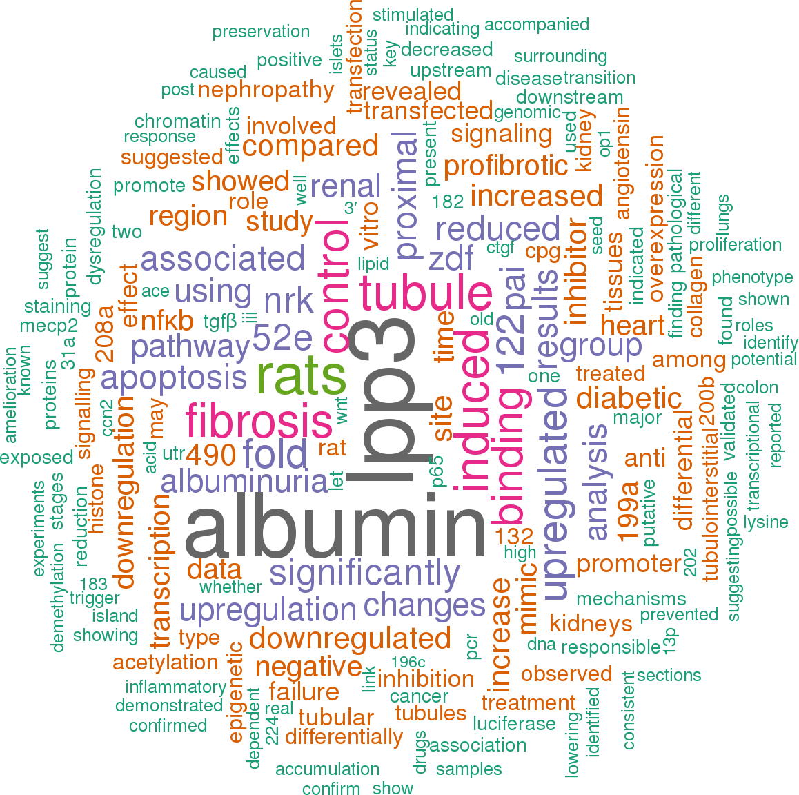
Open access articles that are associated with the species Rattus norvegicus and mention the gene name mir-184. Click the [+] symbols to view sentences that include the gene name, or the word cloud on the right for a summary.
1
[+] score: 271
This results in miR-184 upregulation and repression of the downstream target LPP3, which in turn upregulates Pai-1 transcription through the β-catenin (β-Cat)–TCF signalling pathway in a cAMP response-element binding protein (CREB) binding protein (CBP) -dependent fashion In conclusion, this study provides the novel finding that miR-184 is predominantly expressed in the renal tubules of ZDF rats and plays a role in tubulointerstitial fibrosis through downregulation of LPP3. [score:14]
Treatment of cells overexpressing miR-184 with ICG-001, a small-molecule inhibitor of TCF/β-catenin transcription [30], prevented Pai-1 mRNA upregulation (Fig. 4f), indicating the involvement of the β-catenin–TCF signalling pathway in the profibrotic effect of LPP3 downregulation. [score:11]
Anti-miR-184 counteracted albumin -induced LPP3 downregulation and overexpression of plasminogen activator inhibitor-1. In ZDF rats, ACE -inhibitor treatment limited albuminuria and reduced miR-184, with tubular LPP3 preservation and tubulointerstitial fibrosis amelioration. [score:10]
Renal miRNA profiling of young and old rodents revealed upregulated miR-184 in old kidneys [41, 42], suggesting that epigenetic regulation of renal ageing likely occurs through inhibition of miR-184 targeted genes encoding antioxidant-, ECM-degrading- and longevity-related proteins [41]. [score:9]
On the other hand, the finding that miR-184 was upregulated in TGF-β1-stimulated cells suggests that this miRNA can likely mediate the cytokine profibrotic effects but, unlike other miRNAs [46], does not contribute to TGF-β auto-upregulation, at least in proximal tubule cells. [score:7]
Although the present study did not prove a direct link between LPP3 downregulation and miR-184 -induced fibrosis, in vitro experiments suggested β-catenin/TCF signalling as one of the intracellular mechanisms through which miR-184/LPP3 dysregulation may induce profibrotic genes. [score:6]
Here, we suggest a link between abnormal tubular miR-184 and tubulointerstitial fibrosis in the diabetic kidneys through inhibition of the target LPP3, which plays a key role in regulating biosynthesis of lipid phosphates involved in multiple organ fibrosis as well as in cell signal transduction [23– 25]. [score:6]
Unlike albumin, angiotensin II, a critical mediator of proteinuria and fibrosis [10], failed to induce miR-184 expression in NRK-52E cells, known to express angiotensin II type 1 receptors [50], indicating that angiotensin II lacks a direct effect on miR-184. [score:6]
To prove that decrease in LPP3 in response to albumin was dependent on miR-184 upregulation, cells were transfected with anti-miR-184 or with miRNA inhibitor negative control, before albumin incubation. [score:6]
Moving to diabetic nephropathy, here we demonstrated that miR-184 was the most upregulated miRNA in the kidneys of ZDF rats at an advanced phase of the disease. [score:6]
By contrast, 4-phenylbutyric acid significantly upregulated miR-184 expression, which was further enhanced by the combined treatment. [score:6]
Our data showed that in NRK-52E cells with reduced LPP3 after miR-184 mimic transfection, inhibition of β-catenin–TCF signalling by ICG-001 did prevent Pai-1 mRNA upregulation. [score:6]
Compared with negative control treatment, anti-miR-184 prevented albumin -induced LPP3 downregulation (Fig. 6a) and limited Pai-1 overexpression (Fig. 6b), indicating that albumin exerted a profibrotic effect through miR-184/LPP3. [score:5]
Albumin and FA-free albumin (Fig. 5c) caused a 4.8- and 3.5-fold increase in miR-184 expression compared with the control, respectively, suggesting a partial contribution of FA to albumin -induced miR-184 upregulation. [score:5]
Albuminuria Diabetic nephropathy Fibrosis miR-184 Zucker diabetic fatty rats Diabetic nephropathy is one of the major microvascular complications of diabetes and the leading cause of chronic kidney disease and end-stage renal disease (ESRD) throughout the world [1, 2]. [score:5]
Scale bars, 50 μm To identify potential miR-184 target genes, miRNA target prediction algorithms miRanda and EIMMo were used in conjunction with the microRNA body map web tool. [score:5]
Scale bars, 50 μm We used NRK-52E, a rat proximal tubule cell line, for in vitro mechanistic studies to establish a causal role of miR-184 on LPP3 downregulation and renal fibrosis. [score:4]
miR-184 is upregulated in the kidneys of ZDF rats. [score:4]
Consistent with Lpp3 mRNA downregulation, protein levels were almost halved in the miR-184 mimic -transfected cells (Fig. 4b). [score:4]
In vitro miR-184 causes LPP3 downregulation accompanied by a profibrotic phenotype of tubule epithelial cells. [score:4]
Moreover, miR-184 mimic caused a transient increase in Ctgf/ Ccn2 mRNA and a sustained upregulation of Pai-1 mRNA (Fig. 4c,d), while it did not affect Tgf-β (Fig. 4e). [score:4]
Our data show that albumin has a role in the dysregulation of epigenetic mechanisms like DNA demethylation and histone lysine acetylation, which ultimately lead to miR-184 overexpression in tubule cells. [score:4]
Albumin -induced miR-184 expression in tubule cells was epigenetically regulated through DNA demethylation and histone lysine acetylation and was accompanied by binding of NF-κB p65 subunit to miR-184 promoter. [score:4]
miR-184 showed the strongest differential upregulation (18-fold). [score:4]
Albumin -induced miR-184 expression translated into a 47% reduction in LPP3, compared with control cells (Fig. 5d); a 32% reduction was observed after FA-free albumin. [score:4]
An interesting observation is that while Pai-1 and Ctgf/ Ccn2 mRNA levels increased in miR-184 -overexpressing cells, no changes were observed in Tgf-β transcripts. [score:3]
Fig. 7Epigenetic mechanisms involved in albumin -induced miR-184 expression in proximal tubule cells. [score:3]
Tubular localisation of miR-184 was associated with reduced expression of lipid phosphate phosphatase 3 (LPP3) and collagen accumulation. [score:3]
Of the putative candidates, we focused on Lpp3 because it was the only mRNA predicted as a target of miR-184 by all databases. [score:3]
These data were corroborated by in vitro experiments demonstrating that tubule cells showing reduced LPP3 after miR-184 overexpression acquired a profibrotic phenotype documented by enhanced Ctgf/ Ccn2 and Pai-1 mRNAs. [score:3]
Bioinformatic analysis of miR-184 target genes. [score:3]
In ZDF rats, miR-184 showed the strongest differential upregulation compared with lean rats (18-fold). [score:3]
Human AD-293 cells (Agilent, Santa Clara, CA, USA; negative for mycoplasma contamination) were co -transfected with a construct containing the human LPP3–3′ untranslated region (UTR) downstream of the Firefly luciferase gene, the co-reporter vector pRL-TK encoding the Renilla luciferase and rat miR-184 mimic or control mimic using Lipofectamine 2000 (Life Technologies). [score:3]
Among mediators of fibrosis, albumin was the most potent stimulus of miR-184, consistent with the putative role of albuminuria in exacerbating disease progression in diabetic nephropathy [40]. [score:3]
Fig. 8ACE inhibition limits albuminuria in association with modulation of miR-184/LPP3 and amelioration of fibrosis. [score:3]
This was accompanied by reduction in renal miR-184 and preservation of LPP3 in the tubular epithelium (Fig. 8b,c), along with attenuation of fibrosis, as indicated by reduced expression of α-SMA (Fig. 8d), type III collagen (Fig. 8e) and Pai-1 mRNA (Fig. 8f). [score:3]
To identify triggers of miR-184 expression, NRK-52E cells were exposed to angiotensin II, human serum albumin, holo-transferrin, TGF-β1 or fatty acid (FA)-free albumin (Sigma-Aldrich). [score:3]
ch/EIMMo3/) were used to identify potential targets of miR-184. [score:3]
Albuminuria is the main instigator for miR-184 expression in tubule cells under the control of epigenetic mechanisms. [score:3]
NRK-52E cells were transfected with anti-miR-184 or miRNA inhibitor negative control (NC), followed by albumin stimulation (10 mg/ml, 72 h). [score:3]
Fig. 2In vitro validation of Lpp3 as a target of miR-184. [score:3]
Little information is available regarding miR-184 expression in the kidney. [score:3]
Serial kidney sections showed that in ZDF rats miR-184 overexpression was associated with reduced LPP3 and accumulation of interstitial type III collagen, used as a marker for fibrosis (Fig. 3b). [score:3]
These data may offer a new opportunity for targeting miR-184 in association with albuminuria-lowering drugs to achieve fully protective anti-fibrotic effects in diabetic nephropathy. [score:3]
Treating NRK-52E cells with 5-aza-2′-deoxycytidine caused a non-significant increase in miR-184 expression (Fig. 7a). [score:3]
Limiting albuminuria with an ACE inhibitor reduces renal fibrosis in ZDF rats associated with miR-184/LPP3 modulation. [score:3]
LPP3 is a target of miR-184. [score:3]
Albumin was a major trigger of miR-184 expression. [score:3]
Importantly, in ZDF rats, lowering albuminuria using an ACE inhibitor was associated with reduced miR-184, preservation of tubular LPP3 and amelioration of tubulointerstitial fibrosis. [score:3]
Our data, showing that albumin -induced miR-184 in cultured tubule cells was associated with reduced LPP3 and abnormal Pai-1 mRNA (both prevented by miR-184 antagomir treatment), suggest a functional link between miR-184 dysregulation and albumin load -induced renal fibrosis. [score:2]
miR-184 is epigenetically regulated in proximal tubule epithelial cells. [score:2]
This suggests the synergistic effect of DNA demethylation and histone acetylation on chromatin, switching from a silent compact state to an active relaxed state to regulate miR-184 transcription [34, 35]. [score:2]
To confirm that LPP3 is a true miR-184 target, we performed luciferase reporter assays using a plasmid carrying the full-length 3′ UTR of human LPP3 downstream of the luciferase gene. [score:2]
Epigenetic regulation of miR-184 was assessed in NRK-52E cells exposed to the chromatin-modifying drugs 5-aza-2′-deoxycytidine and 4-phenylbutyric acid for of miR-184. [score:2]
Fig. 5Albumin is the primary regulator of miR-184 in proximal tubule cells. [score:2]
Methyl-CpG binding proteins, together with chromatin histone modifications, are involved in the regulation of miR-184 in mouse brain cells [37– 39] and human T cells [39]. [score:2]
RNA was isolated from NRK-52E cells for analysis of miR-184 expression and Lpp3 and Pai-1 mRNA using specific TaqMan assays. [score:2]
Next, we investigated whether FA bound to albumin, rather than albumin itself, was responsible for miR-184 upregulation. [score:2]
Albumin is a major regulator of miR-184 in proximal tubule cells. [score:2]
Fig. 9Hypothetical pathways through which albumin overload promotes renal fibrosis via epigenetic regulation of miR-184. [score:2]
In keeping with this, in the kidneys of ZDF rats, dysregulation of the miR-184/LPP3 pathway would possibly result in increased bioactive lipid phosphate availability, which would activate profibrotic signalling with a consequent accumulation of ECM proteins. [score:2]
NF-κB binding domain, putative CpG island and transcription site of miR-184 are indicated. [score:1]
Notably, the binding site for miR-184 in the 3′ UTR of Lpp3 (Fig. 2a) is evolutionarily conserved among species. [score:1]
Starting from the evidence of the presence of an NF-κB binding domain 958 bp upstream of the transcriptional site of miR-184 (Fig. 7b), we performed ChIP assay for p65, the NF-κB subunit primarily responsible for transcriptional activation of target genes, and demonstrated that NF-κB was recruited to the miR-184 promoter in albumin -treated cells compared with control cells (Fig. 7e). [score:1]
These results suggest that miR-184 may act as a downstream effector of albuminuria through LPP3 to promote tubulointerstitial fibrosis, and offer the rationale to investigate whether targeting miR-184 in association with albuminuria-lowering drugs may be a new strategy to achieve fully anti-fibrotic effects in diabetic nephropathy. [score:1]
By contrast, albumin was a potent inducer of miR-184, causing a 2.6-fold increase over control cells at 24 h and a 6-fold increase at 48 h. The marked stimulatory effect of albumin was not shared by transferrin, another component of proteinuria known to be toxic to proximal tubule cells [32, 33]. [score:1]
Next, through computational analysis of the rat genomic region [36] surrounding the miR-184 gene, we identified a putative CpG island located 731 bp upstream of the transcription site of miR-184 (Fig. 7b), suitable for binding methyl-CpG binding proteins, such as MeCP2 and MBD1, known to act as transcriptional repressors [15]. [score:1]
Our finding that p65 was recruited to miR-184 promoter in albumin-stimulated tubule cells indicates that the chromatin modification events observed in response to albumin make the miR-184 promoter region more accessible to NF-κB, thereby inducing miR-184 transcription (Fig. 9). [score:1]
In albumin -treated cells, ChIP-qPCR using MeCP2 antibody revealed a reduction in MeCP2 binding to the genomic region surrounding miR-184 (R1–6), which reached statistical significance within the CpG island (R2) and in the region upstream of the miR-184 transcription site (R4) (Fig. 7c). [score:1]
The seed-recognising site (position 1272–1278 of rat Lpp3 3′ UTR) binding to miR-184 RNA is indicated. [score:1]
Data from Multiplex PCR were validated by, and confirmed a 23-fold increase of miR-184 in ZDF rats (Fig. 1a), mainly localised at the tubular epithelium (Fig. 1b), in proximal and distal tubules, as revealed by aquaporin1 and Tamm–Horsfall protein staining (Fig. 1c). [score:1]
Rat proximal tubule NRK-52E cells (DSMZ, Braunschweig, Germany, negative for mycoplasma contamination) [20] were transfected with miR-184 mimic or control mimic using Lipofectamine 2000. [score:1]
Proximal (arrow heads) and distal (arrows) tubules positive for miR-184 are shown. [score:1]
Angiotensin II did not stimulate miR-184. [score:1]
Insets show high magnification of a miR184 -positive tubule with reduced LPP3 staining and surrounded by interstitial type III collagen deposition. [score:1]
In albumin-stimulated cells, H3K9 acetylation was enriched only in the genomic region upstream of the miR-184 transcription site (R4) (Fig. 7d), whereas H3K4me3 did not show significant enrichment (ESM Fig. 3b). [score:1]
A 2.6-fold increase in miR-184 was induced by TGF-β at 48 h. The increase in miR-184 in cells exposed to albumin was dose dependent, starting with a dose as low as 1 mg/ml (Fig. 5b). [score:1]
In 8-month-old ZDF rats, miR-184 focally localised in areas of damaged proximal tubules. [score:1]
No signal for miR-184 was detected in the kidneys of lean rats (Fig. 1b). [score:1]
Albumin reduces binding of MeCP2 to miR-184 promoter and fosters histone lysine acetylation (Ac), favouring accessibility of NF-κB-p65 to its recognition sequence on the miRNA promoter. [score:1]
NRK-52E cells were transfected with anti-miR-184 or miRNA inhibitor negative control before albumin stimulation for Lpp3 and Pai-1 mRNA evaluation. [score:1]
Using serial kidney sections, we did document increased type III collagen staining in areas surrounding tubules that were positive for miR-184 and exhibited reduced LPP3 staining. [score:1]
To investigate whether and which of these pathogenic insults could be a trigger for miR-184 expression in renal tubule cells, NRK-52E cells were exposed to the different stimuli for 6–48 h (Fig. 5a). [score:1]
Transfection of NRK-52E cells with miR-184 mimic reduced LPP3, promoting a profibrotic phenotype. [score:1]
One key finding of the present study is that albumin is a major trigger for miR-184 in tubule cells. [score:1]
Importantly, an NF-κB binding site is present in the promoter of miR-184. [score:1]
miR-184 hybridisation was assessed on paraffin-embedded kidney sections using the double-digoxigenin -labelled LNA miRCURY probe (Exiqon, Vedbaek, Denmark). [score:1]
[1 to 20 of 87 sentences]
2
[+] score: 77
There were 49 differentially expressed miRNAs in the colon tissues of the UC group compared to the NC group, of which 47 were upregulated and 2 were downregulated; miR-184 and miR-18a-5p were both downregulated (Figure 6). [score:11]
miR-184 was downregulated by the UC pathological status; after the HM intervention, its expression was upregulated. [score:9]
miR-184 expression was also significantly downregulated in inflammatory tissues from the colons of DSS -induced UC to participate in the regulation of immune inflammation. [score:7]
Target gene predictions for miR-184 and miR-490-5p and the pathway analysis of the target genes of these 2 small RNAs showed that potential target genes of miR-184 were involved in some pathways that were closely associated with UC, such as the TNF signaling pathway, Ras signaling pathway, pathway in cancer, NF- κB signaling pathway, miRNAs in cancer, colorectal cancer, and AMPK signaling pathway. [score:7]
However, the expression of miR-184 was significantly increased in tumor tissues, and inhibition of miR-184 inhibited tumor growth [48], which was completely different from its function in inflammatory tissues. [score:7]
miR-184 was directly associated with the level of inflammation, and its expression was significantly downregulated in inflammatory tissues [46, 47]. [score:7]
The miR-184 expression level increased and the miR-490-5p expression level decreased in the UCHM group compared to the UC group (both P < 0.05) (Figures 9(a) and 9(b)). [score:4]
The miR-184 expression level decreased and the miR-490-5p expression level increased in the UC group compared to the NC group (both P < 0.05). [score:4]
The upregulated miRNAs in the colon tissues of UC rats changed by HM were miR-149-5p, miR-351-5p, let-7d-5p, miR-98-5p, let-7a-5p, miR-3559-5p, let-7f-1-3p, miR-3596b, miR-224-5p, miR-411-3p, miR-184, miR-26b-3p, and miR-92b-3p. [score:4]
The results suggested that HM could reverse the expression of miR-184 and miR-490-5p, which changed with the UC pathological status. [score:3]
miRNA-184 had 1130 target genes. [score:3]
HM altered the expression of 15 miRNAs, of which 2 were associated with UC pathological changes (miR-184 and miR-490-5p). [score:3]
There were two common differentially expressed miRNAs in the two heat maps shown in Figures 6 and 7 (miR-184 and miR-490-5p). [score:3]
The results in this study suggested that changes in miR-184 and miR-490-5p expression levels in colon tissues under the UC pathological status were reversed in the UCHM group. [score:3]
The samples were amplified, and 2 miRNAs (miR-184 and miR-490-5p) were validated using quantitative real-time PCR. [score:1]
U6, miR-184, and miR-490-5p were synthesized and purified by RiboBio (Guangzhou, China). [score:1]
[1 to 20 of 16 sentences]
3
[+] score: 74
To confirm the results of the miRNA sequencing data, 4 upregulated miRNAs (rno-miR-122-5p, rno-miR-199a-5p, rno-miR-184 and rno-miR-202-5p) and 4 downregulated miRNAs (rno-miR-208a-3p, rno-miR-208a-5p, rno-miR-6314 and rno-miR-22-3p) were chosen to be further examined using real-time quantitative PCR. [score:7]
Differential miR-184 expression has been reported to be responsible for the aberrant activation of Wnt signaling in ischemia -induced retinal neovascularization[29], which is consistent with our results that Wnt signaling pathway was significantly up-regulated in the HF group. [score:6]
Wang et al[31] proved that oxidative modification of miR-184 participated in apoptosis through downregulating Bcl-xL and Bcl-w, which were not the native target of miR-184. [score:6]
Among them, 12 miRNAs (rno-miR-10b-5p, rno-miR-122-5p, rno-miR-184, rno-miR-1843-5p, rno-miR-196c-5p, rno-miR-199a-5p, rno-miR-202-5p, rno-miR-206-3p, rno-miR-208b-5p, rno-miR-224-5p, rno-miR-298-5p and rno-miR-31a-5p) were significantly upregulated(p<0.01, fold-change >1) compared to the control group and only rno-miR-208a-3p were significantly downregulated (p<0.01, fold-change <-1) (Fig 3). [score:6]
Among the top 13 differentially expressed miRNAs, the five most abundantly expressed miRNAs were rno-miR-122-5p, rno-miR-184, rno-miR-31a-5p, rno-miR-199a-5p and rno-miR-208a-3p. [score:5]
Our results suggested that miR-184 expression was gradually increased during the post-infarction heart failure, which indicate that miR-184 regulated the pathogenesis of heart failure. [score:4]
0160920.g005 Fig 5 (A) Relative expression levels of miR-122-5p and miR-184 after transfection. [score:3]
Differential profile and expression patterns of miRNAs in the rats mo del of post-infarction heart failure were found, and the pro-apoptotic roles of rno-miR-122-5p and rno-miR-184 were revealed. [score:3]
Time-course analysis revealed different expression patterns of 4 miRNAs: rno-miR-122-5p, rno-miR-199a-5p, rno-miR-184 and rno-miR-208a-3p. [score:3]
The results showed that the expression of rno-miR-199a-5p and rno-miR-184 gradually increased with time after MI operation. [score:3]
To examine the effects of rno-miR-122-5p and rno-miR-184, cells were transfected with rno-miR mimics, rno-miR inhibitors, or scrambled controls (Guangzhou Ruibo biology Science & Technology Co,; Ltd; China). [score:3]
Meanwhile, rno-miR-10b-5p, rno-miR-184, rno-miR-1843-5p, rno-miR-196c-5p, rno-miR-202-5p, rno-miR-206-3p, rno-miR-224-5p, rno-miR-298-5p and rno-miR-31a-5p were reported for the first time to be differentially expressed in HF tissue. [score:3]
Transfection of miR-184 inhibitor had no obvious effect on apoptosis (p>0.05) (Fig 5). [score:3]
MicroRNA-184 modulates canonical Wnt signaling through the regulation of frizzled-7 expression in the retina with ischemia -induced neovascularization. [score:3]
To further confirm the results of the miRNA sequencing and analyze the dynamic expression pattern of specific miRNAs in HF rats, the dynamic changes of miR-208a-3p (Fig 4B), miR-184 (Fig 4C), miR-122-5p (Fig 4D) and miR-199a-5p (Fig 4E) in the process of post-infarcted heart failure were analyzed. [score:3]
The miRNA signature of failing myocardium in our study indicated for the first time that 9 miRNAs were associated with HF, including rno-miR-184 and rno-miR-31a-5p, which were the most significantly differentially expressed between the two groups. [score:3]
It was possible that miR-184 might play an important role in the regulatory networks of heart failure through apoptosis. [score:2]
In addition, miR-122-5p and miR-184 have been reported to be involved in the regulation of apoptosis under particular conditions, so we speculated that these two miRNAs could play important roles in the pathogenesis of heart failure through apoptosis. [score:2]
Additionally, rno-miR-122-5p and rno-miR-184 were proved to promote apoptosis in vitro. [score:1]
However, the roles of non-oxidative miR-184 in apoptosis and heart failure have not been studied. [score:1]
Finally, gain- and loss-of-function experiments of rno-miR-122-5p and rno-miR-184 were analyzed in H [2]O [2] treated H9c2 cells. [score:1]
In vitro results also found that miR-184 could promote apoptosis in H [2]O [2] treated H9c2 cells. [score:1]
Time course analysis of miR-208a-3p (B), miR-184 (C), miR-122-5p (D) and miR-199a-5p (E) were studied. [score:1]
Effect of rno-miR-122-5p and rno-miR-184 on cell apoptosis. [score:1]
[1 to 20 of 24 sentences]
4
[+] score: 24
Analysis at P21 revealed 74 mature miRNAs with differential expression between LPD -induced IUGR rat lungs and control lungs (Fig 1): 10 showed more than twofold differential expression: miR-184, miR-127-3p, miR-378a-5p and miR541-5p were downregulated, and miR-30e-5p, miR-23b-5p, miR-451-5p, miR-1839-5p, miR-449a-5p, and miR-19b-3p were upregulated in LPD -induced IUGR versus control lungs. [score:11]
At P21, the differential expression of 5 of 10 of the miRNAs was statistically confirmed, with miR378a-5p, miR127-3p, and miR184 downregulated and miR30e-5p and miR449a-5p upregulated. [score:9]
Among the other miRNAs found deregulated in our study, miR-34c-5p, miR-128-3p miR-184, miR127-3p, miR-30e-5p, and miR-23b-5p were also previously described as “tumour suppressors”[22– 26], although many miRNA studies previously dealt with cancer or oncogenic proliferation states and the current analysis depended on previous reports. [score:4]
[1 to 20 of 3 sentences]
5
[+] score: 21
down-regulated mRNAs (Fig.   6A), e. g., up-regulated miR-184 interacted with 9 down-regulated genes (Abcg8, Dhcr7, Hmgcr, Tmem255a, etc. [score:10]
In addition to the down-regulated miRNAs, we obtained 7 up-regulated miRNAs (miR-200b, miR-200b-3p, miR-200b-5p, miR-200c-3p, miR-378b, miR-184, and miR-349) and 114 correlated miRNA and mRNA regulatory pairs. [score:8]
We observed that the intersection genes Abcg8, Cyp1a1 and Tmem255a were the targets of miR-349, miR-378b, miR-200b-3p, miR-200c-3p, miR-200b-5p and miR-184 (Fig.   6B). [score:3]
[1 to 20 of 3 sentences]
6
[+] score: 18
Other miRNAs from this paper: hsa-mir-16-1, hsa-mir-17, hsa-mir-20a, hsa-mir-21, hsa-mir-23a, hsa-mir-100, hsa-mir-103a-2, hsa-mir-103a-1, hsa-mir-107, hsa-mir-16-2, mmu-mir-1a-1, mmu-mir-23b, mmu-mir-125b-2, mmu-mir-130a, mmu-mir-9-2, mmu-mir-145a, mmu-mir-181a-2, mmu-mir-184, mmu-mir-199a-1, hsa-mir-199a-1, mmu-mir-205, mmu-mir-206, hsa-mir-181a-2, hsa-mir-181b-1, hsa-mir-199a-2, hsa-mir-205, hsa-mir-181a-1, hsa-mir-214, hsa-mir-219a-1, hsa-mir-223, mmu-mir-302a, hsa-mir-1-2, hsa-mir-23b, hsa-mir-125b-1, hsa-mir-130a, hsa-mir-145, hsa-mir-9-1, hsa-mir-9-2, hsa-mir-9-3, hsa-mir-125b-2, hsa-mir-184, hsa-mir-206, mmu-mir-16-1, mmu-mir-16-2, mmu-mir-20a, mmu-mir-21a, mmu-mir-23a, mmu-mir-103-1, mmu-mir-103-2, rno-mir-338, mmu-mir-338, rno-mir-20a, hsa-mir-1-1, mmu-mir-1a-2, hsa-mir-181b-2, mmu-mir-107, mmu-mir-17, mmu-mir-100, mmu-mir-181a-1, mmu-mir-214, mmu-mir-219a-1, mmu-mir-223, mmu-mir-199a-2, mmu-mir-9-1, mmu-mir-9-3, mmu-mir-181b-1, mmu-mir-125b-1, hsa-mir-302a, hsa-mir-219a-2, mmu-mir-219a-2, hsa-mir-302b, hsa-mir-302c, hsa-mir-302d, hsa-mir-367, hsa-mir-372, hsa-mir-338, mmu-mir-181b-2, rno-mir-9a-1, rno-mir-9a-3, rno-mir-9a-2, rno-mir-16, rno-mir-17-1, rno-mir-21, rno-mir-23a, rno-mir-23b, rno-mir-100, rno-mir-103-2, rno-mir-103-1, rno-mir-107, rno-mir-125b-1, rno-mir-125b-2, rno-mir-130a, rno-mir-145, rno-mir-181a-2, rno-mir-181b-1, rno-mir-181b-2, rno-mir-199a, rno-mir-205, rno-mir-206, rno-mir-181a-1, rno-mir-214, rno-mir-219a-1, rno-mir-219a-2, rno-mir-223, hsa-mir-512-1, hsa-mir-512-2, rno-mir-1, mmu-mir-367, mmu-mir-302b, mmu-mir-302c, mmu-mir-302d, rno-mir-17-2, hsa-mir-1183, mmu-mir-1b, hsa-mir-302e, hsa-mir-302f, hsa-mir-103b-1, hsa-mir-103b-2, rno-mir-9b-3, rno-mir-9b-1, rno-mir-9b-2, rno-mir-219b, hsa-mir-23c, hsa-mir-219b, mmu-mir-145b, mmu-mir-21b, mmu-mir-21c, mmu-mir-219b, mmu-mir-219c, mmu-mir-9b-2, mmu-mir-9b-1, mmu-mir-9b-3
The highest upregulated miRNA (miR-184) at the GP-early OP transition shows a strong conserved 8mer binding site to BCL2-like 1 (Bcl2l1), a gene highly expressed in astrocytes [36]. [score:6]
The highest upregulated miRNA at this transition, miR-184, shows a high likelihood of binding to Bcl2l1, a gene which has been shown to be co-expressed with GFAP in various samples of astrocyte tissue [36]. [score:6]
On the other hand, the top upregulated miRNAs at the OP3-OL transition included miRNAs (miR-181a, miR-181b, miR-125b, and miR-184) that are associated with decreased proliferation in maturing CNS cells and decreased malignancy in glioma stem cells [49], [50], [51], [52], [53], [54], [55]. [score:4]
Therefore, it is possible that transfection of miR-184 and/or miR-1183 analogs to GPs may help reduce astrocyte differentiation and promote the production of oligodendrocyte cultures with increased purity. [score:1]
The key miRNAs discussed in this manuscript were validated by conducting real-time qRT-PCR for samples from the appropriate stages, including the following: miR-199a and miR-145 at the OP1, OP2, OP3, and OL stages; miR-214 at the OP1 and OP2 stages; miR-184 and miR-1183 at the GP and OP1 stages (Table 1 ). [score:1]
[1 to 20 of 5 sentences]
7
[+] score: 11
Other miRNAs from this paper: hsa-let-7a-1, hsa-let-7a-2, hsa-let-7a-3, hsa-let-7b, hsa-let-7c, hsa-let-7d, hsa-let-7e, hsa-let-7f-1, hsa-let-7f-2, hsa-mir-16-1, hsa-mir-17, hsa-mir-21, hsa-mir-23a, hsa-mir-24-1, hsa-mir-24-2, hsa-mir-25, hsa-mir-26a-1, hsa-mir-26b, hsa-mir-30a, hsa-mir-31, hsa-mir-96, hsa-mir-99a, hsa-mir-16-2, hsa-mir-30c-2, hsa-mir-30d, hsa-mir-182, hsa-mir-183, hsa-mir-211, hsa-mir-217, hsa-mir-218-1, hsa-mir-218-2, hsa-mir-221, hsa-mir-222, hsa-let-7g, hsa-let-7i, hsa-mir-15b, hsa-mir-23b, hsa-mir-30b, hsa-mir-125b-1, hsa-mir-132, hsa-mir-143, hsa-mir-145, hsa-mir-191, hsa-mir-125a, hsa-mir-125b-2, hsa-mir-126, hsa-mir-184, hsa-mir-190a, hsa-mir-195, rno-mir-322-1, rno-let-7d, rno-mir-335, rno-mir-342, rno-mir-135b, hsa-mir-30c-1, hsa-mir-299, hsa-mir-30e, hsa-mir-26a-2, hsa-mir-379, hsa-mir-382, hsa-mir-342, hsa-mir-135b, hsa-mir-335, rno-let-7a-1, rno-let-7a-2, rno-let-7b, rno-let-7c-1, rno-let-7c-2, rno-let-7e, rno-let-7f-1, rno-let-7f-2, rno-let-7i, rno-mir-15b, rno-mir-16, rno-mir-17-1, rno-mir-21, rno-mir-23a, rno-mir-23b, rno-mir-24-1, rno-mir-24-2, rno-mir-25, rno-mir-26a, rno-mir-26b, rno-mir-30c-1, rno-mir-30e, rno-mir-30b, rno-mir-30d, rno-mir-30a, rno-mir-30c-2, rno-mir-31a, rno-mir-96, rno-mir-99a, rno-mir-125a, rno-mir-125b-1, rno-mir-125b-2, rno-mir-126a, rno-mir-132, rno-mir-143, rno-mir-145, rno-mir-183, rno-mir-190a-1, rno-mir-191a, rno-mir-195, rno-mir-211, rno-mir-217, rno-mir-218a-2, rno-mir-218a-1, rno-mir-221, rno-mir-222, rno-mir-299a, hsa-mir-384, hsa-mir-20b, hsa-mir-409, hsa-mir-412, hsa-mir-489, hsa-mir-494, rno-mir-489, rno-mir-412, rno-mir-543, rno-mir-542-1, rno-mir-379, rno-mir-494, rno-mir-382, rno-mir-409a, rno-mir-20b, hsa-mir-542, hsa-mir-770, hsa-mir-190b, hsa-mir-543, rno-mir-466c, rno-mir-17-2, rno-mir-182, rno-mir-190b, rno-mir-384, rno-mir-673, rno-mir-674, rno-mir-770, rno-mir-31b, rno-mir-191b, rno-mir-299b, rno-mir-218b, rno-mir-126b, rno-mir-409b, rno-let-7g, rno-mir-190a-2, rno-mir-322-2, rno-mir-542-2, rno-mir-542-3
Thus, it is possible that the down-regulation of miRNAs (rno-miR-770, rno-miR-466c, rno-miR-31, rno-miR-183, rno-miR-96, rno-miR-132, rno-miR-182, rno-miR-384-3p and rno-miR-184) observed in this study could be associated with promoted thecal hyperandrogenesis [37, 38]. [score:4]
MiRNAs found to be primarily down-regulated in DHT -treated rats includes rno-miR-770, rno-miR-466c, rno-miR-21, rno-miR-31, rno-miR-182, rno-miR-183, rno-miR-96, rno-miR-132, rno-miR-182, rno-miR-384-3p and rno-miR-184. [score:4]
Among the fourteen miRNAs mapped to the ingenuity databases, twelve (rno-let-7d, rno-miR-132, rno-miR-182, rno-miR-183, rno-miR-184, rno-miR-21, rno-miR-221, rno-miR-24, rno-miR-25, rno-miR-26b, rno-miR-31 and rno-miR-96) had 171 experimentally validated targets. [score:3]
[1 to 20 of 3 sentences]
8
[+] score: 9
rno-miR-184, rno-miR-347, rno-miR-181a-2-3p, rno-miR-204-5p, rno-miR-132-3p and rno-miR-328b-3p are significantly up-regulated while other 10 miRNAs are significantly down-regulated Fig. 16 Hierarchical clusterings analysis of miRNA expressionprofile for intrauterine infection group and control at P3. [score:9]
[1 to 20 of 1 sentences]
9
[+] score: 8
A comparison between these identified microRNAs and those showing expression changes in the present microarray analysis revealed similar expression changes for miR-21 24 hours after SCI as well as for 4 other microRNAs, namely, miR-184, miR-340-5p, miR-369-3p and miR-466b, at 7 dpo (Table 2). [score:5]
These analyses identified four microRNAs with expression changes at 7 dpo; these included miR-340-5p and miR-369-3p, which are both involved in proliferation and adipogenic differentiation, as well as the proapoptotic miR-184 [109] and miR-466b. [score:3]
[1 to 20 of 2 sentences]
10
[+] score: 7
While miR-184 and miR-204 which were down-regulated in at least one mouse strain in their study, also showed down-regulation in our mo del. [score:7]
[1 to 20 of 1 sentences]
11
[+] score: 5
al, silence of miR-184, another miRNA that significantly up-regulated after SE, led to an increase in the counts of apoptotic neurons, suggesting a negative regulation of miRNA on neuronal apoptosis [14]. [score:5]
[1 to 20 of 1 sentences]
12
[+] score: 5
Overexpression of miR-132 and inhibition of miR-184 protects beta cells against palmitate- or cytokine -induced apoptosis [7]. [score:5]
[1 to 20 of 1 sentences]
13
[+] score: 3
As demonstrated in Fig. 6A, rno-miR-184, rno-miR-484 and rno-miR-138-1-3p were the most significantly dysregulated miRNAs in GHS rats, and the results were comparable with our microarray data. [score:2]
control rats (Table 1 and Table S2); of these rno-miR-184, rno-miR-21-3p and rno-miR-672-5p had the largest positive fold changes, while rno-miR-484, rno-miR-138-1-3p and rno-miR-201-3p had the largest negative fold changes. [score:1]
[1 to 20 of 2 sentences]
14
[+] score: 3
These were rno-miR-184 and rno-miR-6325, expressed 9.5-fold and 2.8-fold higher in HA than LA rats, respectively. [score:3]
[1 to 20 of 1 sentences]
15
[+] score: 3
For example, mutation in the seed region of miR-184 causes familial keratoconus with cataract [14] and mutations in the seed region of miR-96 are responsible for nonsyndromic progressive hearing loss [15]. [score:3]
[1 to 20 of 1 sentences]
16
[+] score: 3
We confirmed the expression changes (> 2-fold) of mir-184 and mir-1250 in brain and of mir-1281, mir-551b, mir-3185, mir-3162-5p and mir-1228 in heart in mechanical asphyxia cases (Figure 2A and 2C). [score:3]
[1 to 20 of 1 sentences]
17
[+] score: 1
Moreover, the level of miRNA-375, together with miRNA-127-3p and miR-184 is positively correlated to insulin mRNA levels in islets from human donors and the association between these miRNAs and β-cell function was deranged in islets from glucose intolerant donors [18]. [score:1]
[1 to 20 of 1 sentences]
18
[+] score: 1
Other miRNAs from this paper: rno-mir-130a, rno-mir-130b, rno-mir-132, rno-mir-152, rno-mir-212
In db/ db and high-fat diet fed mice, miR-132 and miR-184 in the pancreatic islets were suggested to be induced by hyperglycaemic and hyperlipidaemic conditions typically encountered in prediabetic and diabetic states 37. [score:1]
[1 to 20 of 1 sentences]