sort by

88 publications mentioning rno-mir-124-1

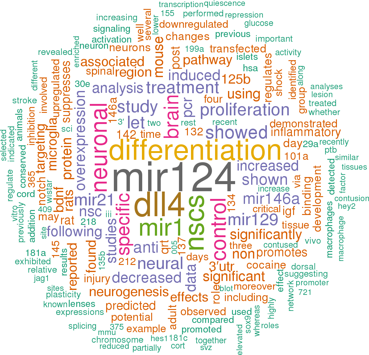
Open access articles that are associated with the species Rattus norvegicus and mention the gene name mir-124-1. Click the [+] symbols to view sentences that include the gene name, or the word cloud on the right for a summary.
1
[+] score: 351
In the present study, we demonstrated that miR-124 expression was up-regulated and DLL4 expression was down-regulated in NSC differentiation. [score:11]
demonstrated that exogenous expression of miR-124 remarkably inhibited DLL4 protein level, while DLL4 overexpression partially restored miR-124 -mediated DLL4 expression inhibition (Fig.   6a). [score:11]
Also, miR-124 overexpression promoted the expression of neuron-specific marker β-tubulin-III (Fig.   4g, i) and suppressed the expression of astrocyte-specific marker GFAP (Fig.   4h, j) at mRNA and protein levels, while miR-124 suppression showed the reverse effects. [score:11]
Moreover, increased expression of miR-124 promotes β-tubulin-III expression and inhibits GFAP expression in NSC cells, suggesting that miR-124 overexpression promotes neural differentiation in NSCs. [score:11]
Moreover, miR-124 overexpression or DLL4 down-regulation improved β-tubulin III expression but decreased GFAP expression in NSCs. [score:10]
Elevated expression of miR-124 suppressed the expressions of HES1, HEY2, and CCND1 in NSCs, while these effects were attenuated following the enhancement of DLL4 expression. [score:9]
As shown in Fig.   7a, b, forced expression of miR-124 notably repressed the protein levels of HES1, HEY2, and CCND1 in NSCs, whereas overexpression of DLL4 abated the inhibitory effects of miR-124 on the expressions of HES1, HEY2, and CCND1. [score:9]
As shown in Fig.   3c, d, ectopic expression of miR-124 significantly inhibited the expression of DLL4 at mRNA and protein levels in NSCs, while miR-124 inhibition exerted the reverse effects. [score:9]
Furthermore, increased expression of DLL4 prominently alleviated the inductive effects of miR-124 on Nestin expression (Fig.   6e), neurospheres formation (Fig.   6f) and β-tubulin-III expression (Fig.   6g, i). [score:7]
Inhibition of endogenous miR-124 in NPCs in SVZ suppresses neuronal differentiation, whereas overexpressing miR-124 leads to acquisition of precocious and increased neuron formation [14, 38]. [score:7]
miR-124 overexpression inhibited the Notch pathway by targeting DLL4. [score:7]
a qRT-PCR analysis of miR-124 expression in NSCs incubated with differentiation medium at day 0, 1, 3, and 7. qRT-PCR and western blot analyses of DLL4 at mRNA (b) and protein (c) levels in NSCs incubated with differentiation medium at day 0, 1, 3, and 7. * P < 0.05, n = 3 TargetScan algorithms were performed to search for the potential targets of miR-124. [score:7]
miR-124 mimic (miR-124) (5′-UAA GGC ACG CGG UGA AUG CC-3′), miRNA scrambled control (miR-control) (5′-UUC UCC GAA CGU GUC ACG UTT-3′), miR-124 inhibitor (anti-miR-124) (5′-UAA GGC ACG CGG UGA AUG CC-3′), inhibitor scrambled control (anti-miR-control) (5′-CAG UAC UUU UGU GUA GUA CAA-3′), siRNA targeting DLL4 (si-DLL4), and siRNA scrambed control (si-control) were purchased from the GenePharma (Shanghai, China). [score:7]
Moreover, miR-124 was found to target DLL4 and inhibit its expression. [score:7]
miR-124 was up-regulated and DLL4 was down-regulated during NSC differentiation. [score:7]
Consistent with the previous study, our findings indicate that miR-124 expression is upregulated during NSC differentiation. [score:6]
As shown in Fig.   2a, a marked up-regulation of miR-124 expression was observed over time during NSC differentiation. [score:6]
Recent studies have revealed that miR-124, the most highly expressed miRNA in the CNS, is closely associated with the development of some CNS diseases. [score:6]
In addition, qRT-PCR and western blot analyses revealed that enforced expression of miR-124 notably elevated and miR-124 inhibition effectively repressed the expression of proliferation marker protein ki-67 at both mRNA (Fig.   4c) and protein (Fig.   4d) levels in NSCs as compared with that in corresponding control group. [score:6]
miR-124 overexpression promoted proliferation and neural differentiation in NSCs by targeting DLL4. [score:5]
miR-124 expression was discovered to be overexpressed in neuronal differentiation, both prenatal and post-natal [12]. [score:5]
Consistently, enhanced expression of miR-124 also increased the mRNA expression of NSC-specific marker Nestin and promoted neurospheres formation relative to miR-control group, whereas miR-124 antagomirs exerted the opposite effects (Fig.   4e, f). [score:5]
However, further studies are still needed to explore whether miR-124 inhibits the Notch pathway by targeting other Notch ligands in NSCs, such as Jag1, Jag2, DLL1, and DLL3, and comprehensively elucidate the molecular mechanism of miR-124 involved in NSC differentiation. [score:5]
In conclusion, our study elucidated for the first time that miR-124 promoted the proliferation and neural differentiation of NSCs by targeting DLL4 through suppressing the Notch pathway, contributing to the understanding of molecular mechanism of miR-124 in NSCs. [score:5]
qRT-PCR analysis demonstrated that miR-124 transfection apparently increased miR-124 expression and anti-miR-124 introduction obviously decreased miR-124 expression in NSCs (Fig.   4a), confirming the transfection efficiency of miR-124 and anti-miR-124. [score:5]
These data indicated that miR-124 overexpression inactivated the Notch pathway by targeting DLL4. [score:5]
Moreover, miR-124-3p, one of the subtypes of miR-124, may play a neuroprotective role in 6-hydroxydopamine -induced cell mo del of Parkinson’s disease (PD) by targeting annexinA5 (ANXA5) [35]. [score:5]
More importantly, transfection of exogenous miR-124 inhibits the Notch pathway in NSCs, which is partially alleviated by DLL4 overexpression. [score:5]
Mechanistic analysis revealed that overexpression of miR-124 promoted the proliferation and neural differentiation of NSCs by targeting DDL4 through inactivation of Notch pathways. [score:5]
Therefore, we detected the expressions of downstream targets for Notch including HES1, HEY2, and CCND1 in NSCs after introduction with miR-124 alone or together with pcDNA-DLL4. [score:5]
Collectively, these results demonstrated that miR-124 suppressed DLL4 expression in NSCs by binding to its 3′UTR. [score:5]
, qRT-PCR and western blot analysis confirme that miR-124 suppresses the expression of DLL4 by binding to its 3′UTR region in NSCs. [score:5]
We also observed that there was a remarkable increase of ki-67 expression at mRNA (Fig.   6c) and protein (Fig.   6d) levels in miR-124 -transfected NSCs, which was partially reversed by DLL4 overexpression. [score:5]
Several lines of evidence have suggested that overexpression of miR-124 inhibited proliferation in medulloblastomas and adult neural precursors [13, 14], and induced neuronal differentiation of both progenitor cells [15] and HeLa cells [16]. [score:5]
DLL4 was a direct target of miR-124 in NSCs. [score:4]
MTT assay revealed that cell growth was significantly improved in NSCs following transfection with miR-124 when compared with miR-control -transfected cells but strikingly suppressed by miR-124 downregulation in NSCs relative to control cells (Fig.   4b). [score:4]
Ectopic expression of miR-124 or knockdown of DLL4 promoted the proliferation and the formation of NSCs to neurospheres. [score:4]
MTT assay showed that ectopic expression of DLL4 dramatically attenuated the promotive effect of miR-124 overexpression on cell proliferation in NSC cells (Fig.   6b). [score:4]
To further verify the target reaction between miR-124 and DLL4, gain- or loss-of-function experiments of miR-124 in NSCs were performed. [score:3]
In our study, according to the bioinformatics analysis, DLL4, one of the Notch ligands, was predicted to be a potential target of miR-124. [score:3]
These results suggested that miR-124 overexpression promoted neural differentiation in NSCs. [score:3]
DLL4 was identified as a target of miR-124 in NSCs. [score:3]
The expression of DLL4 at mRNA (c) and protein (d) levels were assessed by qRT-PCR and western blot in NSC cells transfected with miR-124, anti-miR-124, or matched controls. [score:3]
Ectopic expression of miR-124 promotes NSC proliferation and increases NSC formation to neurospheres. [score:3]
These data indicated that miR-124 overexpression facilitated NSC proliferation. [score:3]
Our study identifies a novel target of miR-124 in NSCs and suggests that manipulating miR-124 might be a novel therapeutic strategy for relevant neurological disorders. [score:3]
miR-124, Delta-like 4 (DLL4), ki-67, Nestin, β-tubulin III, glial fibrillary acidic protein (GFAP), HES1, HEY2, and cyclin D1 (CCND1) expressions were detected by qRT-PCR and western blot. [score:3]
a Online TargetScan website predicted the putative binding sequences of miR-124 in the 3′UTR of DLL4. [score:3]
Jag1, Jag2 and Notch down-stream effector Sox9 have been identified as the targets of miR-124 by previous studies [14, 22– 24]. [score:3]
a qRT-PCR analysis of miR-124 expression in transfected NSCs. [score:3]
NSCs were transfected with si-DLL4 or si-control and cultured for 48 h. a miR-124 expression in transfected NSCs was detected by qRT-PCR. [score:3]
Therefore, these results indicate that miR-124 promotes proliferation and neural differentiation of NSCs by targeting DLL4 via inactivating Notch pathway. [score:3]
Bioinformation prediction showed that Jag1, Jag2 and DLL4 were the potential targets of miR-124. [score:3]
Xiao HJ Ji Q Yang L Li RT Zhang C Hou JM In vivo and in vitro effects of microRNA-124 on human gastric cancer by targeting JAG1 through the Notch signaling pathwayJ Cell Biochem. [score:3]
Fig.  2Expressions of miR-124 and DLL4 during NSC differentiation. [score:3]
Together, these data elucidated that miR-124 overexpression contributed to proliferation and neural differentiation in NSCs. [score:3]
Furthermore, enforced expression of DLL4 partially reversed the effects of miR-124 on NSCs proliferation and differentiation. [score:3]
Moreover, elevated expression of DLL4 reverses the promotive effects of miR-124 on NSC proliferation and differentiation. [score:3]
miR-124 expression was increased and DLL4 was decreased during NSC differentiation. [score:3]
To delineate whether DLL4 was a target of miR-124, the 3′UTR sequences of DLL4 with wild-type or mutated miR-124 binding sites were cloned into luciferase reporter plasmids, and the reporter constructs were cotransfected with miR-124 or miR-control into NSCs. [score:3]
miR-124 overexpression promoted proliferation and neural differentiation in NSCs. [score:3]
Additionally, the decrease of GFAP (Fig.   6h, j) at mRNA and protein levels triggered by miR-124 was partially relieved by exogenous expression of DLL4. [score:3]
Therefore, these findings indicated that DLL4 overexpression alleviated the promotive effects of miR-124 on NSC proliferation and differentiation. [score:3]
n = 3 To determine the roles of miR-124 and DLL4 in NSCs differentiation, the expressions of miR-124 and DLL4 at mRNA and protein levels during NSC differentiation were detected by qRT-PCR and western blot. [score:3]
Therefore, our study focused on DLL4 and firstly investigated whether miR-124 could directly target DLL4. [score:2]
Ponomarev ED Veremeyko T Barteneva N Krichevsky AM Weiner HL MicroRNA-124 promotes microglia quiescence and suppresses EAE by deactivating macrophages via the C/EBP-alpha-PU. [score:2]
It was also reported that miR-124 balances the choice between neuronal and astrocyte-specific differentiation in embryonic mouse NSCs by fine-turning a critical epigenetic regulator enhancer of zeste homolog 2 (Ezh2) [36]. [score:2]
For miR-124 expression detection, aliquots of total RNA (0.5 μg) was reversely transcribed into cDNA using the one-step Primescript miRNA cDNA synthesis kit (Takara, Dalian, China) and RT-PCR was performed using the TaqMan MicroRNA Assay kit (Applied Biosystems Inc. [score:2]
However, the exact role of miR-124 in the development of NSCs and its underlying mechanism remain to be explored. [score:2]
a, b was performed to determine the protein levels of HES1, HEY2, and CCND1 in NSCs after introduction with miR-124, miR-control, miR-124 + vector, or miR-124 + pcDNA-DLL4. [score:1]
In this study, we reporte the role and molecular mechanism of miR-124 during NSC proliferation and differentiation. [score:1]
The wild-type or mutant 3′UTR of DLL4 containing the putative miR-124 binding sites was synthesized and inserted into pGL3-luciferase reporter plasmids (Promega, Madison, WI, USA), named as DLL-3′UTR-WT or DLL-3′UTR-MUT. [score:1]
NSCs were transfected with miR-124, anti-miR-124, or respective controls. [score:1]
miR-124 Proliferation Differentiation NSCs Notch pathway Neurogenesis in the mammalian central nervous system (CNS) is a complex process that occurs throughout life. [score:1]
Then, NSCs were transfected with DLL-3′UTR-WT or DLL-3′UTR-MUT reporter, pRL-TK (Promega), and miR-124 or miR-control using Lipofectamine 2000 (invitrogen). [score:1]
NSCs (5 × 10 [4] cells/well) were seeded into 96-well plates and transfected with miR-control, miR-124, si-DLL4, si-control, miR-124 + vector, or miR-124 + pcDNA-DLL4. [score:1]
For instance, miR-124 participates in the neural protection of ischemic stroke by activating the PI3K/Akt signaling pathway [34]. [score:1]
Relative gene expression levels of miR-124 and DLL4 were calculated using the 2 [−ΔΔCt] method, with U6 and GAPDH as respective internal control. [score:1]
miR-124 is one of the most abundant and the best characterized miRNA specifically expressed in the adult brain [11]. [score:1]
However, the molecular mechanisms of miR-124 associated with proliferation and differentiation of NSCs are still unknown. [score:1]
miR-124 promoted proliferation and differentiation of NSCs through inactivating Notch pathway. [score:1]
NSCs were transfected with miR-124, miR-control, miR-124 + vector, or miR-124 + pcDNA-DLL4. [score:1]
According to the prediction results, a putative binding site for miR-124 was found in the 3′UTR of DLL4 (Fig.   3a). [score:1]
Specially, the involvement of miR-124 has been previously documented in neuronal differentiation of mouse inner ear neural stem cells [37]. [score:1]
After introduction with miR-124, miR-control, si-DLL4, si-control, miR-124 + vector, or miR-124 + pcDNA-DLL4, dissociated neurospheres were seeded at a density of 5000 cells in 96-well plates in NSC medium. [score:1]
These data suggested that miR-124 and DLL4 may participate in NSC differentiation. [score:1]
demonstrated that cotransfection of miR-124 and DLL4-3′UTR-WT markedly restrained the luciferase activity, while cotransfection of miR-124 and DLL4-3′UTR-MUT had no obvious effect on luciferase activity (Fig.   3b). [score:1]
[1 to 20 of 87 sentences]
2
[+] score: 132
A contusion injury down-regulated expression of miR1, miR124, mi129-2, and up-regulated miR21, 1–7 days after treatment. [score:9]
Interestingly, shock treatment shifted the regional distribution miRNA expression in contused rats, leading to decreased expression of miR124 in the dorsal region and at 7 days, a region-specific up-regulation of miR1 (ventral) and miR129-2 (dorsal) relative to unshocked animals. [score:8]
At 7 days, only shocked-contused rats exhibited a significant down-regulation of miR1 and miR124 in the dorsal region, whereas miR129-2 was down-regulated in both regions. [score:7]
This overall pattern replicates key components of our earlier study (Strickland et al., 2011), which showed that a contusion injury down-regulates miR1, miR129, and miR124, and up-regulates miR21 and miR146a. [score:7]
A step-wise linear regression analysis revealed that variation in miR124, miR21, and miR146 accounted for a significant proportion of the variation in IGF-1 mRNA expression, and that miR21 and miR124 accounted for variation in BDNF mRNA expression, suggesting that both BDNF and IGF-1 regulation involves a network of miRNAs. [score:6]
In addition, differences in expression of miR124 were accounted for by variation in the expression of miR1, miR129-2, and miR146a, suggesting that these miRNAs may be co-regulated. [score:6]
Similarly, there was a significant increase in the expression of miR129-2 in dorsal/sensory tissue at 7 days following SCI [shock] treatment relative to SCI [unshock] treatment (p < 0.05; Figure 3), and a significant decrease in the expression of miR124 in dorsal/sensory tissue at 1 day following SCI [shock] relative to SCI [unshock] treatment (p < 0.01; Figure 2). [score:5]
As BDNF and IGF-1 can be mRNA targets of miR1, we hypothesized that a statistical relationship would exist between expression changes in miR1 and that of BDNF and IGF-1. Pearson’s product–moment correlations (combining data for dorsal and ventral spinal cord and for day 1 and 7 post-shock) indicated two groups of significant correlations between SCI-sensitive miRNAs, BDNF, and IGF-1. There were significant correlations between BDNF and both miR21 and miR124, and between IGF-1 and miR1, miR124, and miR129-2 (Table 4). [score:5]
When we examined the effect of shock treatment on miRNA expression in contused rats, we found that shock significantly decreased expression of miR124 in dorsal/sensory tissue at 1 day. [score:5]
BDNF mRNA expression was largely explained by miR21 and miR124 expression (both Fs > 7.57, p < 0.001), which together explained 24.8% of the variance. [score:5]
FIGURE 4 Correlation analyses to assess the relationship between miR146a miRNA expression and expression of miR1, miR21, and miR124 following SCI. [score:5]
The SCI [shock] rats alone exhibited a significant down-regulation of miR1 and miR124 in the dorsal region of the spinal cord at 7 days posttreatment. [score:4]
For example, miR124 suppresses activation of resting microglia and macrophages prior to injury, and both miR21 and miR146a have been shown to negatively regulate astrocyte activation following SCI (Ponomarev et al., 2011; Bhalala et al., 2012; Iyer et al., 2012; Willemen et al., 2012; Kynast et al., 2013). [score:4]
Our previous study showed that miR1, miR124, and miR129 were significantly down-regulated following a spinal cord contusion, while miR146a and miR21 were transiently induced (Strickland et al., 2011), and that these miRNAs were sensitive to opioid analgesics like morphine (Strickland et al., 2014). [score:4]
At 1 day, the contusion injury down-regulated miR1, miR124, and miR129-2 in the ventral region of unshocked subjects. [score:4]
Shocked rats also showed a down-regulation of miR124 (dorsal and ventral) and miR129-2 (ventral) at 1 day. [score:4]
FIGURE 3 Bar graphs depicting qRT-PCR analysis of miRNA expression of miR1, miR21, miR124, miR129-2, and miR146a at the lesion site for sham animals and after unshocked or shock treatment in contused animals at 7 days following tailshock treatment. [score:3]
There was also a significant interaction effect of time and treatment on miR124 expression, and of time, treatment, and spinal region on miR1, miR129-2, and miR146a (all Fs > 3.68, p < 0.05). [score:3]
MiR124 is an especially important candidate, as it is involved in the regulation of both BDNF and IGF-1, is sensitive to uncontrollable intermittent tailshock, and has been shown to inhibit nociceptive behavior associated with neuropathic pain (Kynast et al., 2013). [score:3]
Alternatively, BDNF and IGF-1 may also influence miRNA expression and the potential role of their down-stream signaling cascades in influencing the expression of mR21 and miR124 need further investigation. [score:3]
FIGURE 2 Bar graphs depicting qRT-PCR analysis of miRNA expression of miR1, miR21, miR124, miR129-2, and miR146a at the lesion site for sham animals and after unshocked or shock treatment in contused animals at 1 day following tailshock treatment. [score:3]
For example, though the expression of miR124 is often associated with early neuronal differentiation (Visvanathan et al., 2007), and neurite outgrowth (Yu et al., 2008a), miR124 is also important for promoting neuronal survival in the adult (Doeppner et al., 2013). [score:3]
We found that miR1, miR124, and miR129-2 expression was significantly decreased following spinal cord trauma, irrespective of exposure to uncontrollable intermittent tailshock (p [miR1] < 0.001, p [miR124] < 0.001, and p [miR129-2] < 0.001; Figures 2 and 3). [score:3]
FIGURE 1 Bar graphs depicting qRT-PCR analysis of miRNA expression of miR1, miR21, miR124, miR129-2, and miR146a at the lesion site for sham animals and after unshocked or shock treatment in contused animals at 1 h following tailshock treatment. [score:3]
Moreover, miR124 may play an important role in neuropathic pain by inhibiting neuro-inflammation (Ponomarev et al., 2011; Kynast et al., 2013) and inflammatory hyperalgesia (Willemen et al., 2012) in the adult. [score:3]
MicroRNA miR-124 regulates neurite outgrowth during neuronal differentiation. [score:2]
MicroRNA-124 promotes microglia quiescence and suppresses EAE by deactivating macrophages via the C/EBP-alpha-PU. [score:2]
The microRNA miR-124 antagonizes the anti-neural REST/SCP1 pathway during embryonic CNS development. [score:2]
We previously reported that miR1, miR21, miR124, miR129-2, and miR146a were significantly affected by a spinal cord contusion (Strickland et al., 2011). [score:1]
Post hoc univariate ANOVAs indicated a main effect of time on miR1, miR21, miR124, and miR146a, a main effect of treatment on miR1, miR21, miR124, and miR129-2, and a main effect of spinal region on miR1 and miR146a (all Fs > 9.14, p < 0.005). [score:1]
mRNA Primers Forward Reverse BDNF TGGACATATCCATGACCAGAAA CACAATTAAAGCAGCATGCAAT IGF-1 CCGCTGAAGCCTACAAAGTC GGGAGGCTCCTCCTACATTC GAPDH AGTATGTCGTGGAGTCTACTG TGGCAGCACCAGTGGATGCAG miRNA Primers/cat# Target Sequence Sequence reference hsa-miR-1/#204344 UGGAAUGUAAAGAAGUAUGUAU MIMAT0000416 hsa-miR-21-5p/#204230 UAGCUUAUCAGACUGAUGUUGA MIMAT0000076 hsa-miR-124-3p/#204319 UAAGGCACGCGGUGAAUGCC MIMAT0000422 hsa-miR-129-2-3p/# 204026 AAGCCCUUACCCCAAAAAGCAU MIMAT0004605 hsa-miR-146a-5p/# 204688 UGAGAACUGAAUUCCAUGGGUU MIMAT0000449 U6 snRNA/# 203907 All data were analyzed using SPSS software version 18 (SPSS; Chicago, IL, USA). [score:1]
In combined analysis of data obtained from both dorsal and ventral spinal cord, and at both 1 and 7 days, there were significant correlations between miR1 and miR21, miR124, miR129-2, and miR146a (Figure 4A), between miR124 and miR1, miR129-2, and miR146a, and between miR146a and miR1, miR21, and miR124 (for all Pearson’s rs, p < 0.05, see Table 3; Figure 4B). [score:1]
miR1 miR21 miR124 miR129-2 miR146a BDNF mRNA Pearson correlation 0.069 0.332** -0.289* -0.164 0.154 Sig. [score:1]
Pearson’s correlations indicated a significant correlation between miR1 (black diamonds), miR21 (white squares), and miR124 (gray triangles), and miR146a (Pearson’s r = 0.59, P < 0.001, Pearson’s r = 0.69, P < 0.001, and Pearson’s r = 0.39, P < 0.005, respectively). [score:1]
MiR1, miR124, and miR129-2 were not significantly altered at the lesion site, either by contusion or by intermittent tail-shock at 1 h post-stimulation. [score:1]
Intermittent tailshock produces a selective increase in mR1/miR129-2 and a decrease in miR124 in contused rats. [score:1]
Three main clusters of miRNAs correlate significantly together: miR1 correlates with miR21, miR124, miR129-2, and miR146a, miR124 correlates with miR1, miR129-2, and miR146a, and miR146a correlates with miR1, miR21, and miR124. [score:1]
For IGF-1, the analyses revealed that miR124, miR21, and miR146a (in that order) each accounted for an independent proportion of the variance (all Fs > 6.36, p < 0.05), and together explained 42.3% of the variance. [score:1]
miR1 miR21 miR124 miR129-2 miR146a miR1 Pearson correlation 0.282* 0.575** 0.349** 0.589** Sig. [score:1]
[1 to 20 of 39 sentences]
3
[+] score: 80
To replicate the findings obtained using TLDA array plate, we selected three miRNAs that were upregulated (mir-124, miR-218, miR-29a) and three, miRNAs that were downregulated (miR-146a, miR-200c, miR-155) based on their highest degree of significance by chronic CORT treatment and re-analyzed their expression individually by qPCR. [score:9]
Interestingly its target gene BDNF, whose expression is compromised in depressed brain [58] and whose expression and functions are regulated by CORT, [59] is predicted to be regulated by miR-124 and miR-30e. [score:9]
Since expression of NR3C1 gene is reduced after CORT administration, [74] our finding of upregulated miR-124 suggests that decreased GR by CORT could be associated with increased expression of miR-124. [score:8]
When most significantly upregulated or downregulated miRNAs (for example, miR-124, miR-218, miR-146a and miR-155) were further analyzed, we found that these miRNAs had at least three simple GR elements (Supplementary Figure 1). [score:7]
In this study, we found that miR-124 was the most significantly upregulated miRNA in the PFC of CORT -treated rats. [score:4]
Further functional network analysis based on regulatory relationship between miR-124 and miR-218 target genes showed critical genes that have earlier been reported to be the stress-related pathology (Figure 2b). [score:4]
It has been shown that acute stress regulates miR-124 in amygdala of mice in a negative manner which is inversely correlated with mineralocorticoid receptor expression. [score:4]
[71] As with our present study, Cao et al. [75] reported upregulation of miR-124 in hippocampus of rats subjected to unpredictable chronic mild stress, another mo del of depression. [score:4]
[63] A large number of cyclic AMP-specific PDEs are also targets of multiple miRNAs (miR-101a, miR-124, miR-721, miR-137, miR-19b, miR-30e, miR-365). [score:3]
miR-124 is specifically expressed in brain [70] and has a significant role in maintaining neuronal cell identity [71] and synaptic plasticity. [score:3]
[50] MiR-101a, miR-124, miR-721, miR-181c and miR-365 target ERK/MAPK1, a gene involved in several physiological functions in brain including cell proliferation, differentiation and cell survival. [score:3]
As listed in Supplementary Table 6, these genes were predicted targets of miR-124, miR-101, miR-29a, miR-30e, miR-181c, miR-365 and miR-218. [score:3]
Several components of PI3 kinase signaling such as AKT3, PTEN, PIK3C2A and PIK3C2, which play critical roles in neurotrophin -mediated signaling and cell survival, [62] are targets of miR-29a, miR-101a, miR-124, miR-181c and miR-678. [score:3]
[72] Earlier, Vreugdenhil et al. [73] showed that miRNA-124 binds to the 3′ untranslated region of GR (NR3C1) and decreases its activity. [score:3]
Not only GR but mineralocorticoid receptor is also a target of miR-124. [score:3]
Interestingly, these investigators also found that physiological miRNA-124 expression levels in several brain areas exceeds that of what is needed to reduce GR protein levels, suggesting a critical role for miRNA-124 in regulating GR activity. [score:2]
Interestingly, in Aplysia, serotonin rapidly and robustly regulates miRNA-124. [score:2]
For example, miR-218, miR-324-5p, miR-365 and miR-146a were localized on chromosome 10; miR-764-5p and miR-351 on chromosome X; miR-101 and miR-30e on chromosome 5; miR-582 and miR-137 on chromosome 2; miR-153 and miR-203 on chromosome 6; miR-124 and 181a on chromosome 3 and miR-135a*/miR-135a-3p and let-7i on chromosome 7. Some of the miRNAs that were localized on the chromosome and in close proximity showed the same direction of changes. [score:2]
[72] In future, it will be interesting to examine whether manipulation of miR-124 can lead to depression phenotype. [score:1]
We confirmed this finding by analyzing the six most significant CORT -induced altered miRNAs (miR-218, miR-124, miR-29a, miR-146a, miR-200c, miR-155) by qPCR. [score:1]
We next selected two specific miRNAs—miR-124 and miR-218—which showed the most statistically significant changes in the CORT -treated group. [score:1]
These include: AKT1 (miR-101a, miR-124, miR-181c, 29a, 365), BCL2 (153, 30e, 365), BDNF (124, 30e, 365), CREB (101a, 124, 721, 181c, 203, 218, 582-5p, 351, 155, 200c), DNMT3A (101a, 29a, 30e), ETS (351, 155, 200c), GABAR1 (101a, 721, 137, 181c, 155, 203), GRIA4 (124, 137, 218), GSK3B (155, 101a, 124, 137, 19b, 218, 29a), MAPK1 (miR-101a, 124, 721, 181c, 365), NR3C1(29a, 30e, 365, 582-5p, 124), phosphodiesterase 4 (PDE4a) (101A, 124, 137, 19B) and PDE4D (101a, 124, 721, 137, 30e, 365). [score:1]
[1 to 20 of 22 sentences]
4
[+] score: 63
In this study, we found that D-4F decreases MMP9, TLR4 and TNFa inflammatory factor expression; however, inhibition of miR-124 only partially attenuates D-4F -induced decreasing MMP9 expression in cultured microglia. [score:7]
To test effects of miR-124 in regulation of anti-inflammatory and M2 macrophage polarization, miR-124 inhibitor was employed to knockdown of PCN and microglia miR-124 expression. [score:7]
In vitro, miR-124 expression is down-regulated by activated microglia, and in the CNS, miR-124 levels correlate inversely with the state of microglial activation and macrophages [11]. [score:6]
Inhibition of miR-124 in cultured microglia significantly decreases D-4F induced M2 macrophage polarization and attenuated the decreased MMP9 expression. [score:5]
MiR-124 is not only expressed in neurons, but is also expressed in microglia and is a key regulator of microglial quiescence in the CNS [11]. [score:5]
In patients with stroke, a significant decrease in serum miR-124 expression was found within 24 hours post stroke, and decreased miR-124 correlated with increased infarct volume and neuroinflammatory factor matrix metalloproteinase 9 (MMP9) expression [13]. [score:5]
In addition, miR-124 regulates many target genes as well as inflammatory factors. [score:4]
MiR-124 up-regulation also decreases other neuroinflammatory factors such as tumor necrosis factor-α (TNFα) [14] and toll-like receptors (TLR) [15]. [score:3]
Briefly, a mixture of 100 μl Ingenio Electroporation Solution (Mirus) and 5 nmol rno-miR-124-3p inhibitor or mimic (GE Dharmacon) was prepared. [score:3]
Increasing miR-124 expression in the brain of mice subjected to stroke via intravenous injection of miR-124 agomir significantly decreases infarct volume and attenuates neuronal cell death and late apoptosis [53]. [score:3]
Gene expression using PCR and miR-124 expression was measured. [score:3]
Cultured OGD-PCN and OGD-microglia were treated with: 1) non -treated control; 2) D-4F (50 ng/ml and 100 ng/ml); 3) miR-124-knockdown (miR-124-/-)+D-4F treatment. [score:2]
Experimental treatment group in vitro Cultured OGD-PCN and OGD-microglia were treated with: 1) non -treated control; 2) D-4F (50 ng/ml and 100 ng/ml); 3) miR-124-knockdown (miR-124-/-)+D-4F treatment. [score:2]
MiR-124 is expressed in neurons in the developing and adult nervous systems, and promotes neurite outgrowth during neuronal differentiation [27]. [score:2]
Additionally, miR-124 promotes macrophage polarization by decreasing the proinflammatory M1 phenotype and increasing the anti-inflammatory M2 phenotype [16]. [score:1]
These data indicate that miR-124 may at least partially contribute to D-4F induced anti-inflammatory effect and M2 macrophage polarization. [score:1]
MiR-124 knock down in microglia culture, significantly attenuated D-4F induced M2-macrophage polarization (Figure 5c, [*]p<0.05). [score:1]
To further test whether D-4F treatment induced anti-inflammatory effects are derived from increased miR-124 activity, primary cortical neuron (PCN) and primary microglia cell cultures subjected to high glucose and oxygen glucose deprivation (OGD) conditions were employed. [score:1]
MiR-124 knockdown in PCN and microglia using electroporation. [score:1]
MiR-124 directly or indirectly regulates MMP9, TNFa and TLR4 warranted investigated in future study. [score:1]
[1 to 20 of 20 sentences]
5
[+] score: 62
We demonstrated that REST, HuR, and PTB proteins are expressed predominantly in epithelial cells in mouse and rat lenses, and showed these factors are also replaced by the predominant expression of REST4, HuB/C/D and nPTB in post-mitotic fiber cells, together with miR-124 expression in vertebrate lenses. [score:7]
Thus, in post-mitotic neurons miR-124 is expressed and suppresses PTB and its non-neuronal alternative splicing activities. [score:5]
Conversely, REST repression of miR-124 in non-neural cells permits PTB expression that in turn suppresses nPTB and its neuronal splicing activities, and promotes PTB -dependent non-neuronal alternative splicing in non-neural cells. [score:5]
In post-mitotic neurons, REST repression of miR-124 expression is alleviated, allowing miR-124 to suppress hundreds of non-neuronal transcripts, including PTB. [score:5]
miR-124 was found to interact with hundreds of non-neuronal mRNAs in neuronal cells, as well as in miR-124 transfected non-neural tissue culture cells, and suppresses their expression [36]. [score:5]
Previously, we determined that miR-124 is also uniquely expressed in adult rat and mouse lenses [37], and subsequently others showed miR-124 is highly expressed in other eye tissues, as well as in the regenerating newt lens [38, 39]. [score:5]
We also demonstrated an additional key member of this regulatory network, miR-124, is expressed in fish as well as mammalian lenses. [score:4]
miR-124 was shown to be further integrated into this global regulatory network in studies that demonstrated that miR-124 gene expression is also repressed by REST in non-neuronal cells [15]. [score:4]
In neurons REST/NRSF transcription factors, HuR-HuB/C/D and PTB-nPTB RNA binding proteins, with miR-124 form a network that differentially regulates non-neural and neuron-specific alternative splicing and gene expression. [score:4]
Conaco et al. [15] showed the miR-124 gene is also a target of REST repression in non-neural cells. [score:3]
In post-mitotic neural cells, miR-124 suppresses PTB [8, 29] to allow neuronal alternative splicing (see diagram below) [2, 26]. [score:3]
miR-124 can also trigger neurogenesis due to its suppression of PTB. [score:3]
During neurogenesis these factors are replaced by what has been considered neuron-specific HuB/C/D, nPTB, and alternatively spliced REST (REST4), which work with miR-124 to activate this battery of genes, comprehensively reprogram neuronal alternative splicing, and maintain their exclusive expression in post-mitotic neurons. [score:2]
Here, we used northern blots to extend these findings and demonstrated miR-124 is produced in a wider array of vertebrate lenses (Figure 9). [score:1]
miR-124 in vertebrate lenses. [score:1]
Right: miR-124 detected with radiolabeled miR-124 probe. [score:1]
In brain, several studies had characterized miR-124 as neuron-specific and showed it suppresses hundreds of non-neuronal transcripts in post-mitotic neurons. [score:1]
Lower asterisk: ~22 bp nucleotide miR-124. [score:1]
Right: miR-124 detected with labeled probe. [score:1]
In a previous study, we demonstrated miR-124 is produced in rat and mouse lenses along with other brain-enriched miRNAs, and by contrast, muscle-specific miR-1 is not present in lenses [37]. [score:1]
[1 to 20 of 20 sentences]
6
[+] score: 53
Among the identified miRNAs, miR-124-3p, -125b-5p, -135b-5p, and -199a-5p showed a gradual decrease in expression in either HRECs or RPE cells along with an increase in HG-treatment time, whereas miR-145-5p and -146a-5p exhibited opposite expression changes (downregulation in HRECs and upregulation in RPE cells) in response to HG exposure for increasing durations. [score:11]
Although the expression of miR-124-3p and -125b-5p decreased gradually with an increase in HG-exposure time and concomitant with the early development of DR, miR-135b-5p, -145-5p, -146a-5p, and -199a-5p were upregulated on the first day and then downregulated on the following days (Figure 2(a)). [score:10]
Although miR-124-3p and -125b-5p remained downregulated during the different periods of DR examined, miR-135b-5p, -145-5p, -146a-5p, and -199a-5p exhibited increases in expression along with DR development. [score:7]
MiR-124, which was identified as a tumor suppressor, has been confirmed to be downregulated in cancers and investigated for its role in regulating cell proliferation and apoptosis by targeting different genes in distinct types of cancer [28– 30]. [score:6]
The expression of miR-124-3p, -125b-5p, -135b-5p, and -199a-5p decreased gradually along with an increase in HG-exposure time, whereas the expression of miR-145-5p and -146a-5p was elevated from day 1 to 7 (Figure 2(b)), with the levels of these two miRNAs on day 7 > 2.5-fold higher than those in the NG-exposed group. [score:5]
Unexpectedly, the expression of only miR-124-3p and -125b-5p was lower in the retina of DM rats in relation to DR development. [score:4]
Among the miRNAs studied here, miR-124-3p and -125b-5p were found to be downregulated during DR progression both in vitro and in vivo. [score:4]
Here, based on the results of bioinformatics analysis, we examined the expression of hsa-miR-124-3p, -125b-5p, -135b-5p, -145-5p, -146a-5p, and -199a-5p in HRECs or RPE cells under hyperglycemic conditions or in vivo in diabetic rats. [score:3]
These results and those presented in the preceding subsection together showed that both in vitro and in vivo, the expression of miR-124 and -125b decreased in correspondence with DR progression, but that of miR-135b and -199a decreased in retinal cells under hyperglycemia exposure and increased in the DM retina. [score:3]
[1 to 20 of 9 sentences]
7
[+] score: 44
In addition, an FGF 5′ UTR region and a 3′UTR containing the target sequence for miR124 are added to regulate expression of ICP4 for tumour-specific translation (Figure 1). [score:8]
Significantly downregulated ICP4 expression was observed in the presence of miR124 precursor (Figure 4A). [score:6]
Our results showed that combined with 5′UTR and 3′UTR miR124 translational regulators, survivin promoter -driven ICP4 expression was higher in tumour cells. [score:6]
To select an effective micro RNA target for our glioma-specific oncolytic virus, we studied miR124, miR143 and miR145 expression profiles in a panel of different human tissues and found that the miRNA 124 level is significantly higher in human brain tissue (Figure 3A). [score:5]
C. miR124 expression levels in the indicated gliomas and normal cells were detected by qRT-PCR. [score:3]
To that end, we further tested an HSV-1 vector, SU4-124 HSV-1, of which the ICP4 gene is controlled by the survivin promoter and FGF 5′UTR in addition to the miR124 target in the 3′ regions. [score:3]
Moreover, replication of CMV-124T HSV-1 in which the ICP4 gene is controlled by a 3′UTR region with an miR124 target, drastically decreased in miR124 precursor -transfected cells (Figure 4B). [score:3]
Moreover, its expression profile in different mouse tissues also confirmed the augmentation of miR124 in the brain (Figure 3B). [score:3]
Replication & cytotoxicity of miRNA124 targeted amplicon virus. [score:3]
Data are presented as means ± S. D. To evaluate the specificity of the miR124-regulated ICP4 expression, 293FT cells were co -transfected with different concentrations (20 ng, 50 ng and 200 ng) of miR124 precursor and CMV-124T plasmid. [score:2]
miR124 prevented the replication of miRNA regulated virus. [score:2]
[1 to 20 of 11 sentences]
8
[+] score: 40
Three general trends of miRNA expression trajectories were observed for Wistar islets at 2.8G vs 16.7G: i) increased expression as exhibited by rno-miR-132, rno-miR-212 and rno-miR-409-3p, ii) decreased expression as in the case of rno-miR-124, rno-miR-142-3p, rno-miR-375, rno-miR-335, rno-miR-130a and rno-miR-708 and, iii) no change as seen in rno-miR-376a, rno-miR-142-5p and rno-miR-433. [score:7]
0018613.g004 Figure 4Three general trends of miRNA expression trajectories were observed for Wistar islets at 2.8G vs 16.7G: i) increased expression as exhibited by rno-miR-132, rno-miR-212 and rno-miR-409-3p, ii) decreased expression as in the case of rno-miR-124, rno-miR-142-3p, rno-miR-375, rno-miR-335, rno-miR-130a and rno-miR-708 and, iii) no change as seen in rno-miR-376a, rno-miR-142-5p and rno-miR-433. [score:7]
In GK, more pronounced variation of miRNA expression trajectories (magnitude and direction of expression) were observed upon glucose stimulation at 8.3G, especially for rno-miR-124, rno-miR-142-5p and rno-miR-409-3p (Fig. 3; p<0.01 vs 2.8G in GK). [score:6]
Using assays [24] we validated the expression of ten most upregulated miRNAs from the significant list, and also prioritizing miRNAs which appeared in previous studies on pancreatic islets or insulin-secreting beta cell lines such as miR-124, miR-376a, miR-132 and miR-212. [score:5]
Four of the glucose-regulated miRNAs found in this study, mir-124, mir-212, mir-132, and mir-409-3p, were previously reported to be upregulated in the mouse insulin secreting cell line, MIN6, after 16 h stimulation in 25 mM glucose [31], which is noteworthy despite that tumour-derived cell lines have been shown to generally exhibit significantly different miRNA signature compared to primary cells [6]. [score:4]
In general, aside from more significant changes in expression levels of miRNAs at 24 h incubation compared to 1 h incubation, three trends in terms of expression changes are also observed in the Wistar islet upon stimulation at 16.7G as compared to 2.8G: i) increasing miRNA levels, as displayed by rno-miR-132, rno-miR-212 and rno-miR-409-3p, ii) decreasing miRNA levels as exhibited by rno-miR-124, rno-miR-142-3p, rno-miR-375, rno-miR-335, rno-miR-130a and rno-miR-708, and iii) no significant change as displayed by rno-miR-376a, rno-miR-142-5p and rno-miR-433. [score:3]
Data analysis and presentation are as described for Figure 3. In comparison, the expression trajectories of some miRNAs in the GK islets also follow similar trends as in the Wistar islets, such as rno-miR-124, rno-miR-142-3p and rno-miR-375. [score:3]
In contrast, rno-miR-212 and rno-miR-132 showed the opposite trend leading to decreased miRNA levels upon stimulation at 16.7 G. Thus, for miRNAs that increases with increasing glucose concentrations in the normal Wistar islets, the GK islet miRNAs levels go down (rno-miR-132 and rno-miR-212) or do not change (rno-miR-409-3p), whereas for miRNAs whose levels decrease with increasing glucose concentration in Wistar islets, the GK islet miRNAs is also reduced (e. g. rno-miR-124, rno-miR-142-3p, rno-miR-375) (Fig. 4). [score:1]
However, despite these attempts in the GK islet to attain normal levels of miRNAs, failures were seen in most GK miRNAs wherein the levels at 16.7G either overshoot those of Wistar's as in rno-miR-142-3p, rno-miR-142-5p, rno-miR-375 and rno-miR-124, or completely miss the normal levels as in the case of rno-miR-335 and rno-miR-376a (Fig. 4). [score:1]
[1 to 20 of 10 sentences]
9
[+] score: 30
Despite extensive disagreement, several microRNAs showed concordant changes in expression across studies; these included the upregulation of miR-223 and miR-21 and the downregulation of miR-124 and miR-219, which were observed in most, if not all, of the examined studies. [score:9]
Conversely, changes in microRNA expression may reduce the activation of the inflammatory NF-κB Ρpathway; for example, this may have occurred via the decreased expression of miR-124 and miR-181b at 3 and 7 days after injury and the increased expression of miR-15, miR-223 and miR-146a (Table 8). [score:7]
Nakanishi et al. [5] observed similar changes in miR-223 and miR-124 expression, which were also observed by Liu et al. [6], and these studies also identified coincident expression changes in miR-21, miR-146a, and miR-17, among others. [score:5]
The death of specific cell types may explain the downregulation of microRNAs that are associated with neurons, such as miR-124 and miR-128 [48], and those associated with oligodendrocytes, such as miR-219, miR-138, and miR-338 [49], [50]. [score:4]
Microglia and monocyte activation upon injury may also be a consequence of the downregulation of miR-124 [81], otherwise a well-known neuronal microRNA [82]. [score:4]
These 53 microRNAs include members of the miR-17-92 cluster, miR-21, or the nervous system-specific miR-124 (see the table in file S3). [score:1]
[1 to 20 of 6 sentences]
10
[+] score: 27
Thus, miR-124 (also known as miR-124a) is an exclusively expressed neuronal miRNA and its downregulation through cAMP response element -binding protein (CREB) -dependent transcription may contribute to constrain long-term plasticity. [score:6]
To assess whether cocaine induced long-lasting changes in the expression level of striatal miRNAs that are important for synaptic plasticity, we analyzed the expression pattern of miR-124, miR-132, miR-134, and miR-212 after 14 days of the SA of the drug and after the 10-day extinction training (Table 2). [score:5]
Furthermore, miR-124 is predicted to target of REST complex that itself acts as a negative regulator of miR-124 via RE1 sequences [13]. [score:4]
A recent study showed that chronic passive cocaine treatment downregulates the brain-specific miR-124 transcript in rat striatum and nucleus accumbens (NAc) [4]. [score:4]
Consistent with the above findings, in this study, we analyzed the expression of miR-124, miR-132, miR-134, and miR-212, as well as the levels of the Ago2, Pum2, and REST mRNAs and proteins following cocaine self-administration (SA) and its withdrawal with using extinction training (neither cocaine delivery nor the presentation of the conditioned stimulus) in rats. [score:3]
The expression levels of miR-124 and miR-134 did not exceed the 1.3-fold cutoff threshold in the cocaine SA-1 rats and the YC-1 group compared to the YS-1 group or in the rats that underwent extinction training following cocaine SA-2 or YC-2 versus YS-2. Therefore, these miRNAs were excluded from the subsequent analyses. [score:2]
Real-time PCR was performed with TaqMan [®] MicroRNA assays (Applied Biosystems, USA) to analyze the expression of the following mature miRNAs: miR-124, miR- 132, miR- 134, and miR- 212. [score:2]
Recently, it was demonstrated that four miRNAs (miR-124, miR-132, miR-134, and miR-212) are especially important for neuronal function, plasticity, and/or substance use disorder. [score:1]
[1 to 20 of 8 sentences]
11
[+] score: 25
In addition to its role in neurogenesis, upregulation of miR-124 suppresses development of autoimmune encephalomyelitis by inactivation of macrophages [42]. [score:7]
Previous studies have shown that under non-ischemic conditions, miR-124 targets Sox9, JAG1 and DLX2, and that miR-124 mediates neurogenesis by repressing Sox9 in SVZ cells [14], [25]. [score:3]
Scale bar = 100 µm in D and 20 µm in L and O. Previous studies have shown that under non-ischemic conditions, miR-124 targets Sox9, JAG1 and DLX2, and that miR-124 mediates neurogenesis by repressing Sox9 in SVZ cells [14], [25]. [score:3]
MiR-124, a preferentially expressed miRNA in neurons, has recently been implicated in the positive modulation of the transitory progression of adult SVZ neurogenesis by repressing Sox9 [14], indicating that this specific miRNA is critical for the homeostasis of differentiation versus proliferation of adult neural progenitor cells [14], [15]. [score:3]
Under non-ischemic conditions, miR-124 promotes transition from the transit amplifying SVZ progenitor cells to the neuroblasts by physiologically targeting Sox9 [14], [25]. [score:3]
Whether miR-124 regulates JAG1 at the transcriptional level is warranted for future studies. [score:2]
MiR-124 targets DLX2 [14]. [score:2]
MiR-124 is a neuron-specific miRNA and regulates neurogenesis in SVZ neural progenitor cells of adult normal mice [14], [15], [30], [31]. [score:2]
[1 to 20 of 8 sentences]
12
[+] score: 20
As well characterized examples, miR-9 has been shown to regulate embryonic neurogenesis by targeting the transcription factor TLX [8]; miR-219 [9] and miR-338 [10] have been identified as regulators of oligodendrocyte differentiation; miR-124 have been shown to promote neuronal differentiation and regulate adult neurogenesis [11, 12]; and miR-134 have been shown to regulate dendritic spine morphology through inhibiting the local translation of Limk1 [13]. [score:9]
As positive control, the expression of three known miRNAs, miR-134, miR-124, and the newly identified miR-344b-5p, was significantly reduced in Dicer knockout brain. [score:4]
C. Expression level of three known miRNAs, miR-344b-3p, miR-124, and miR-134, in P0 cortical tissue of wide type and Dicer knockout mice revealed by qPCR. [score:4]
Recently, exogenous expression of miR-9/9* and miR-124 in human fibroblasts was shown to convert these cells into neurons [51, 52], suggesting the wide application potential of miRNAs. [score:3]
[1 to 20 of 4 sentences]
13
[+] score: 19
They found that chronic cocaine administration suppressed the expression of miR-124 and let-7d, but induced miR-181a in the mesolimbic dopaminergic system. [score:5]
Another brain-enriched miRNA, miR-124, has been shown to play a critical role in the transition of progenitor neuronal cells to adult neurons by inhibiting networks of non-neuronal genes, thereby facilitating the expression of the neuronal identity (Conaco et al., 2006; Makeyev et al., 2007). [score:5]
Chandrasekar and Dreyer identified another set of miRNAs (miR-181a, let-7d, and miR-124) whose expression is sensitive to cocaine (Chandrasekar and Dreyer, 2009). [score:3]
Regulation of MiR-124, Let-7d, and MiR-181a in the accumbens affects the expression, extinction, and reinstatement of cocaine -induced conditioned place preference. [score:3]
microRNAs miR-124, let-7d and miR-181a regulate cocaine -induced plasticity. [score:2]
The MicroRNA miR-124 promotes neuronal differentiation by triggering brain-specific alternative pre-mRNA splicing. [score:1]
[1 to 20 of 6 sentences]
14
[+] score: 18
From P4 to P28, miRNAs displayed very similar expression between both progeny: 0–22 (<5%) miRNAs displayed differential expression and 0–7 of them (<2%) had expression differences higher than 3. Ratios of miR-7a-5p, miR-24-3p, miR-29-3p, miR-137-3p or miR-1843-5p expressions relatively to miR-124-3p expression calculated from RT-qPCR at P28 data matched those calculated from HTS data (Fig. 3B). [score:7]
As the U6 snRNA was excluded from small RNA fractions and could not been used as an internal reference to quantify miRNA expressions, we quantified the expression of miR-7a-5p, miR-24-3p, miR-29-3p, miR-137-3p and miR-1843-5p relatively to that of miR-124-3p, at P4, P8, P14.4 and P21.4 relatively to P28, from RT-qPCR amplification or sequencing data (Fig. 3A). [score:5]
Our data established that miR-124, miR-145 and miR-219 are steadily expressed in ARC/ME from stages P4 to P28 (see Supplemental Table S4). [score:3]
Expression of miR-7a-5p, miR-24-3p, miR-29-3p, miR-137-3p and miR-1843-5p were quantified relatively to those of miR-124-3p, and relatively to those of stage P28, by using the ΔΔCt method 25 and experimentally ascertained amplification efficiencies. [score:3]
[1 to 20 of 4 sentences]
15
[+] score: 17
Other miRNAs from this paper: hsa-let-7a-1, hsa-let-7a-2, hsa-let-7a-3, hsa-let-7b, hsa-let-7c, hsa-let-7d, hsa-let-7e, hsa-let-7f-1, hsa-let-7f-2, hsa-mir-16-1, hsa-mir-17, hsa-mir-21, hsa-mir-22, hsa-mir-28, hsa-mir-29b-1, hsa-mir-16-2, mmu-let-7g, mmu-let-7i, mmu-mir-1a-1, mmu-mir-29b-1, mmu-mir-124-3, mmu-mir-9-2, mmu-mir-133a-1, mmu-mir-145a, mmu-mir-150, mmu-mir-10b, mmu-mir-195a, mmu-mir-199a-1, hsa-mir-199a-1, mmu-mir-200b, mmu-mir-206, mmu-mir-143, hsa-mir-10a, hsa-mir-10b, hsa-mir-199a-2, hsa-mir-217, hsa-mir-218-1, hsa-mir-223, hsa-mir-200b, mmu-let-7d, hsa-let-7g, hsa-let-7i, hsa-mir-1-2, hsa-mir-124-1, hsa-mir-124-2, hsa-mir-124-3, hsa-mir-133a-1, hsa-mir-133a-2, hsa-mir-143, hsa-mir-145, hsa-mir-9-1, hsa-mir-9-2, hsa-mir-9-3, hsa-mir-150, hsa-mir-195, hsa-mir-206, mmu-mir-200a, mmu-let-7a-1, mmu-let-7a-2, mmu-let-7b, mmu-let-7c-1, mmu-let-7c-2, mmu-let-7e, mmu-let-7f-1, mmu-let-7f-2, mmu-mir-16-1, mmu-mir-16-2, mmu-mir-21a, mmu-mir-22, mmu-mir-29c, rno-let-7d, rno-mir-329, mmu-mir-329, rno-mir-331, mmu-mir-331, rno-mir-148b, mmu-mir-148b, rno-mir-135b, mmu-mir-135b, hsa-mir-200c, hsa-mir-1-1, mmu-mir-1a-2, mmu-mir-10a, mmu-mir-17, mmu-mir-28a, mmu-mir-200c, mmu-mir-218-1, mmu-mir-223, mmu-mir-199a-2, mmu-mir-124-1, mmu-mir-124-2, mmu-mir-9-1, mmu-mir-9-3, mmu-mir-7b, mmu-mir-217, hsa-mir-29c, hsa-mir-200a, hsa-mir-365a, mmu-mir-365-1, hsa-mir-365b, hsa-mir-135b, hsa-mir-148b, hsa-mir-331, mmu-mir-133a-2, mmu-mir-133b, hsa-mir-133b, rno-let-7a-1, rno-let-7a-2, rno-let-7b, rno-let-7c-1, rno-let-7c-2, rno-let-7e, rno-let-7f-1, rno-let-7f-2, rno-let-7i, rno-mir-7b, rno-mir-9a-1, rno-mir-9a-3, rno-mir-9a-2, rno-mir-10a, rno-mir-10b, rno-mir-16, rno-mir-17-1, rno-mir-21, rno-mir-22, rno-mir-28, rno-mir-29b-1, rno-mir-29c-1, rno-mir-124-3, rno-mir-124-2, rno-mir-133a, rno-mir-143, rno-mir-145, rno-mir-150, rno-mir-195, rno-mir-199a, rno-mir-200c, rno-mir-200a, rno-mir-200b, rno-mir-206, rno-mir-217, rno-mir-223, dre-mir-7b, dre-mir-10a, dre-mir-10b-1, dre-mir-217, dre-mir-223, hsa-mir-429, mmu-mir-429, rno-mir-429, mmu-mir-365-2, rno-mir-365, dre-mir-429a, hsa-mir-329-1, hsa-mir-329-2, hsa-mir-451a, mmu-mir-451a, rno-mir-451, dre-mir-451, dre-let-7a-1, dre-let-7a-2, dre-let-7a-3, dre-let-7a-4, dre-let-7a-5, dre-let-7a-6, dre-let-7b, dre-let-7c-1, dre-let-7c-2, dre-let-7d-1, dre-let-7d-2, dre-let-7e, dre-let-7f, dre-let-7g-1, dre-let-7g-2, dre-let-7h, dre-let-7i, dre-mir-1-2, dre-mir-1-1, dre-mir-9-1, dre-mir-9-2, dre-mir-9-4, dre-mir-9-3, dre-mir-9-5, dre-mir-9-6, dre-mir-9-7, dre-mir-10b-2, dre-mir-16a, dre-mir-16b, dre-mir-16c, dre-mir-17a-1, dre-mir-17a-2, dre-mir-21-1, dre-mir-21-2, dre-mir-22a, dre-mir-22b, dre-mir-29b-1, dre-mir-124-1, dre-mir-124-2, dre-mir-124-3, dre-mir-124-4, dre-mir-124-5, dre-mir-124-6, dre-mir-133a-2, dre-mir-133a-1, dre-mir-133b, dre-mir-133c, dre-mir-143, dre-mir-145, dre-mir-150, dre-mir-200a, dre-mir-200b, dre-mir-200c, dre-mir-206-1, dre-mir-206-2, dre-mir-365-1, dre-mir-365-2, dre-mir-365-3, dre-let-7j, dre-mir-135b, rno-mir-1, rno-mir-133b, rno-mir-17-2, mmu-mir-1b, dre-mir-429b, rno-mir-9b-3, rno-mir-9b-1, rno-mir-9b-2, rno-mir-133c, mmu-mir-28c, mmu-mir-28b, hsa-mir-451b, mmu-mir-195b, mmu-mir-133c, mmu-mir-145b, mmu-mir-21b, mmu-let-7j, mmu-mir-21c, mmu-mir-451b, mmu-let-7k, rno-let-7g, rno-mir-29c-2, mmu-mir-9b-2, mmu-mir-124b, mmu-mir-9b-1, mmu-mir-9b-3
For example, in mouse [14], miR-10b is highly expressed in spinal cord; miR-124 is wi dely expressed in brain tissues; miR-200b, miR-128a, miR-128b, miR-429 are specifically expressed in olfactory bulb; miR-200a is highly expressed in olfactory bulb; miR-7b is highly expressed in hypothalamus. [score:11]
Moreover, miR-200b is enriched in zebrafish olfactory bulb; miR-124 and miR-9 expression are detected throughout adult brain [16]. [score:3]
In them, four miRNAs (miR-9, miR-124, miR-128a and miR-128b) were previously reported to be specifically expressed in the cortex and hippocampus in rat [18]. [score:3]
[1 to 20 of 3 sentences]
16
[+] score: 17
Expression of miR1 and Let7f is elevated in middle-aged (10–12 month) females as compared to adults females (5–7 month), while age does not affect the expression of miR124, a brain-specific miRNA not associated with IGF-1. *: p<0.05. [score:4]
In the following experiments, we therefore compared the effects of suppressing miR1 and Let7f (as a prototype member of the Let7 family, conserved throughout vertebrate evolution, [43]), with the effects of suppressing miR124 on functional recovery from stroke. [score:4]
MiR124 expression was not altered with age. [score:2]
Our two presumptive IGF pathway interacting miRNAs, Let-7 and miR1 as well as our control, miR124, represent three members of a small family of five miRNAs that have been conserved throughout bilaterian evolution (from invertebrates to mammals) [47], and the functions of the Let7 family, in particular, exhibit strong evolutionary conservation [43]. [score:1]
In contrast, both published literature and bioinformatics analysis did not implicate miR124, a brain-specific miRNA, in IGF signaling. [score:1]
As an additional control, we also tested miR124, which has no predicted binding site on the 3′ UTR of the IGF-1 gene or in other IGF signaling components, but is known to promote neuronal differentiation [45]. [score:1]
Four hours after ET-1 injection, animals received an intracerebroventricular (ICV) injection of either scrambled oligonucleotides, anti-miR1, anti-Let7f, or anti-miR124 oligonucleotides (LNA -modified, Exiqon, Vedbaek, Denmark). [score:1]
Antagomirs to miR124 did not affect either cortical or striatal infarct volume (Fig. 2C). [score:1]
Four hours post-stroke, animals were administered intracerebroventricular (ICV) injections (ICV; coordinates −1.0 mm anterior posterior, +1.4 mm medial lateral, −3.5 mm relative to the dural surface [75] of either scrambled miR (control), anti-Let 7f, anti-miR1 or an unrelated, anti-miR124 antisense LNA-oligonucleotide sequences, (termed “antagomirs”, Exiqon, Vedbaek, Denmark). [score:1]
C. injected with anti-miR124 had infarct volumes that were similar to the control group. [score:1]
[1 to 20 of 10 sentences]
17
[+] score: 17
This alteration could explain the down-regulation of the neuron-specific miRNA, miR-124 [21]; similarly, astrocyte activation upon injury might be a consequence of the up-regulation of miR-21 [22]. [score:7]
These comparisons revealed that the highly expressed miRNAs from our study were also reported in the study by Timo Brandenburger et al. [20]; they showed that miR-124, the let-7 family and miR-34b-3p belonged to the group of highly expressed miRNAs in the rat spinal cord. [score:5]
After normalizing the signal intensities for all miRNA expression levels, miR-124-3p, miR-9a-3p, miR-34a-5p, miR-9a-5p, miR-125b-5p, miR-let-7c-5p, miR-29a-3p, miR-23b-3p, miR-451-5p, and miR-30c-5p were the miRNAs expressed at the highest levels (Figure  1). [score:5]
[1 to 20 of 3 sentences]
18
[+] score: 16
Similarly, among the miR-124 target genes was the Rho -associated, coiled-coil containing protein kinase 2. Recent evidence suggests repression of ROCK proteins to prevent hepatic steatosis by reducing lipid synthesis while pharmacological inhibition of ROCK prevented severe ischemia/reperfusion injury in steatotic fatty liver allografts 28. [score:5]
Additional miRNAs involved in the regulation of the AhR include has-mir-26a-5p, hsa-mir-130b-3p, has-mir-124-3p, has-miR-625-5p and has-miR-98-5p with proven experimental evidence for their participation in the regulation of genes coding for lipid transport most notable CD36, fatty acid binding proteins FABP1, FAB6, FAB7, low density lipoprotein receptor, RXRß and others based on miRTarBase data analysis and PubMed searches. [score:3]
The frequency by which individual miRNAs target steatosis related genes was considered; Fig. 3a depicts hsa-mir-335-5p and has-miR-124-3p as top ranking. [score:2]
Alike, individual networks for top regulated miRNAs were considered and as an example Fig. 6 depicts hsa-miR-124-3p and hsa-miR-335 regulated DIS genes after single and repeated treatment of rats with 17 drugs for up to 28 days. [score:2]
For its role in the regulation of the Ah-receptor miR-124-3p is of great importance. [score:2]
Green and orange colored bars refer to transcriptional repression of genes induced by has-miR-335-5p and hsa-miR-124-3p after single and repeat treatment of rats with N = 17 steatotic drugs. [score:1]
Transcriptional repression of lipid droplet associated genes by has-miR-3355p and has-miR-124-3p. [score:1]
[1 to 20 of 7 sentences]
19
[+] score: 15
However, during spinal cord development, neither inhibition nor overexpression of miR-124 significantly altered the acquisition of neuronal fate, suggesting that it is not acting as a primary determinant of neural differentiation [70]. [score:6]
control, respectively), while miR-124 expression was unchanged (Fig. 6F). [score:3]
The miScript PCR system (Qiagen) was used to analyze the expression of miRNAs, including rno-miR-9, rno-miR-29a, rno-miR-124 and rno-miR-181a, according to the manufacturer’s instructions. [score:3]
It is known that miR-124 antagonizes the anti-neural Small c-terminal domain phosphatase 1 and induces neurogenesis in P19 cells [69]. [score:1]
Although KWV is a potent neurogenic factor, we did not observe significant increase of miR-124 suggesting that miR-124 is not involved in KWV mediated neuronal differentiation. [score:1]
The most abundant and best studied miRNA in the brain is miR-124 [68]. [score:1]
[1 to 20 of 6 sentences]
20
[+] score: 15
Figure 1Prediction of potential miRNAs targeting at the 3′UTR of CCL2 gene(A and B) The potential target sites of miR-124 (A) or miR-33 (B) in the 3′UTR of CCL2 gene were conserved in human, mouse and rat species. [score:5]
Indeed, miR-124 was a well-documented suppressor of CCL2 in recent studies [20, 21]. [score:3]
We found that the potential binding sites for miR-124 (Figure 1A) and miR-33 (Figure 1B) were conserved in multiple species, indicating that those two miRNAs might be functional in CCL2 suppression. [score:3]
Most recently, CCL2 is reported to be regulated by miR-124 [20, 21]. [score:2]
The high-throughput screening technology should be used to select out those functional miRNAs besides miR-124 and miR-133 in the future. [score:1]
In the present study, we predicted and selected out the potential miRNAs of CCL2 by choosing the conserved ones, miR-124 and miR-133. [score:1]
[1 to 20 of 6 sentences]
21
[+] score: 12
We performed network analyses using top 10 identified miRNAs (up-regulated: let-7i, let-7c, let-7a, miR-124, miR -145, miR-143, miR-34a, miR-466; down-regulated: miR-21, miR-146b) to predict their potential target transcripts. [score:9]
B: microRNAs let-7i, let-7a, let-7c, miR-34a, miR-124, miR-145, and miR-143 were up regulated; miR-21 was down regulated 12 weeks post-irradiation vs. [score:3]
[1 to 20 of 2 sentences]
22
[+] score: 12
The upregulation of miR-21 and miR-223 for all tissues examined indicates the possibility they are ubiquitously upregulated in all tissues affected by PAH, while other miRNAs may have a more tissue-specific dysregulation, for example miR-124 which is predominantly dysregulated in adventitial fibroblasts. [score:9]
Among these the miR-17–92 cluster, miR-21, miR-145, miR-204 and miR-210 are dysregulated in PASMCs, miR-124 is primarily dysregulated in fibroblasts in the adventitia, and miR-17, miR-21, miR-424, and miR-503 appear to play important roles in PAECs. [score:3]
[1 to 20 of 2 sentences]
23
[+] score: 10
Other miRNAs from this paper: rno-mir-124-3, rno-mir-124-2, rno-mir-132
As mentioned earlier, a recent study showed that a miRNA important for inhibiting OD plasticity in the visual cortex (mir132) and the miRNA which produced a transgenerational “giant” phenotype (mir124) was upregulated in the mPFC of P14 mice following maternal-separation (130). [score:6]
The evidence for this comes from experiments which show that injection of a miRNA critical for brain development [mir124; (119, 143)] directly into cell embryos resulted in offspring which exhibited a much faster growth rate (increased by 30%) than non -injected offspring (144). [score:3]
Importantly, this “giant” phenotype was transmitted across multiple generations via alterations of mir124 in the spermatozoa. [score:1]
[1 to 20 of 3 sentences]
24
[+] score: 10
Other miRNAs from this paper: hsa-let-7a-2, hsa-let-7c, hsa-let-7e, hsa-mir-15a, hsa-mir-16-1, hsa-mir-21, hsa-mir-22, hsa-mir-23a, hsa-mir-24-2, hsa-mir-100, hsa-mir-29b-2, mmu-let-7i, mmu-mir-99b, mmu-mir-125a, mmu-mir-130a, mmu-mir-142a, mmu-mir-144, mmu-mir-155, mmu-mir-183, hsa-mir-196a-1, mmu-mir-199a-1, hsa-mir-199a-1, mmu-mir-200b, hsa-mir-148a, mmu-mir-143, hsa-mir-181c, hsa-mir-183, hsa-mir-199a-2, hsa-mir-199b, hsa-mir-181a-1, hsa-mir-200b, mmu-mir-298, mmu-mir-34b, hsa-let-7i, hsa-mir-124-1, hsa-mir-124-2, hsa-mir-130a, hsa-mir-142, hsa-mir-143, hsa-mir-144, hsa-mir-125a, mmu-mir-148a, mmu-mir-196a-1, mmu-let-7a-2, mmu-let-7c-1, mmu-let-7c-2, mmu-let-7e, mmu-mir-15a, mmu-mir-16-1, mmu-mir-21a, mmu-mir-22, mmu-mir-23a, mmu-mir-24-2, rno-mir-148b, mmu-mir-148b, hsa-mir-200c, hsa-mir-155, mmu-mir-100, mmu-mir-200c, mmu-mir-181a-1, mmu-mir-29b-2, mmu-mir-199a-2, mmu-mir-199b, mmu-mir-124-1, mmu-mir-124-2, mmu-mir-181c, hsa-mir-34b, hsa-mir-99b, hsa-mir-374a, hsa-mir-148b, rno-let-7a-2, rno-let-7c-1, rno-let-7c-2, rno-let-7e, rno-let-7i, rno-mir-21, rno-mir-22, rno-mir-23a, rno-mir-24-2, rno-mir-29b-2, rno-mir-34b, rno-mir-99b, rno-mir-100, rno-mir-124-2, rno-mir-125a, rno-mir-130a, rno-mir-142, rno-mir-143, rno-mir-144, rno-mir-181c, rno-mir-183, rno-mir-199a, rno-mir-200c, rno-mir-200b, rno-mir-181a-1, rno-mir-298, hsa-mir-193b, hsa-mir-497, hsa-mir-568, hsa-mir-572, hsa-mir-596, hsa-mir-612, rno-mir-664-1, rno-mir-664-2, rno-mir-497, mmu-mir-374b, mmu-mir-497a, mmu-mir-193b, mmu-mir-466b-1, mmu-mir-466b-2, mmu-mir-568, hsa-mir-298, hsa-mir-374b, rno-mir-466b-1, rno-mir-466b-2, hsa-mir-664a, mmu-mir-664, rno-mir-568, hsa-mir-664b, mmu-mir-21b, mmu-mir-21c, rno-mir-155, mmu-mir-142b, mmu-mir-497b, rno-mir-148a, rno-mir-15a, rno-mir-193b
The predicted genomic coordinates of pri-mir-124-1 are given in Additional file 1. We also identify promoter -associated regulatory features and DNase1 hypersensitive sites in the region 9800.9–9801.8 kb upstream of human mir-124a-1, overlapping with predicted TSS/CpG, and the corresponding region is found to be conserved in mouse and rat (Figure 5). [score:2]
The lower panel shows the promoter region and UCSC conservation scores along the length of predicted pri-hsa-mir-124-1. We annotate the boundaries of four pri-miRNAs conserved in 2 of the 3 genomes. [score:1]
The human has 15 tightly clustered 5'CAGE tags within 90 bp of the predicted TSS, strongly supporting the 5' end of pri-miRNA of hsa-mir-124-1 (Figure 5). [score:1]
mir-124-1 has the highest number of TSS predictions of all miRNAs in this study, the majority falling within 3,500 bp upstream of mir-124-1 in all 3 species. [score:1]
Figure 5 Transcription features mapped in the flanking regions surrounding mir-124-1 in human, mouse and rat. [score:1]
miR-23a~27a~24-2. miR-124-1. Group II pri-miRNAs. [score:1]
The lower panel shows the promoter region and UCSC conservation scores along the length of predicted pri-hsa-mir-124-1. The miRNA is conserved in all 3 species, located on chromosomes 12, 10 and 7 in human, mouse and rat respectively. [score:1]
In mouse, the predicted TSS/CpG is further supported by 3 overlapped ESTs (accessions: BY712882.1, BE994895.1 and AV159961.1) and 1 cDNA (accession AK132065.1), which are located 3900 bp upstream of mmu-miR-124-1 (Figure 5). [score:1]
The lower panel shows the promoter region and UCSC conservation scores along the length of predicted pri-hsa-mir-124-1. We annotate the boundaries of four pri-miRNAs conserved in 2 of the 3 genomes. [score:1]
[1 to 20 of 9 sentences]
25
[+] score: 10
It has been shown that miR-29, miR-15 and miR-107 are upregulated; while miR-124, miR-34 and miR-153 are downregulated in patients with AD (Delay et al., 2012; Lau et al., 2013). [score:7]
MicroRNA miR-124 controls the choice between neuronal and astrocyte differentiation by fine-tuning Ezh2 expression. [score:3]
[1 to 20 of 2 sentences]
26
[+] score: 10
The majority of these progression -associated miRNAs, including miR-10a, miR10b, miR-124, miR-125b, miR-126, miR-145, were increasingly downregulated with increasing lesion severity, while 2 miRNAs, miR-21 and miR-200a, were upregulated with advancing tumor progression. [score:7]
The 8 miRNAs we found from our profiling (specifically, miR-10a, miR-10b, miR-21, miR-124, miR-125b, miR-126, miR-145, and miR-200a) each showed progressive changes in expression with advancing lesion grade. [score:3]
[1 to 20 of 2 sentences]
27
[+] score: 10
Furthermore, as a number of the differentially expressed genes (e. g. FOXO1, NUMBL, MIR24-1) or molecules predicted by the IPA software (e. g. MIR124) have reported roles in neurogenesis these highlight a potential role of gene expression in LTP-stimulated neurogenesis. [score:5]
Within 24 h-Network 1 (Figure 9A; all interactions; Score = 41), the microRNA MIR-1 and MIR-124 form central hubs, with the majority of the genes in this network downregulated. [score:4]
MIR-124 has been implicated in the long-term plasticity of synapses in the mature nervous system and as a central controller of neurogenesis. [score:1]
[1 to 20 of 3 sentences]
28
[+] score: 10
Bish et al. [14] reported that strong cardiac shPLB expression in canines altered the expression of several miRs and induced toxic side effects such as transiently increased serum levels of troponin I. To investigate whether scAAV6-shPLBr and scAAV6-amiR155-PLBr treatment might affect the expression levels of selected cardiac miRs that had been analyzed in the in vivo study of Bish et al. [14], the levels of miR-1, miR-21, miR-124, miR-195 and miR-199a were determined in CM at day 14 after transduction with 25×10 [3] vg/c of scAAV6-shPLBr, scAAV6-amiR155-PLBr, scAAV6-shCon or scAAV6-amiR155-Con. [score:5]
To analyse the expression of miR-1, miR-21, miR-124, miR-195, and miR-199a, 10 ng of total RNA, isolated from CM, were reverse transcribed using the TaqMan MicroRNA Reverse Transcription Kit (Life Technologies, Applied Biosystems Inc. [score:3]
Expression levels were determined by real-time PCR using the TaqMan MicroRNA Assays rno-miR1, hsa-miR21, hsa-miR124, hsa-miR195 and hsa-miR199a (Life Technologies, Applied Biosystems Inc. [score:2]
[1 to 20 of 3 sentences]
29
[+] score: 9
Other miRNAs from this paper: rno-mir-27a, rno-mir-124-3, rno-mir-124-2
[28] MicroRNA-124 also downregulates pro-inflammatory cytokines and M1 markers (e. g., TNF- α, CD86, and nitric oxide) and upregulates anti-inflammatory cytokines and M2 markers (e. g., TGF- β1, arginase 1, and FIZZ1). [score:6]
26, 27 Furthermore, microRNA-124 promotes microglia quiescence and suppresses experimental autoimmune encephalomyelitis. [score:3]
[1 to 20 of 2 sentences]
30
[+] score: 9
As shown in Figure 1E, F, miR-153 expression was elevated and miR-222 expression was decreased at 24 hours post-CCI; expression of both miR-135a and miR-135b was reduced at 7 days post-CCI; miR-124 expression was comparable in control and CCI samples at both the 24-hour and 7-day time points. [score:9]
[1 to 20 of 1 sentences]
31
[+] score: 9
The abnormal expression of multiple miRNAs was also reported to be associated with the occurrence of neural tube defects [13], abnormal expression of miR-219 led to N-methyl-D-aspartate (NMDA) receptor- associated neurobehavioral disorders [11], and abnormal expression of miR-124 was observed in Alzheimer's disease [14]. [score:9]
[1 to 20 of 1 sentences]
32
[+] score: 7
Although it has been reported that brain-specific miR-124, miR-125b and let-7 are expressed in mouse and rat eye lenses [27– 30], no studies have involved the spatial and temporal expression profiles of miRNAs in lens development and cataractogenesis. [score:6]
In previous research [27, 29, 30], several miRNAs such as miR124, miR7, miR125b and let7b have been detected in rat lens and in regeneration of new lens by transdifferentiation of pigment epithelial cells of the dorsal iris. [score:1]
[1 to 20 of 2 sentences]
33
[+] score: 7
Drosophila miR-124 regulates neuroblast proliferation through its target anachronism. [score:4]
miR-124 activity is required to support proliferation of neuroblasts in the larval brain by limiting expression of Anachronism (Weng and Cohen, 2012). [score:3]
[1 to 20 of 2 sentences]
34
[+] score: 6
Just very recently, Morel et al. [31] suggested that miRNA-124 can broadly modulate astroglial gene expression. [score:3]
Mor et al. [13] have demonstrated a global shift in miRNA expression in astrocytes treated with LPS and IFN- γ. Although miRNA-124 is the most abundant brain-specific miRNA, there is a limited data about the importance of this miRNA in astrocyte function. [score:3]
[1 to 20 of 2 sentences]
35
[+] score: 6
Dysregulated miR-124 and miR-200 expression contribute to cholangiocyte proliferation in the cholestatic liver by targeting IL-6/STAT3 signalling. [score:6]
[1 to 20 of 1 sentences]
36
[+] score: 6
Importantly, human AKT2 was previously described as a validated target for miR-124-3p [37], and its binding sequence in both human and rat mRNAs is preserved and shares complete homology. [score:3]
Expression of miR-34a-5p, miR-34c-5p, miR-124-3p, and miR-150-5p (respectively, 52%, 56%, 47% and 20% lower than CTL; P < 0.05) but not miR-449a was reduced in the liver of 60-hour fasted rats born to DEX -treated mothers (Fig.   5G). [score:3]
[1 to 20 of 2 sentences]
37
[+] score: 6
miR-124 was used as normalization control because previous studies showed that miR-124 is neuron-specific and stably expressed in SNs and homogenates obtained from adult mouse forebrain [53]. [score:3]
We similarly observed equivalent expression of miR-124 in SNs and lysate samples. [score:3]
[1 to 20 of 2 sentences]
38
[+] score: 5
Other miRNAs from this paper: rno-mir-124-3, rno-mir-124-2
The miR-124 inhibits the tumor necrosis factor-alpha converting enzyme (TACE) causing a reduced concentration of TNFα despite of an increased TNFα mRNA expression. [score:5]
[1 to 20 of 1 sentences]
39
[+] score: 5
Other miRNAs from this paper: rno-mir-124-3, rno-mir-124-2
Acetylcholine activation of α7 nicotinic receptor triggers a complex intracellular signaling pathway including the inhibition of the NF-kB, JAK/STAT3 pathway and the miR124 14, 15. [score:3]
Recent studies suggest that the efficacy of the cholinergic anti-inflammatory pathway is based on its potential to regulate several critical factors including MAPKs, JAK/stat3 and miR124 [14]. [score:2]
[1 to 20 of 2 sentences]
40
[+] score: 5
A recent study has shown that the in vivo administration of miR-124 suppresses experimental autoimmune encephalitisby affecting macrophages, suggesting that miRNA delivery could be used to treat some inflammatory diseases associated with microglial activation [39]. [score:5]
[1 to 20 of 1 sentences]
41
[+] score: 5
A total of 2518 genes were targeted by 29 consistently expressed microRNAs, including two star microRNAs (miR-28-star and miR-124-star). [score:5]
[1 to 20 of 1 sentences]
42
[+] score: 4
In fact, these autophagy linked genes’ mRNA includes, the target sequence for miRNAs related to diverse families 5, 6. The gene networks regulating autophagy pathway were determined using a system biology and unrevealed miR-130, miR-98, miR-124, miR-204, and miR-142 as presumed posttranscriptional modulators of this pathway at different levels [6]. [score:4]
[1 to 20 of 1 sentences]
43
[+] score: 4
The results showed that miR-208, miR-124, miR-146a-5p, miR-103, and miR-21 were all expressed abnormally in spinal tissue of ASCI rats (Figure 2). [score:3]
In the ASCI group, the levels of miR-208, miR-124, and miR-146a-5p were decreased, while the levels of miR-103 and miR-21 were increased. [score:1]
[1 to 20 of 2 sentences]
44
[+] score: 4
In an independent study, asynergistic effectof miR-9 and miR-124 has been reportedin the regulation of dendritic branching via the AKT/GSK3β pathway by targeting the Rap GTP -binding proteinRap2a [37]. [score:4]
[1 to 20 of 1 sentences]
45
[+] score: 4
We recently showed that methyl donor deficiency (folate and B12) during the embryofetal period in the rat causes overexpression of miR-124, resulting in the loss of Stat3 signaling and in a substantial impairment of brain development [14]. [score:4]
[1 to 20 of 1 sentences]
46
[+] score: 4
The relative expression was normalized to that of miR-10a-3p and miR-124-3p, the two miRNAs showing the least variability across the samples according to Normfinder. [score:3]
For miRNA data normalization, miR-10a-3p and miR-124-3p were both used as endogenous controls because they present the least variability across the samples according to Normfinder [15]. [score:1]
[1 to 20 of 2 sentences]
47
[+] score: 4
And 17 miRNAs are downregulated as shown in the lower part of this figure, let-7d, miR-665, miR-125b*, let-7b*, miR-124*, miR-770, miR-383, miR-29b-2*, miR-760-3p, miR-324-3p, miR-135b, miR-21, miR-409-5p, let-7f-1*, miR-28, miR-499*,let-7i* (Table 2). [score:4]
[1 to 20 of 1 sentences]
48
[+] score: 3
Lin and colleagues confirmed that MSCs transferred exosomal miR-124 to astrocytes, enhanced their anti-inflammatory effects, and benefited neurite remo deling and functional recovery by increasing the expression of glutamate transporters [20]. [score:3]
[1 to 20 of 1 sentences]
49
[+] score: 3
Sun Y. Li Q. Gui H. Xu D. P. Yang Y. L. Su D. F. Liu X. MicroRNA-124 mediates the cholinergic anti-inflammatory action through inhibiting the production of pro-inflammatory cytokinesCell Res. [score:2]
Sun Y. Qin Z. Li Q. Wan J. J. Cheng M. H. Wang P. Y. Su D. F. Yu J. G. Liu X. MicroRNA-124 negatively regulates LPS -induced TNF-α production in mouse macrophages by decreasing protein stabilityActa Pharmacol. [score:1]
[1 to 20 of 2 sentences]
50
[+] score: 3
After adjusting p-values using the Benjamini and Hochberg correction 27, three miRNAs were differently expressed which included miR-182, miR-10a-5p and miR-124-3p (FDR p < 0.05). [score:3]
[1 to 20 of 1 sentences]
51
[+] score: 3
We identified 78 miRNA and 150 mRNA transcripts that were differentially expressed (fdr adjusted p < 0.05, absolute log2 fold change >0.5); these included genes not previously associated with addiction (miR-125a-5p, miR-145 and Foxa1), loci encoding receptors related to drug addiction behaviors and genes with previously recognized roles in addiction such as miR-124, miR-181a, DAT and Ret. [score:3]
[1 to 20 of 1 sentences]
52
[+] score: 3
Expression patterns of miR-124, miR-134, miR-132, and miR-21 in an immature rat mo del and children with mesial temporal lobe epilepsy. [score:3]
[1 to 20 of 1 sentences]
53
[+] score: 3
In Aplysia californica, miR-124 was rapidly down regulated on stimulus of serotonin constrained synaptic facilitation through regulation of transcription factor, CREB [22]. [score:3]
[1 to 20 of 1 sentences]
54
[+] score: 3
Other miRNAs from this paper: rno-mir-124-3, rno-mir-124-2, rno-mir-210
Similar approaches have been applied to identify other miRs that target the GR transcript, including miR-124, -142, -101a, -433, and -96 [42– 45]. [score:3]
[1 to 20 of 1 sentences]
55
[+] score: 3
MicroRNA-124 -mediated regulation of inhibitory member of apoptosis-stimulating protein of p53 family in experimental stroke. [score:3]
[1 to 20 of 1 sentences]
56
[+] score: 3
In addition, a therapeutic approach was used to improve memory performance in MS patients after miR-124 inhibition in hippocampal neurons[34]. [score:3]
[1 to 20 of 1 sentences]
57
[+] score: 3
Other miRNAs from this paper: rno-mir-124-3, rno-mir-124-2
Chronic morphine -induced microRNA-124 promotes microglial immunosuppression by modulating P65 and TRAF6. [score:3]
[1 to 20 of 1 sentences]
58
[+] score: 3
Moreover, altered expressions of miR-18a, miR-124, miR-343-3p, miR-16, miR-141, miR-182, and miR-200a in the brain tissue of suicidal patients have been reported [6]. [score:3]
[1 to 20 of 1 sentences]
59
[+] score: 3
Many of the best-studied miRNAs contained TFBS (e. g., mir-200a, b, c; mir-125a; let-7b), including those that have wide tissue expression patterns (e. g. mir-16-2) and others enriched in specific organs such as brain (mir-124-1,2) or liver (mir-122) [7]. [score:3]
[1 to 20 of 1 sentences]
60
[+] score: 2
Recently, some miRNAs in the nucleus accumbens have been reported to be involved in behavioral changes in cocaine CPP, such as miR-181a, miR-124 and let-7b [7, 8]. [score:1]
For example, miR-181, miR-124 and let-7d are suggested to be involved in cocaine -induced nervous plasticity and cocaine -induced conditioned place preference (CPP) [7, 8]. [score:1]
[1 to 20 of 2 sentences]
61
[+] score: 2
MicroRNA miR-124 regulates neurite outgrowth during neuronal differentiation. [score:2]
[1 to 20 of 1 sentences]
62
[+] score: 2
MiR-98, miR-124, miR-130, miR-142, and miR-204, might regulate autophagy [25, 26]. [score:2]
[1 to 20 of 1 sentences]
63
[+] score: 2
Other miRNAs from this paper: rno-mir-124-3, rno-mir-124-2
In addition, palmitoylation and miR-124 have been proven to participate in the regulation of GLT-1 [32]. [score:2]
[1 to 20 of 1 sentences]
64
[+] score: 2
Recent research showed that hypothalamus let-7, miR-148a, miR-124, miR-107 and miR-370 were confirmed to be related to EA tolerance (Cui et al., 2017). [score:1]
EA pretreatment had a protective effect on ischemia/reperfusion injury via miR-214 (Liu et al., 2014) and miR-124 (Chen S. H. et al., 2016). [score:1]
[1 to 20 of 2 sentences]
65
[+] score: 2
For example, during neurogenesis, the levels of both miR-124 and miR-9 are greatly increased, and both of them were indicated involving in neuronal differentiation in vitro experiments [44, 48]. [score:1]
Definitive proof of the role of miR-124 in neurogenesis has now been achieved in vivo, revealing its critical role in the differentiation of neurons from neural precursors [49]. [score:1]
[1 to 20 of 2 sentences]
66
[+] score: 2
MiR-137, miR-124, miR-34a, and miR-34c have also been implicated in ketamine -induced neurotoxicity in various in vivo and in vitro mo dels [20– 23]. [score:1]
MiR-124 and miR-137 affect early neurogenic response through cooperative control of caspase-3 activity [13]. [score:1]
[1 to 20 of 2 sentences]
67
[+] score: 2
These include: let-7a, miR-124, miR-125a-5p, and miR-132. [score:1]
miR-124 is involved in neurogenesis and is associated with the differentiation status of neuronal cells in mouse brain (26). [score:1]
[1 to 20 of 2 sentences]
68
[+] score: 2
Mef2 binding sites within a 5 Kb upstream region of isoforms of the brain-enriched microRNAs miR-9 and miR-124 are enriched as compared to a random set of expressed microRNA upstream regions. [score:2]
[1 to 20 of 1 sentences]
69
[+] score: 2
The results of miRror2.0 for the input of mmu miR-98, mmu miR-124, mmu miR-153 and mmu miR-361 are shown. [score:1]
Figure 2A shows the difference in the mapping of the four selected mouse miRNAs (mmu-miR-124, mmu-miR-153, mmu-miR-361 and mmu-miR-98; only four miRNAs were selected for simplicity). [score:1]
[1 to 20 of 2 sentences]
70
[+] score: 2
MicroRNA-124 promotes microglia quiescence and suppresses EAE by deactivating macrophages via the C/EBP-alpha-PU. [score:2]
[1 to 20 of 1 sentences]
71
[+] score: 2
MicroRNA-124 promotes microglia quiescence and suppresses EAE by deactivating macrophages via the C/EBP-alpha-PU. [score:2]
[1 to 20 of 1 sentences]
72
[+] score: 2
Name Sequence miR-125b-5p-F ACTGATAAATCCCTGAGACCCTAAC miR-125b-5p-R TATGGTTTTGACGACTGTGTGAT U6-F ATTGGAACGATACAGAGAAGATT U6-R GGAACGCTTCACGAATTTG BDNF-F GCGCGAATGTGTTAGTGGTTACCT BDNF-R AACGGCACAAAACAATCTAGGCTAC GAPDH-F GCCCATCACCATCTTCCAGGAG GAPDH-R GAAGGGGCGGAGATGATGAC mGluR6-F GTGCTAGGTCAACCCTCAAA mGluR6-R CTAGAAGAGATCCCAGAGGAGAA miR-9a-3p-F GGCGCGGAAATAAAGCTAGATA miR-9a-3p-R TATGGTTGTTCACGACTCCTTCAC miR-124-5p-F ACTTTCAACGTGTTCACAGCG miR-124-5p-R TATGCTTGTTCTCGTCTCTGTGTC miR-134-5p-F CCTCTATTCTGTGACTGGTTGACC miR-134-5p-R AAAGGTTGATCTCGTGACTCTGTT miR-219a-5p-F CTGATTCCCTGATTGTCCAAAC miR-219a-5p-R TATGCTTGTTCTCGTCTCTGTGTC miR-379-5p-F GCGGCGGGTGGTAGACTATG miR-379-5p-R GTGCAGGGTCCGAGGT In situ hybridization and immunostaining In situ RNA hybridization was performed using Basescope technology (Advanced Cell Diagnostics, Hayward, California) following the manufacturer’s protocol with minor modifications. [score:1]
Name Sequence miR-125b-5p-F ACTGATAAATCCCTGAGACCCTAAC miR-125b-5p-R TATGGTTTTGACGACTGTGTGAT U6-F ATTGGAACGATACAGAGAAGATT U6-R GGAACGCTTCACGAATTTG BDNF-F GCGCGAATGTGTTAGTGGTTACCT BDNF-R AACGGCACAAAACAATCTAGGCTAC GAPDH-F GCCCATCACCATCTTCCAGGAG GAPDH-R GAAGGGGCGGAGATGATGAC mGluR6-F GTGCTAGGTCAACCCTCAAA mGluR6-R CTAGAAGAGATCCCAGAGGAGAA miR-9a-3p-F GGCGCGGAAATAAAGCTAGATA miR-9a-3p-R TATGGTTGTTCACGACTCCTTCAC miR-124-5p-F ACTTTCAACGTGTTCACAGCG miR-124-5p-R TATGCTTGTTCTCGTCTCTGTGTC miR-134-5p-F CCTCTATTCTGTGACTGGTTGACC miR-134-5p-R AAAGGTTGATCTCGTGACTCTGTT miR-219a-5p-F CTGATTCCCTGATTGTCCAAAC miR-219a-5p-R TATGCTTGTTCTCGTCTCTGTGTC miR-379-5p-F GCGGCGGGTGGTAGACTATG miR-379-5p-R GTGCAGGGTCCGAGGT In situ RNA hybridization was performed using Basescope technology (Advanced Cell Diagnostics, Hayward, California) following the manufacturer’s protocol with minor modifications. [score:1]
[1 to 20 of 2 sentences]
73
[+] score: 2
For example, studies in mutant mice have shown that, miR-183/96/182 clusters and miR132/212 were essential for the synaptic development in retina [16- 18], and miR-124 was critical for the maturation of cone cells [19] in retina. [score:2]
[1 to 20 of 1 sentences]
74
[+] score: 2
Some studies have shown an involvement of miRNAs (e. g. miR-9a and miR-124) in neuronal development and differentiation [8]– [10]. [score:2]
[1 to 20 of 1 sentences]
75
[+] score: 2
FXR1P but not FMRP regulates the levels of mammalian brain-specific microRNA-9 and microRNA-124. [score:2]
[1 to 20 of 1 sentences]
76
[+] score: 1
Among these miRNAs, miRNAlet-7a, miRNA-124, and miRNA-137 were reported to induce neuroprotection after cerebral ischemia, while miRNA-34a, microRNA-181c, and miRNA-17–92 were reported to exacerbates brain injury in ischemic Stroke (Szulwach et al., 2010; Liu et al., 2013; Hamzei Taj et al., 2016; Liang and Lou, 2016; Ma et al., 2016; Wang et al., 2016). [score:1]
[1 to 20 of 1 sentences]
77
[+] score: 1
To validate the accuracy and reliability of the microarray profiling data, some transcripts, including four circRNAs (rno_circRNA_001555, rno_circRNA_010684, rno_circRNA_01398, and rno_circRNA_017759), four miRNAs (rno-miR-181a-2-3p, rno-miR-124-3p, rno-miR-136-3p, and rno-miR-206-3p), and four mRNAs (IGF2, IGFBP2, S100a8, and IGF1) were randomly selected for quantitative real-time polymerase chain reaction (qRT-PCR) analysis in nine samples including those used for microarray analysis. [score:1]
[1 to 20 of 1 sentences]
78
[+] score: 1
For hybridization, 0.15nmol/ml of hsa-mir17-5p miRCURY LNA detection probe (88084-15) or positive control mir-124 miRCURY LNA detection probe and negative scramble control probe (90004) were incubated in hybridization buffer at 55 degree Celsius for one hour in an hybridization oven. [score:1]
[1 to 20 of 1 sentences]
79
[+] score: 1
Several studies have suggested the important roles of miRNAs in I/R injury, such as miR-122, miR-124, miR-146a, miR-223, miR-370 [11– 15]. [score:1]
[1 to 20 of 1 sentences]
80
[+] score: 1
Converging miRNA functions in diverse brain disorders: a case for miR-124 and miR-126. [score:1]
[1 to 20 of 1 sentences]
81
[+] score: 1
Gascon E Alterations in microRNA-124 and AMPA receptors contribute to social behavioral deficits in frontotemporal dementiaNat. [score:1]
[1 to 20 of 1 sentences]
82
[+] score: 1
Baudet ML miR-124 acts through CoREST to control onset of Sema3A sensitivity in navigating retinal growth conesNat. [score:1]
[1 to 20 of 1 sentences]
83
[+] score: 1
Other miRNAs from this paper: rno-mir-124-3, rno-mir-124-2
Dynamic modulation of microglia/macrophage polarization by miR-124 after focal cerebral ischemia. [score:1]
[1 to 20 of 1 sentences]
84
[+] score: 1
These include miR-103 [18], miR-124 [20], miR-23b [21] and miR-7a [22]. [score:1]
[1 to 20 of 1 sentences]
85
[+] score: 1
In addition to let-7 and miR-23b, other miRNAs involved in morphine tolerance include miR-124, miR-190, miR-103 and miR-93-5p [14– 17]. [score:1]
[1 to 20 of 1 sentences]
86
[+] score: 1
miR-124, known as tissue injury plasma biomarker, is also increased in I/R hearts [24]. [score:1]
[1 to 20 of 1 sentences]
87
[+] score: 1
Other miRs which were more abundant in CDC-EVs vs MSC-EVs included miR-124, miR-210, miR-92 and miR-320. [score:1]
[1 to 20 of 1 sentences]
88
[+] score: 1
Other miRNAs from this paper: rno-mir-124-3, rno-mir-124-2
Taming of macrophage and microglial cell activation by microRNA-124. [score:1]
[1 to 20 of 1 sentences]